Abstract
Using models helps students learn from a “whole systems” perspective when studying the cell. This paper describes a model that employs guided inquiry and requires consensus building among students for its completion. The model is interactive, meaning that it expands upon a static model which, once completed, cannot be altered and additionally relates various levels of biological organization (molecular, organelle, and cellular) to define cell and organelle function and interaction. Learning goals are assessed using data summed from final grades and from images of the student’s final cell model (plant, bacteria, and yeast) taken from diverse seventh grade classes. Instructional figures showing consensus-building pathways and seating arrangements are discussed. Results suggest that the model leads to a high rate of participation, facilitates guided inquiry, and fosters group and individual exploration by challenging student understanding of the living cell.
INTRODUCTION
While the fundamental major themes of cell biology have been outlined (12), there is no end to exciting developments that can become part of a biology curriculum. For secondary science teachers, this creates a need to balance the time devoted to teaching core material and cutting edge discoveries, while always including the cell. Unfortunately, secondary biology classes often begin by reducing the cell to individual components, memorizing organelles and their functions, and then learning anatomical differences between plant, animal, and bacterial cells. While this approach builds familiarity, it does not enable students to understand the cell as an interconnected system. The question that arises, then, is how a systems-based approach can be taught so as to challenge students while encouraging a deeper understanding of cellular structures and functions.
At the same time, emphasis is also shifting from “students obtaining a base of scientific facts to students developing a deep understanding of important concepts” (4). This transformation requires a significant shift in approach on the part of both instructors and students. But just how free are secondary teachers to consider such options when curricular standards dictate the information required? While curricular freedom is a hallmark of university education, it is not necessarily so in secondary education, where standards of learning promise greater unification of direction, if not unifying lessons themselves, and where professional development funding is reserved for requirements such as student learning objectives.
One way to expand upon rote memorization of cellular differences and functions is the use of scientific models (8). Using models as project-based instruction can provide such benefits as those mentioned above. For example, as instructors and students combine their focus on content with participatory project-based lessons, it is possible to achieve “a clearly defined focus on communication, leadership, teamwork, and other skills needed for lifelong success” (18).
In addition, learning that incorporates inquiry places “higher demands on students in terms of participation, personal responsibility for learning, and intellectual effort (7). Thus, teachers must decide how much specific advice or instruction to offer versus how much independence to allow for student investigations. Such concerns were weighed for the model presented here, resulting in the adoption of a guided-inquiry approach.
Given the importance of standards to secondary schooling, despite how thoughtfully prepared a project or model may be, it is more quickly accepted when it aligns with local, state, or national standards. For this reason, this lesson, titled A Cellular Encounter (CE), is aligned to learning objectives taken from the Next Generation Science Standards (NGSS) (10). Thus, CE addresses the NGSS focus question, “How can one explain the ways cells contribute to the function of living organisms?” Going beyond middle school, the article also considers how the CE model can be positioned for post-secondary and university education.
Intended audience
Parkland Magnet Middle School for Aerospace Technology is a public whole-school science magnet. Its most advanced life science course is a seventh grade course that includes advanced, on-level, and inclusion classes. The gender and racial/ethnic composition for the 2013–14 school term (grades 6–8) was: 49% female and 51% male; 10% Caucasian, 18% Asian American, 24% African American, 44% Hispanic, and 4% of other ethnicity. Those whose first language was not English accounted for 10% while 51% of the students received free or subsidized meals. The participants in this study were from both honors and basic life science classes. The total number of students varies slightly from year to year, as shown in Table 1. On average, there were 25 to 34 students per class, with about a 55:45 ratio of males to females. The students were required to pass an introductory ecology and earth science course.
TABLE 1.
Final model assessment grade averages by class (on-grade level and advanced) over four school terms.
| School Year | 2013–14 | 2012–13 | 2011–12 | 2010–11 | Mean of All Years | Mean of All Years |
|---|---|---|---|---|---|---|
| Student population number | 95 | 128 | 147 | 124 | Students in basic or inclusion classes* | Students in advanced level classes |
| Average assessment grades: | ||||||
| Class 1 | 79 | 78* | 81* | 86 | ||
| Class 2 | 79 | 89 | 86 | 82 | ||
| Class 3 | 72* | 74* | 84 | 82 | ||
| Class 4 | 83 | 85 | 71* | 78* | ||
| Class 5 | 90 | |||||
| Mean | 78 | 82 | 82 | 82 | 76 | 84 |
| Standard error | 2.3 | 1.7 | 3.2 | 1.6 | 1.08 | 1.61 |
| T Value at p < 0.01 | 4.48** |
basic (as opposed to advanced) classes.
While this model was tested with middle school students, it is equally applicable to high school and undergraduate education. Modifications, as mentioned in the final section, can add diversity and increasingly advanced topics, making the model relevant and more challenging for older students.
Learning time
As presently taught, this lesson requires four to five 90-minute classroom periods. The amount of time may vary depending on the preparation given before the lesson, the amount of time available in the overall curriculum, and the level of detail expected on the final model. The first period begins with an overview, followed by a discussion of student learning goals, objectives, and the final model rubric. In the remaining time, students begin work on the packets. In the second period, the focus is on completing the first part of the packets and preparing the draft of the model to be discussed interactively with the instructor. Period three is used to transition from the draft cell models to the final model on engineering drafting paper. In periods four and, if needed, five, students complete the final models, hold interactive reviews with the instructor, and complete evidence-based questions at the end of the packet. Once graded, packets and models are returned for review, which can take a short time or an entire period if students have not grasped the learning objectives.
Prerequisite student knowledge
As this is a culminating project, students first require a basic introduction to cells, organelles, energy reactions, and the fundamental chemical compounds of life. Second, they must review functions of the membrane, including as a gate keeper to allow certain compounds in and wastes out, protecting internal chemical reactions, and separating the cell from its external environment. The model also follows other labs and previous lessons on cell structure and function.
Specific lessons and laboratory practicums include the following:
basic microscope safety, procedures, and observation of microorganisms
lessons and worksheets on fundamentals of cell structure and function
organelles, their inter-dependence and chemical reactions
culture of yeast and preparation of wet-mount slides to observe budding
recognizing compounds that form the basic building blocks of life.
Learning objectives
By the end of the lesson, students have drawn their final cell model, showing eight organelles, passage of molecules through the cell membrane, locating glucose (food) molecules at the correct organelle, and gathering other needed compounds from matching cellular table groups. These activities address NGSS performance expectations encouraging students to:
MS-LS1-1: conduct an investigation to provide evidence that living things are made of cells
MS-LS1-2: develop and use a model to describe the function of a cell as a whole and ways parts of cells contribute to the function
MS-LS1-7: develop a model to describe how food is rearranged through chemical reactions forming new molecules that support growth and/or release energy (10).
PROCEDURE
Materials
The following materials (with possible alterations depending on resources) are needed to complete the lesson: (i) 8 sheets of KELVIN DesignGrid paper, measuring 17 by 22 inches, per class of 32 students, (ii) pencils, crayons, colored pencils, and heavy permanent markers for completing the final model, (iii) computers attached to printers for student inquiry, (iv) cell posters around the room illustrating plant, animal, and bacteria cell types, (v) teacher’s flipchart/PowerPoint instructions for the lesson, (vi) student instruction packet, differentiated for different learners, containing directions, questions, rubric, and symbols for compounds, (vii) text books, glossaries, and other sources for background research, and (viii) 10- by 14-inch paper for the first draft of the model.
Student instructions
The cell model is produced by students in draft form, presented to the instructor, and revised accordingly. Through group consensus, a final model of the cell is then produced on engineering draft paper (Fig. 1). Students work individually and in groups to complete the model. A maximum grade of 50 points is awarded for the cell model, and up to 50 points for the completed packet, totaling to the final grade of 100 points.
FIGURE 1.
Pathway and steps used for guided inquiry and consensus-building while constructing model cells.
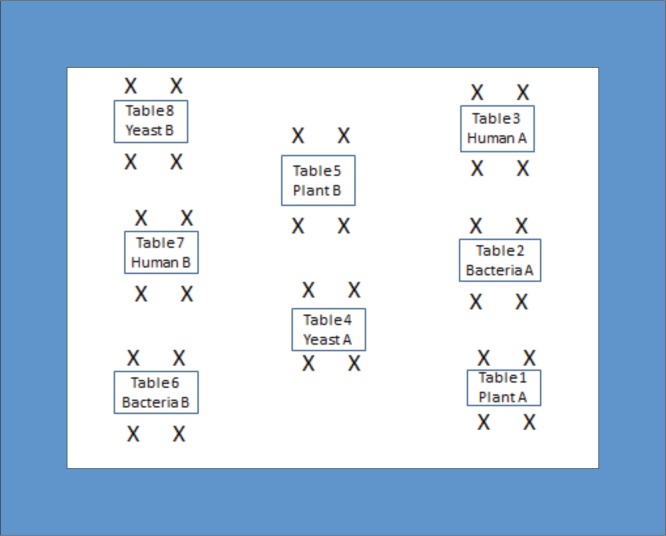
On the first day, the students, sitting in table groups of four (Fig. 2), are each given an instruction packet, which will be completed and submitted individually with the final cell model. Full student packets and instructions for four possible cell types (human, bacteria, yeast, and plant) are found in Appendix I. The students first determine the particular cell they will model by using clues in the packet (described later). They then find their matching cellular table group (Fig. 2), with whom they will later pursue simulated “cellular communication” and exchange of compounds. Once this is done, tasks are appointed for each member of the group, and construction of the draft cell model begins (Fig. 1).
FIGURE 2.
Eight table groups of four students each, identified by organism, and showing their matching tables used in participatory cellular communication and exchange.
On day two, groups continue drafting, while some members answer specific portions of the packet. Once the draft cell model is done, it is brought to the teacher by each group, and reviewed together by focusing on the model rubric (Fig. 1, C and D). Corrections are made as needed, or approval is given to start the final model on KELVIN DesignGrid paper. On the third day, everyone should be working on the final model, incorporating molecules and compounds showing the number of their partner table group (as shown in Fig. 2, using Tables 1 and 5 as an example). This takes a full period in itself. By day four, student consensus building needed to complete the packets and model drawings should be complete and finalized for submission (Fig. 2, step F).
Faculty instructions
As a first step, faculty should ensure familiarity with key organelles, their function and structure, respective reactants and products, and an understanding of how molecules transport across a membrane. This model supports a constructivist view of learning, and students are therefore expected to actively learn by integrating their work on the model with their prior understanding (1). Finally, teachers should note that, “students also have difficulty conceptualizing the relative and absolute sizes of cells that, in turn, results in confusion between cells, atoms, and molecules and has been shown to interfere with students’ development of a robust understanding of biological processes” (15). This is important as this model incorporates molecular reactions. Of course, there are other options for teaching middle school cell biology, as presented in Table 2.
TABLE 2.
A limited selection of cell projects illustrating both static and interactive platforms.
| Static Models | Format of Model | Model Rubric Included | Unique Features | Reference |
|
| ||||
| 1. Analytical model comparing cells to city functions | Paper sketch, followed by full model construction | Yes | Building analogies between cell and organelles, and an external environment of their choosing | Grady & Jeanpierre (6) |
| 2. A model of the ultrastructure of a cell | Plasticine model | No | Ability to make models to scale; includes starch and glycogen | Bushell (2) |
| 3. Cells as molecular factories | Factory function analogy table | No | How eukaryotic cell organelles cooperate to function as a protein-producing factory | Waldron (16) |
| 4. Silk batik cell model, using art and science | Batik model relies on assumption that abstract thinking supports better understanding of nature phenomena | Yes | Students depict organelles’ function through artwork | Dambekalns & Medina-Jerez (3) |
|
| ||||
| Interactive Models | ||||
|
| ||||
| 1. Cell exploration program | Uses haptic computer technology allowing manipulation of a virtual cell | No | Computer-based model allows for individual manipulations of the cell | Mingoue et al., (9) |
| 2. An inquiry-driven cell culture project | Use of living cells as a model for cell division and response of cells to various environments | No | Use cultured fibroblast cells to explore cell division/responses of cultured cells to environmental changes, student experiments | Palombi & Jagger (11) |
| 3. Using “The Cell: An Image Library” | Uses student cell drawings, and digital library explored on own by students | Digital presentation checklist | Enhanced output in revisions to original drawing; digital images for cells/organelles | Saunders & Taylor (13) |
Suggestions for determining student learning
Course grades were determined as follows: production of final cell model for one of four specific cell/organismal types (50%) and completion of student packet with individual responses to short- and long-response questions (50%). The percentage allotted for each table group to produce the final scale model includes up to 1% for individual student participation by taking an active role in consensus building. Successful completion of the model cell, positive participation, and evidence-based answers for the summative questions were used as indicators of learning. In addition, on the model rubric, there is a space for evaluating a “demonstrated understanding” of the cell, which is done orally at each table. Taken together, these grades provide evidence of student progress according to the three learning goals mentioned above.
Sample data
Models represent a simplified system focusing on features that explain certain scientific phenomena (14). Student participation was used to measure students’ active role in consensus building while designing the model. The extent to which the model helped students explain the functions of a living cell was determined from this student participation, the results of the model’s summative assessment, and the ability to sustain interest in the assignment over several class periods. Notes taken by the instructor on involvement in individual and group work were used to measure student participation, graded as a score of 10, 7, 5, 3, or 0, and forming a part of each student’s final grade as shown on the model’s rubric.
The participation rate was very high in this portion of the project, with 93% of students actively, productively, and collegially working either independently or together. The remaining 7% were often off task, sat back and let others do more of the work, or did much less than their partners. While these assessments demanded close attention by the teacher, roaming the tables also provided additional opportunities for answering questions, for individual discussions, and for helping those who were not working effectively. This task increased teacher interaction with individual students at their table groups, which was one of the factors identified in stimulating engagement. The percentages given above were averaged over three periods, indicating that the model was capable of sustaining a high level of interest and participation over several days.
Data from models and packets. The three learning goals set for this investigation are to provide evidence that living things are made of cells, to use a model to describe cell function as a system, and to understand the function of chemical reactions in food and energy transfer. These goals are evaluated by student activities and responses to questions found in both the student packet and the final model drawings. The first section below summarizes depictions of the model cells, their chemical reactions, and organelle functions. This is followed by a grade analysis of the packet as a whole, in which specific questions address the above goals not covered by the model drawings.
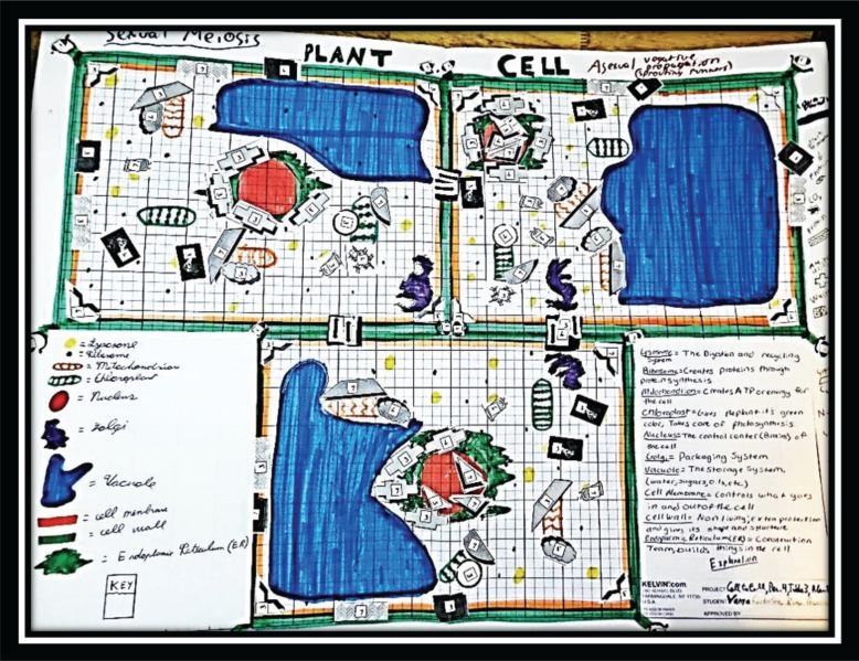
Assessing cell representations. Figures 3 to 5 present final cell models made for cell types covered in the 2013–14 school term. They include yeast, plant, and bacteria. As can be seen, the models display originality, for example, in the way the model was drawn and constructed, the way that organelles were identified, how chemical molecules were affixed to the correct organelle, and the way external and internal structures or molecules were added to the membrane. The placement of gap junctions or plasmodesmata was an example of modifications encouraged after students automatically first drew the entire membrane, and then went back and decided how to construct the junctions and the signal receptors. Each decision regarding the placement of organelles and molecules is the result of a group consensus process to improve the initial draft of the model.
FIGURE 3.
Student-generated cell model of plant cells.
FIGURE 5.
Student-generated model of bacterial cells.
The students carefully checked and revised their models to exclude organelles that cannot exist or chemical reactions that cannot take place in a particular cell type. While clues to the reactions were in the project’s student packet, whether or not they belong to a particular cell is not provided, leaving each student group to debate and decide on what reactions belong in their particular cell. Each group of students was also required to determine the mode of reproduction for their cell’s organism. This would include asexual budding (as in yeast), sexual reproduction (plant and animal), binary fission (as in bacteria), or vegetative propagation (as seen in the plant example of strawberry).
The organelles must have the correct molecules or reactants attached to them. For example, in the plant cell (Fig. 3), some of the chloroplasts show the symbols for the inputs (sunlight, water, and carbon dioxide), while others show the symbols for the outputs, oxygen and glucose. For the mitochondria, glucose and oxygen are shown as the inputs, and energy is the output. Protein synthesis occurs on the rough endoplasmic reticulum, where amino acids appear, followed by proteins. Numerous chemical molecules are shown entering or being exchanged between cells, coming through the plasmodesmata and the selectivity of the membrane.
As can be seen for the yeast model (Fig. 4), cells were separated from one another, having just split apart after budding. This is consistent with yeast being a unicellular organism. The same is true of bacteria, while in the animal and plant cells (Fig. 3), we see cells attached to one another, sharing material through their plasmodesmata. This furthers student understanding of cell-to-cell transport and recognition of both multicellular and unicellular organisms. The yeast cell also demonstrated respiratory reactions in the mitochondrion, placement of the cell wall, and clearly placed protein receptors on the membrane.
FIGURE 4.
Student-generated model of yeast cells.
The membrane receptors caused the greatest degree of confusion, as students searched for matching proteins that bind to receptors. Each figure reveals a different approach to this, with some of the figures failing to have clear signal receptors, others with the receptor inverted, some where the receptor had to be moved from floating off of the cell entirely, to being attached to the membrane. With practice, students became more comfortable with interrupting the membrane and inserting the transfer and communication junctions. At first, it was hard for students to erase work they had done to construct the cell membrane but their reluctance diminished once they saw how compounds flowed through these gaps.
Figure 5 depicts the model for the bacteria cell. Cells were shown individually, not attached to one another, as was the case for the unicellular organisms studied previously through the microscope. In this model students also drew the cell’s flagellum and pilus, and made clear that the DNA was not contained in a nucleus, thus recognizing it as a prokaryotic cell. They were careful to show the ribosomes, but did not include any membrane-bound organelles, such as the mitochondrion. Gap junctions allowing materials to flow through the cell wall and membrane are depicted as lines between the three cells.
Packet grade analysis. Data were summed from four to five classes per year (Table 1), with distinctions between on-level and advanced classes. While the average scores across five years for all classes were not statistically different, there was a significant difference (4.48) between those in honor or advanced classes and those in the grade level courses. Class grades shown include both advanced and grade level (indicated by an * in Table 1) science classes. Thus, while the project is equally amenable to both groups, there was a significant difference (p < 0.01) between the averages of the on-grade level/inclusion classes and the advanced classes. This suggests further scaffolding and modifications are required.
DISCUSSION
Field testing
Including the spring term of 2014, CE has now been taught for six years. Each year, substantial changes have been made, based on evaluations of student comments, advent of NGSS expectations, and student learning as observed in their packets and final models. The packets presented in the appendix are the most current, benefitting from extensive review and classroom use. Much has also been learned in terms of sequencing the CE lesson with those coming before and after it. The data presented here are compiled from earlier versions, as the packets available now were used for the first time in the spring term of 2014.
Evidence of student learning
Data, observations, and comparisons with other models attest to the validity of the CE model as an interactive representation of the living cell. This is especially evident in the quality, diversity, and organismal representation of the final cell models shown in Figures 3 to 5. Its value as a tool for learning was also seen in participation and grades from the model. As one example of such, students successfully placed the correct compounds needed either as reactants or products next to the correct organelle or membrane passageway as warranted by their specific cell type. A second example can be seen in the grade analysis, where many high grades were awarded, indicating successful completion of the longer-response questions dealing with the cell functioning as a system and what happens if an organelle is missing entirely from the cell.
It was also clear from the final scale models that seventh graders can rise to the occasion when challenged. Only if students are challenged by a curriculum do we know whether they can achieve above grade level. Given this fact, the author would have to carefully consider whether the CE model should be “scaled back” to be completely compliant with, rather than exceeding, student understanding as expressed by NGSS. Such scaling back would severely limit a comprehensive understanding of the parts of a cell, including its molecular reactions.
Potential modifications
Advancing curricular change poses challenges beyond just those of content selection. When new lessons or projects are added, others lessons and their sequencing must be changed or replaced. In this article, curricular change was effected to engage students with interactive models, respond to reform efforts of the NGSS, increase student inquiry, and incorporate a systems approach to the study of the cell. Some of the changes reviewed would be the same for introducing any type of interactive model, while some are specific to the needs of the CE model.
In our curriculum schedule, different approaches were noted for teaching photosynthesis, for example, from those for teaching organelles. Such reactions are mentioned briefly when learning organelles, with more exacting details in separate chapters on energy transfer. Structure and function of a chloroplast are introduced in the first quarter, while chemical reactions and photosystems are explained later, sometimes months later.
The same holds true for cellular respiration. While these reactions take place in the mitochondrion, varying levels of chemical detail for this reaction, as determined by student age and grade level, occur in different chapters, such as those tied to chapters on the human respiratory organ system. Here, the structure of the mitochondrion is related to specific respiration reactions, but little emphasis is placed on these reactions in relation to the cell as a system, its interacting organelles, and its neighboring cells.
This separation of structure, chemical reactions, and functions of organelles does not reinforce student understating of the different levels of organization and how they work together. As stated by Waldron (17), a student’s conception of cells is often “as a static structure consisting of multiple independent parts. Students often do not understand how the parts of the cell work together to accomplish the multiple functions of a dynamic living cell. Students also often confuse different levels of organization such as molecules, organelles and cells.”
When cell anatomy is separated from its chemical reactions, students often miss the opportunity to relate and manipulate these things together. Other research has shown that “students have difficulty in making the connection between molecular and cellular organization” (5). This separation of organization is reflected in the majority of models available for studying the cell (Table 2). The model described in this paper is an explicit attempt to interrelate these two levels of organization, plus that of the organelles (Table 3).
TABLE 3.
Separation of features of the Cell Encounter model project into static and interactive portions.
| Level of Cell Encounter Topic | Static portion of cell model | Interactive portion of cell model |
|---|---|---|
| 1. Organelles | Placement of organelles | Cell chemistry: reactants and products for specific organelles, with molecules coming from companion cell |
| 2. Membrane | Drawing membrane to scale on model drafting paper | Opening channels in the membrane for either gap junctions or plasmodesmata Attaching communication receptors molecules and finding their corresponding protein from companion cell |
| 3. Nucleus | Drawing nucleus and DNA as appropriate to cell type | Addition of nucleic acids for DNA replication Addition of mRNA to move out of nucleus to ribosome |
| 4. Ribosome, Endoplasmic Reticulum | Drawing rough ER to scale, attached to nucleus | Addition of amino acids to form proteins, with molecules coming from companion cell |
| 5. Biochemical processes | A. Placement of chloroplasts in correct cell type B. Placement of mitochondrion |
Affixing the inputs required for photosynthesis and then the outputs from the reaction, with both obtained from companion cell Affixing the inputs for cellular respiration and then ATP molecules as the energy output |
| 6. Cell wall | Drawing cell wall, where appropriate, outside of the membrane | Placement of molecules of cellulose for cell wall repair and growth. |
| 7. Waste removal | Drawing lysosome | Placing enzymes and water next to lysosome, as the primary molecules it contains. |
ER = endoplasmic reticulum; ATP = adenosine triphosphate
Additional challenges to the model could be added for senior or more advanced students. For example, adding more exacting details on transport, cellular recognition, and recognizing pathogenic agents, are all possible. Next, cells could join one another on a larger scale as tissue, then organs, and organ systems to link the model to the levels of organization. Finally, an oral presentation to the class from each group could be used as a summarizer and to help build needed skills in scientific communication as well.
SUPPLEMENTAL MATERIALS
Appendix 1: Student instructional packets for yeast, bacteria, human, and plant cells, including rubrics
ACKNOWLEDGMENTS
The author would like to acknowledge the generous time devoted to review and improvement of this unit by Dr. Stephen M. Wolniak, Professor, Department of Cell Biology and Molecular Genetics, University of Maryland, College Park. The author declares that there are no conflicts of interest.
Footnotes
Supplemental materials available at http://jmbe.asm.org
REFERENCES
- 1.Ausubel DP. Educational psychology: a cognitive view. Holt, Rinehart and Winston; New York, NY: 1968. [Google Scholar]
- 2.Bushell J. A model of the ultrastructure of a cell. J Biol Educ. 2001;35(3):152–153. doi: 10.1080/00219266.2001.9655765. [DOI] [Google Scholar]
- 3.Dambekalns L, Medina-Jerez W. Cell organelles and silk batik: a model for integrating art and science. Sci Scope. 2012;36(2):44–51. [Google Scholar]
- 4.diCarlo SE. Cell biology should be taught as science is practiced. Nature. 2006;7:290–295. doi: 10.1038/nrm1856. [DOI] [PubMed] [Google Scholar]
- 5.Driver R, Squires A, Rushworth P, Wood-Robinson V. Making sense of secondary science: research into children’s ideas. Routledge; London, UK, and New York, NY: 1994. p. 224. [Google Scholar]
- 6.Grady K, Jeanpierre B. Tried and true: population 75 trillion—cells, organelles, and their functions. Sci Scope. 2011;34(5):64–69. [Google Scholar]
- 7.Harris CJ, Rooks DL. Managing inquiry-based science: challenges in enacting complex science instruction in elementary and middle school classrooms. J Sci Teach Educ. 2010;21:227–240. doi: 10.1007/s10972-009-9172-5. [DOI] [Google Scholar]
- 8.Lehrer R, Schauble L. Cultivating model-based reasoning in science education. In: Sawyer K, editor. Cambridge handbook of the learning sciences. Cambridge University Press; Cambridge, MA: 2006. pp. 371–388. [Google Scholar]
- 9.Minogue J, Jones G, Broadwell B, Oppewal T. Exploring cells from the inside out: new tools for the classroom. Sci Scope. 2006;29(6):28–32. [Google Scholar]
- 10.NGSS Lead States . Next generation science standards: for states, by states. The National Academies Press; Washington, DC: 2013. [Google Scholar]
- 11.Palombi PS, Jagger KS. Learning about cells as dynamic entities: an inquiry-driven cell culture project. Bioscene. 2003;34(2):27–33. [Google Scholar]
- 12.Pollard TD. No question about exciting questions in cell biology. PLoS Biol. 2013;11(12):e1001734. doi: 10.1371/journal.pbio.1001734. [DOI] [PMC free article] [PubMed] [Google Scholar]
- 13.Saunders C, Taylor A. Close the textbook & open “The cell: an image library.”. Am. Biol. Teach. 2014;76(3):201–207. doi: 10.1525/abt.2014.76.3.9. [DOI] [Google Scholar]
- 14.Schwarz C, Passmore C. Preparing for NGSS: developing and using models. NSTA Web Seminars. 2012. Sep 25,
- 15.Tibell LEA, Rundgren C-J. Educational challenges of molecular life science: characteristics and implications for education and research. CBE Life Sci. Educ. 2010 Spring;9(1):25–33. doi: 10.1187/cbe.08-09-0055. [DOI] [PMC free article] [PubMed] [Google Scholar]
- 16.Waldron I. Cells as molecular factories. 2011. p. 2. Department of Biology, University of Pennsylvania, Philadelphia. [Online.] http://serendip.brynmawr.edu/exchange/bioactivities.
- 17.Waldron I. Cell Structure and function—major concepts and learning activities. 2013. pp. 1–6. [Online.] http://serendip.brynmawr.edu/exchange/bioactivities.
- 18.Wright R, Boggs J. Learning cell biology as a team: a project-based approach to upper division cell biology. Cell Biol Educ. 2002;1:145–153. doi: 10.1187/cbe.02-03-0006. [DOI] [PMC free article] [PubMed] [Google Scholar]
Associated Data
This section collects any data citations, data availability statements, or supplementary materials included in this article.
Supplementary Materials
Appendix 1: Student instructional packets for yeast, bacteria, human, and plant cells, including rubrics


