Abstract
The dorsal and ventral hippocampi are functionally and anatomically distinct. Recently, we reported that dorsal Cornu Ammonis area 1 (CA1) neurons have a more hyperpolarized resting membrane potential and a lower input resistance and fire fewer action potentials for a given current injection than ventral CA1 neurons. Differences in the hyperpolarization-activated cyclic nucleotide-gated cation conductance between dorsal and ventral neurons have been reported, but these differences cannot fully account for the different resting properties of these neurons. Here, we show that coupling of A1 adenosine receptors (A1ARs) to G-protein-coupled inwardly rectifying potassium (GIRK) conductance contributes to the intrinsic membrane properties of dorsal CA1 neurons but not ventral CA1 neurons. The block of GIRKs with either barium or the more specific blocker Tertiapin-Q revealed that there is more resting GIRK conductance in dorsal CA1 neurons compared with ventral CA1 neurons. We found that the higher resting GIRK conductance in dorsal CA1 neurons was mediated by tonic A1AR activation. These results demonstrate that the different resting membrane properties between dorsal and ventral CA1 neurons are due, in part, to higher A1AR-mediated GIRK activity in dorsal CA1 neurons.
Keywords: dorsal and ventral hippocampus, A1 adenosine receptors, GIRK
the rodent hippocampus can be divided into dorsal and ventral regions based on biochemical, anatomical, and behavioral observations (Dong et al. 2009; Fanselow and Dong 2010; Moser and Moser 1998; van Groen and Wyss 1990). Until recently, the electrophysiological properties of Cornu Ammonis area 1 (CA1) pyramidal neurons along the longitudinal hippocampal axis were often thought to be uniform. However, growing evidence demonstrates that intrinsic membrane properties of CA1 neurons from the dorsal and ventral hippocampus are significantly different (Dougherty et al. 2012; Marcelin et al. 2012a). For example, dorsal neurons have a more hyperpolarized resting membrane potential (Vm; RMP) and a lower input resistance (Rin). As such, they fire fewer action potentials in response to a given current injection compared with ventral neurons (Dougherty et al. 2012). Although functional hyperpolarization-activated, cyclic nucleotide-gated current (Ih) is different between dorsal and ventral CA1 neurons (Dougherty et al. 2013), the intrinsic membrane properties of these neurons cannot be fully explained by Ih-related differences.
Voltage-gated ion channels and neuronal morphology contribute to the intrinsic membrane properties of neurons (Hoffman et al. 1997; Magee 1998; Spruston 2008; Vetter et al. 2001). Differences in RMP and Rin between dorsal and ventral CA1 neurons may therefore be explained, in part, by the expression of voltage-gated ion channels. Given the more hyperpolarized Vm and lower Rin in dorsal neurons compared with ventral neurons, we hypothesized that dorsal CA1 neurons possess a greater resting potassium (K+) conductance than ventral CA1 neurons. Furthermore, inwardly rectifying K+ (IRK) channels and G-protein-coupled IRK (GIRK) channels are highly expressed in the CA1 pyramidal neurons (Drake et al. 1997; Karschin et al. 1996; Liao et al. 1996; Ponce et al. 1996). GIRK channels are activated by Gi/o-coupled metabotropic receptors and hyperpolarize the Vm and decrease Rin, providing a decrease in neuronal excitability (Andrade et al. 1986; Luscher et al. 1997; Mihara et al. 1987; North 1989).
In this study, we investigated the resting conductances in dorsal and ventral CA1 pyramidal neurons. We found that dorsal neurons have more barium (Ba2+)-sensitive conductance compared with ventral neurons. Application of Tertiapin-Q revealed that this greater Ba2+-sensitive conductance in dorsal CA1 neurons was mediated by GIRK channels. Finally, we found that this higher resting GIRK conductance in dorsal neurons was mediated by tonic activation of A1 adenosine receptors (A1ARs). Together, our results suggest that higher resting GIRK conductance activated by A1ARs is present in dorsal but not ventral CA1 neurons and contributes to intrinsic membrane properties of these neurons.
MATERIALS AND METHODS
Animals.
Hippocampal slices from the dorsal and ventral poles were prepared from 11- to 13-wk-old male Sprague-Dawley rats, housed two to three per cage on a 12-h light schedule (on 7 AM/off 7 PM) with ad libitum access to water and food. All procedures involving animals were approved by the University of Texas at Austin Institutional Animal Care and Use Committee.
Drugs.
Ba2+ chloride was purchased from Fisher Scientific (Pittsburgh, PA). Tertiapin-Q was purchased from Alomone Labs (Jerusalem, Israel). 1,3-Dipropyl-8-cyclopentylxanthine (DPCPX), ZD7288, 2-chloro-2′-C-methyl-N-6-cyclopentyladenosine (2′ME-CCPA), D-2-amino-5-phosphonovaleric acid (D-APV), Gabazine, CGP55845, and 6,7-dinitroquinoxaline-2,3-dione (DNQX) were purchased from Abcam (Cambridge, MA).
Slice preparation.
Rats were anesthetized with a lethal dose of a ketamine/xylazine mixture (90/10 mg/ml) and transcardially perfused with ice-cold artificial cerebral spinal fluid (aCSF) composed of (in mM) 2.5 KCl, 1.25 NaH2PO4, 25 NaHCO3, 0.5 CaCl2, 7 MgCl2, 7 dextrose, 210 sucrose, 1.3 ascorbic acid, and 3 sodium pyruvate, bubbled with 95% O2-5% CO2. The brain was then removed and hemisected along the longitudinal fissure. For dorsal hippocampal slices, a blocking cut was made at 20–30° from the posterior coronal plane and collected at the anterior end of the forebrain. Ventral hippocampal slices were made from a horizontal section. Hippocampal slices (300 μm thick) were made in ice-cold aCSF using a vibrating microtome (Microslicer DTK-Zero1; DSK, Kyoto, Japan). Slices were then transferred to a holding chamber for 30 min at 35°C containing (in mM) 125 NaCl, 2.5 KCl, 1.25 NaH2PO4, 25 NaHCO3, 2 CaCl2, 2 MgCl2, 12.5 dextrose, 1.3 ascorbic acid, and 3 sodium pyruvate, bubbled with 95% O2-5% CO2. Slices were then incubated for at least 45 min at room temperature.
Whole-cell current-clamp recordings.
Whole-cell current-clamp recordings were performed as described previously (Kim et al. 2012). Briefly, hippocampal slices were submerged in a recording chamber continuously perfused with aCSF containing synaptic blockers (D-APV, 25 μM; Gabazine, 2 μM; DNQX, 20 μM), heated to 32–34°C, flowing at a rate of 1–2 ml/min. aCSF contained (in mM) 125 NaCl, 2.5 KCl, 1.25 NaH2PO4, 25 NaHCO3, 2 CaCl2, 1 MgCl2, and 12.5 dextrose, bubbled with 95% O2-5% CO2. CA1 pyramidal neurons were visualized using a microscope (Axioskop; Carl Zeiss Microscopy, Thornwood, NY), fitted with differential interference contrast optics (Stuart et al. 1993). Patch pipettes (4–7 MΩ) were prepared with capillary glass (external diameter 1.65 mm; World Precision Instruments, Sarasota, FL) using a Flaming/Brown Micropipette Puller and filled with an internal solution containing (in mM) 120 K-gluconate, 20 KCl, 10 HEPES, 4 NaCl, 7 K2-phosphocreatine, 4 Mg-ATP, and 0.3 Na-GTP (pH 7.3 with KOH). Neurobiotin (Vector Laboratories, Burlingame, CA) was used (0.1–0.2%) for subsequent histological processing. Somatic whole-cell current-clamp recordings were performed using a BVC-700A amplifier (Dagan, Minneapolis, MN). Electrical signals were filtered at 5 kHz, sampled at 20 kHz, and digitized by an ITC-18 interface (HEKA Instruments, Bellmore, NY) connected to a computer running custom software written in IGOR Pro (WaveMetrics, Lake Oswego, OR). Experiments were terminated if the series resistance exceeded 30 MΩ. Vm were corrected for liquid junction potentials (−8 mV). For tertiapin experiments, we used log-order increases in concentration (0.03, 0.3, and 2 μM).
Immunohistochemistry.
Animals were anesthetized and perfused through the ascending aorta using ice-cold aCSF composed of (in mM) 2.5 KCl, 1.25 NaH2PO4, 25 NaHCO3, 0.5 CaCl2, 7 MgCl2, 7 dextrose, 210 sucrose, 1.3 ascorbic acid, and 3 sodium pyruvate, bubbled with 95% O2-5% CO2, followed by 4% paraformaldehyde (PFA) in PBS, described previously (Kim et al. 2012). The brain was removed and fixed overnight in 4% PFA and then transferred to 30% sucrose in PBS. Dorsal and ventral hippocampal slices were prepared on a cryostat and stored in cryoprotectant (30% sucrose, 30% ethylene glycol, 1% polyvinyl pyrrolidone, 0.05 M sodium phosphate buffer) until processing for immunohistochemistry. Dorsal and ventral slices were briefly rinsed in PBS buffer and incubated in 0.1% Triton X-100 for 30 min. Subsequently, slices were blocked in PBS solution containing 5% normal goat serum and 0.03% Triton X-100 for 1 h and then incubated in primary antibody diluted in blocking solution overnight at 4°C. Slices were rinsed in PBS buffer and then incubated in secondary antibody for 1 h at room temperature. Primary antibodies in this study were used as follows: rabbit anti-IRK channel (Kir)3.1 (1:200; Alomone Labs), rabbit anti-Kir3.2 (1:200; Alomone Labs), rabbit anti-A1ARs (1:200; Alomone Labs), and mouse anti-microtuble-associated protein 2 (1:1,000; Sigma-Aldrich, St. Louis, MO).
Data analysis.
Rin was determined by the slope of the linear fit of the voltage-current (V-I) plot from the steady-state voltage response to the injected current, ranging from +10 to −150 pA (750 ms). Firing rate as a function of input current (FI) curves were determined by plotting the number of action potentials against injected current (30–300 pA, for 750 ms in 30 pA intervals).
Statistical analyses.
All data are expressed as mean ± SE. Statistical comparisons were performed using ANOVA (one-factor or two-factor), followed by Bonferroni post hoc test or Student's t-test (paired or unpaired) with GraphPad Prism software. *P < 0.05 was considered statistically significant.
RESULTS
Contribution of Ba2+-sensitive conductance to intrinsic membrane properties of dorsal CA1 neurons.
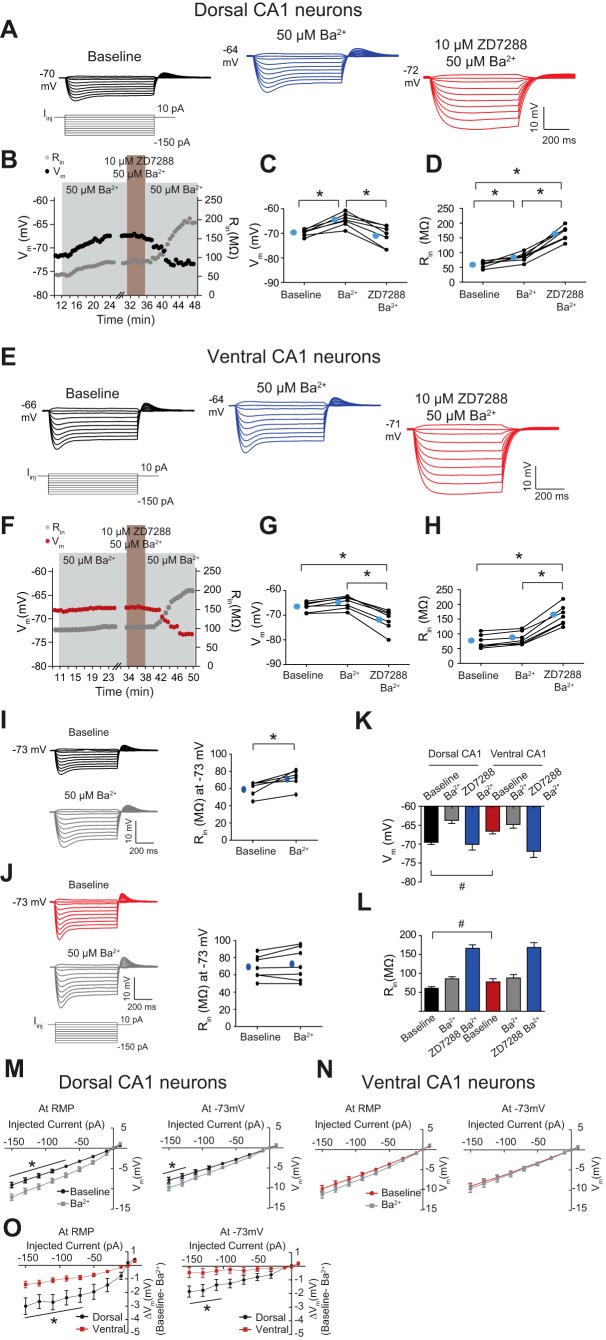
Our initial goal in this study was to explore the resting conductances that contribute to the intrinsic membrane properties of CA1 pyramidal neurons from the dorsal and ventral hippocampus. Although functional differences in Ih and distinct α-subunit expression profiles of hyperpolarization-activated, cyclic nucleotide-gated cations 1 and 2 (HCN1 and HCN2) were reported between dorsal and ventral CA1 neurons (Dougherty et al. 2013), Ih-related properties cannot fully account for the differences in intrinsic membrane properties between dorsal and ventral CA1 neurons (Dougherty et al. 2012). Therefore, we hypothesized that dorsal neurons might have a large, resting K+ conductance, contributed by Kir. A relatively low concentration of Ba2+ (50 μM) is known to block Kir channels but not many other K+ currents (Armstrong and Taylor 1980; Chen and Johnston 2005; Day et al. 2005; Jin et al. 1999; Quayle et al. 1988; Sah 1996; Wang et al. 1998). Because ZD7288 caused a gradual depolarization, 10–15 min after the hyperpolarization of Vm (not shown), we first applied Ba2+ and then added of ZD7288 for 4–5 min (Dougherty et al. 2013) before switching back to the Ba2+-containing aCSF. Vm and Rin at RMP were monitored during baseline application of 50 μM Ba2+ and addition of 10 μM ZD7288 application in dorsal (Fig. 1B) and ventral (Fig. 1F) CA1 pyramidal neurons. Ba2+ (50 μM) significantly depolarized the Vm in dorsal neurons (Fig. 1, A and C, and Table 1) but not in ventral neurons (Fig. 1, E and G, and Table 1). Addition of 10 μM ZD7288 produced a significant hyperpolarization from baseline in ventral neurons (Fig. 1, E and G, and Table 1) but not in dorsal neurons (Fig. 1, A and C, and Table 1). Rin at RMP for dorsal neurons was increased significantly (Fig. 1, A and D, and Table 1) but not for ventral neurons (Fig. 1, E and H, and Table 1) after 50 μM Ba2+ application. Addition of 10 μM ZD7288 led to a significant increase in Rin in dorsal (Fig. 1, A and D, and Table 1) and ventral (Fig. 1, E and H, and Table 1) neurons. For proper comparison between groups, Rin was measured at the same Vm. At a common Vm (−73 mV), 50 μM Ba2+ led to a significant increase in Rin for dorsal neurons (Fig. 1I and Table 2) but not for ventral neurons (Fig. 1J and Table 2). Vm (Fig. 1K and Table 2) and Rin (Fig. 1L and Table 2) were significantly different between dorsal and ventral neurons. This difference was absent when IRKs alone were blocked or were blocked in combination with Ih (Fig. 1, K and L, and Table 2). Because we observed significant changes in voltage response (Vm) to the injected current from RMP and at −73 mV in the presence of Ba2+ in dorsal neurons (Fig. 1, A and I) but not in ventral neurons (Fig. 1, E and J), steady-state V-I curves in control, Ba2+, and the difference between the two were plotted to illustrate better the Ba2+-sensitive conductance in the hyperpolarizing direction (Fig. 1, M–O). The V-I curves before and after Ba2+ application were significantly different in dorsal neurons (Fig. 1M) but not ventral neurons (Fig. 1N) from RMP and at −73 mV, suggesting a greater Ba2+-sensitive conductance in dorsal neurons.
Fig. 1.
Dorsal neurons show greater response to barium (Ba2+) compared with ventral Cornu Ammonis area 1 (CA1) neurons. A and E: representative voltage responses with step current commands at resting membrane potential (Vm; RMP). B and F: time courses of changes in Vm and input resistance (Rin) during successive Ba2+ (50 μM) and ZD7288 (10 μM) wash-in experiments in dorsal (B) and ventral (F) CA1 pyramidal neurons. C and D: dorsal CA1 pyramidal neurons showed a significantly depolarized Vm (C) and increased Rin (D) in the presence of Ba2+. Subsequent ZD7288 application resulted in a significantly hyperpolarized Vm (C) and an increased Rin (D). G and H: ventral CA1 pyramidal neurons showed no significant changes in Vm (G) and Rin (H) in the presence of Ba2+. Addition of ZD7288 led to a significantly hyperpolarized Vm (G) and an increased Rin (H). I and J, left: representative voltage responses with step current commands at −73 mV. Dorsal CA1 neurons showed a significantly increased Rin but not ventral CA1 neurons at a common Vm (−73 mV) in the presence of Ba2+. K and L: dorsal CA1 neurons showed a more hyperpolarized RMP (K) and a lowered Rin at RMP (L) compared with ventral CA1 neurons. There is no difference in Vm (K) and Rin (L) between dorsal and ventral neurons following successive Ba2+ and ZD7288 application. M and N: dorsal neurons showed significant changes in voltage-current (V-I) curve before and after 50 μM Ba2+ application (M) but not ventral neurons (N). O: the differences in V-I curves before and after 50 μM Ba2+ application are indicated. *P < 0.05; #P < 0.05 in dorsal vs. ventral groups. ΔVm, change in Vm; Iinj, injected current. Blue circles throughout figures represent mean number.
Table 1.
Subthreshold properties in successive 50 μM Ba2+ and 10 μM ZD7288 application (related to Fig. 1)
| Baseline | 50 μM Ba2+ wash-in | 10 μM ZD7288 50 μM Ba2+ wash-in | |
|---|---|---|---|
| Dorsal CA1 | |||
| Vm, mV | −69.7 ± 0.5 (n = 7) | −64.4 ± 1.0 (n = 7)* | −71.0 ± 0.7 (n = 7) |
| Rin, MΩ | 60.8 ± 4.1 (n = 7) | 85.7 ± 5.6 (n = 7)* | 166.4 ± 9.0 (n = 7)* |
| Ventral CA1 | |||
| Vm, mV | −66.5 ± 0.7 (n = 7) | −64.8 ± 1.0 (n = 7) | −71.9 ± 1.5 (n = 7)* |
| Rin, MΩ | 73.7 ± 8.3 (n = 7) | 84.8 ± 8.4 (n = 7) | 162.1 ± 12.7 (n = 7)* |
Ba2+, barium; CA1, Cornu Ammonis area 1; Vm, membrane potential; Rin, steady-state input resistance. Statistically significant (
P < 0.05, 1-way ANOVA, followed by Bonferroni post hoc test compared with baseline).
Table 2.
Change in Rin at a common membrane potential (−73 mV) in dorsal and ventral CA1 neurons after 50 μM Ba2+ wash-in experiments (related to Fig. 1)
| Dorsal CA1 | Baseline | 50 μM Ba2+ | Ventral CA1 | Baseline | 50 μM Ba2+ |
|---|---|---|---|---|---|
| Rin, MΩ, at −73 mV | 60.0 ± 3.5 (n = 6) | 72.1 ± 4.2* (n = 6) | Rin, MΩ, at −73 mV | 69.1 ± 5.1 (n = 7) | 72.6 ± 7.2 (n = 7) |
Statistically significant (
P < 0.05, paired t-test compared with baseline).
Comparison of basal GIRK activity between ventral and dorsal CA1 neurons.
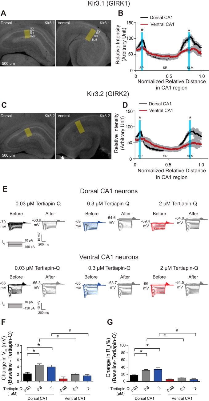
Given the observation that significant changes in Vm and Rin (at RMP and at a common Vm) were observed following blockade of a Ba2+-sensitive conductance in dorsal neurons (Fig. 1), we next examined which ion conductance is responsible for the resting Ba2+-sensitive conductance. Kir are blocked by low concentrations of Ba2+ (Chen and Johnston 2005; Gerber et al. 1991; Standen and Stanfield 1978; Williams et al. 1988), whereas Tertiapin-Q is a specific GIRK channel blocker at nanomolar concentration (Jin et al. 1999). Because GIRK1 (Kir3.1) and GIRK2 (Kir3.2) heterotetrameric channels are expressed predominantly in the brain (Liao et al. 1996), we performed an immunohistochemical staining with antibodies against GIRK1 and GIRK2 in the dorsal and ventral hippocampus. To account for the longer radial axis in the CA1 region of the ventral hippocampus (Dougherty et al. 2012), the quantification of GIRK1 and GIRK2 protein expression was normalized by distance (Fig. 2, A and C). Expression of GIRK1 and GIRK2 was higher in the somatic and distal dendritic regions of dorsal CA1 compared with ventral CA1 (Fig. 2, B and D; P < 0.05, n = 4). Because of these differences, we tested whether dorsal neurons are more responsive to the specific GIRK channel blocker, Tertiapin-Q, compared with ventral neurons. We first used three different concentrations of Tertiapin-Q (0.03, 0.3, and 2 μM) and measured changes in RMP and Rin in dorsal and ventral neurons. Changes in Vm and Rin (at RMP) were significantly affected by bath application of 0.3 μM Tertiapin-Q compared with the 0.03-μM Tertiapin-Q wash-in group in dorsal neurons (Fig. 2, F and G, and Table 3) but not in ventral neurons (Fig. 2, F and G, and Table 3). There were no further changes in Vm (Fig. 2, F and G, and Table 3) and Rin (at RMP; Fig. 2, F and G, and Table 3) following bath application of 2 μM Tertiapin-Q in dorsal and ventral neurons compared with the 0.3-μM Tertiapin-Q wash-in group. Group comparisons showed that there were significant differences in the changes in Vm (Fig. 2, F and G, and Table 3) and Rin (Fig. 2, F and G, and Table 3) between dorsal and ventral neurons after either 0.3 or 2 μM Tertiapin-Q application. Based on these results, all subsequent Tertiapin-Q experiments were performed using 0.3 μM.
Fig. 2.
Dorsal CA1 neurons show a more basal G-protein-coupled inwardly rectifying potassium (GIRK) activity than ventral CA1 neurons. A and C: representative dorsal and ventral hippocampal slices immunolabeled with antibodies against IRK channel (Kir)3.1 (A; GIRK1) and Kir3.2 (C; GIRK2). Yellow boxes depict the region of the slice used for quantification of signal intensity (B and D). Quantification of Kir3.1 (B) and Kir3.2 (D) protein expression from the perisomatic region to the distal dendritic region of CA1 from the dorsal and ventral hippocampus. The protein expression of Kir3.1 (B) and Kir3.2 (D) was increased significantly in the somatic and distal dendritic regions of CA1 from the dorsal hippocampus compared with the ventral hippocampus. Vertical blue bars highlight part of the CA1 radial axis that shows significant differences in Kir3.1 (B) and Kir3.2 (D) protein expression between the dorsal and ventral CA1 region. E: representative voltage responses with step current commands at RMP. F and G: Tertiapin-Q (0.3 μM) significantly changed Vm and Rin in dorsal but not ventral neurons. Tertiapin-Q (2 μM) produced similar effects on Vm and Rin compared with 0.3 μM Tertiapin-Q wash-in in dorsal and ventral neurons. Dorsal CA1 neurons showed significant changes in Vm and Rin in the presence of 0.3 or 2 μM Tertiapin-Q compared with ventral CA1 neurons. *P < 0.05; #P < 0.05 vs. ventral group. SO, stratum oriens; SP, stratum pyramidale; SR, stratum radiatum; SLM, stratum lacunosum moleculare.
Table 3.
Dose-response Tertiapin-Q experiment in dorsal and ventral CA1 neurons (related to Fig. 2)
| 0.03 μM Tertiapin-Q |
0.3 μM Tertiapin-Q |
2 μM Tertiapin-Q |
||||
|---|---|---|---|---|---|---|
| Baseline | Tertiapin | Baseline | Tertiapin | Baseline | Tertiapin | |
| Dorsal CA1 | ||||||
| Vm, mV | −69.9 ± 1.0 (n = 7) | −67.8 ± 1.1 (n = 7) | −69.0 ± 0.5 (n = 7) | −64.4 ± 0.5* (n = 7) | −69.4 ± 0.9 (n = 6) | −65.4 ± 0.9* (n = 6) |
| Rin, MΩ | 60.2 ± 2.8 (n = 7) | 70.5 ± 3.5 (n = 7) | 62.9 ± 1.2 (n = 7) | 82.6 ± 2.2* (n = 7) | 64.5 ± 3.9 (n = 6) | 85.6 ± 4.5* (n = 6) |
| Ventral CA1 | ||||||
| Vm, mV | −65.3 ± 1.0 (n = 6) | −64.7 ± 1.4 (n = 6) | −64.9 ± 0.5 (n = 6) | −63.0 ± 0.7 (n = 6) | −64.1 ± 0.8 (n = 6) | −62.6 ± 0.9 (n = 6) |
| Rin, MΩ | 82.5 ± 4.8 (n = 6) | 87.2 ± 5.8 (n = 6) | 87.3 ± 3.0 (n = 6) | 95.8 ± 2.7 (n = 6) | 85.0 ± 4.8 (n = 6) | 89.1 ± 3.9 (n = 6) |
Statistically significant (
P < 0.0167, 1-way ANOVA with Bonferroni post hoc test compared with baseline).
The Ba2+-sensitive conductance in dorsal CA1 neurons is mostly mediated by GIRKs.
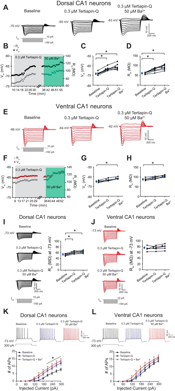
Because Tertiapin-Q changed RMP and Rin in dorsal but not ventral CA1 neurons (Fig. 2), we next sought to determine whether the Ba2+-sensitive conductance in dorsal neurons was mediated by GIRKs. Vm and Rin (at RMP) were monitored during successive 0.3 μM Tertiapin-Q and 50 μM Ba2+ application in dorsal (Fig. 3B) and ventral (Fig. 3F) neurons. Application of Tertiapin-Q significantly depolarized (Fig. 3, A and C, and Table 4) and increased Rin (Fig. 3, A and D, and Table 4) of dorsal neurons. In contrast, Tertiapin-Q had no significant effect on ventral neurons (Fig. 3, G and H, and Table 4). There was no further change in either dorsal or ventral neurons when 50 μM Ba2+ was subsequently added to the bath (dorsal: Fig. 3, C and D; ventral: Fig. 3, G and H, and Table 4). Blockade of both GIRK and Ba2+-sensitive conductances led to a significant depolarization (dorsal: Fig. 3, A and C; ventral: Fig. 3, E and G, and Table 4) and an increased Rin (dorsal: Fig. 3, A and D; ventral: Fig. 3, E and H, and Table 4) in dorsal and ventral neurons. At a common Vm (−73 mV), a change in Rin for dorsal neurons was increased significantly in the presence of 0.3 μM Tertiapin-Q (Fig. 3I and Table 5) but not for ventral neurons (Fig. 3J and Table 5). When subsequent 50 μM Ba2+ was applied after blockade of GIRK conductance, there was no further change in Rin in dorsal (Fig. 3I and Table 5) and ventral (Fig. 3J and Table 5) neurons. When both GIRK and Ba2+-sensitive conductances were blocked, Rin for dorsal neurons (Fig. 3I and Table 5) was increased significantly but not for ventral neurons (Fig. 3J and Table 5). Action potentials evoked in response to depolarizing current steps (30–300 pA in 30 pA increments for 750 ms) were determined during successive 0.3 μM Tertiapin-Q and 50 μM Ba2+ application at a common Vm (−73 mV) in dorsal (Fig. 3K) and ventral (Fig. 3L) neurons. As expected, Tertiapin-Q application increased action-potential firing significantly in dorsal neurons (Fig. 3K) but had no effect on firing in ventral neurons (Fig. 3L) compared with baseline. Addition of 50 μM Ba2+ had no further effects on firing in either dorsal (Fig. 3K) or ventral (Fig. 3L) neurons.
Fig. 3.
Ba2+-Sensitive conductance-mediated changes in Vm and Rin stem from GIRK conductance in dorsal CA1 neurons. A and E: representative voltage responses with step current commands at RMP are shown. B and F: time courses of changes in Vm and Rin during successive 0.3 μM Tertiapin-Q and 50 μM Ba2+ application in dorsal (B) and ventral (F) CA1 pyramidal neurons. C and D: dorsal CA1 neurons showed a significantly depolarized Vm (C) and an increased Rin (D) following bath application of Tertiapin-Q (0.3 μM). Subsequent blockade of Ba2+-sensitive conductance showed no significant changes in Vm (C) and Rin (D) in dorsal CA1 neurons. G and H: ventral CA1 neurons showed no significant changes in Vm and Rin in the presence of Tertiapin-Q (0.3 μM). Further changes in Vm and Rin were not observed following blockade of Ba2+-sensitive conductance in ventral neurons. Blockade of Tertiapin-Q and Ba2+-sensitive conductance produced a significantly depolarized Vm and increased Rin compared with baseline in ventral neurons. I and J, left: representative voltage responses with step current commands at −73 mV. Dorsal CA1 neurons showed a significantly increased Rin (I) but not ventral CA1 neurons (J) at a common Vm (−73 mV) following successive Tertiapin-Q and Ba2+ application. K and L: representative Vm responses with depolarizing current steps (300 pA for 750 ms) at a common Vm (−73 mV). K: dorsal neurons showed significant increases in action-potential (AP) firing after either Tertiapin-Q or Tertiapin-Q + Ba2+ application but not ventral neurons (L). *P < 0.05.
Table 4.
Subthreshold properties in subsequent Tertiapin-Q and Ba2+ wash-in experiments (related to Fig. 3)
| Baseline | 0.3 μM Tertiapin-Q wash-in | 0.3 μM Tertiapin-Q 50 μM Ba2+ wash-in | |
|---|---|---|---|
| Dorsal CA1 | |||
| Vm, mV | −68.0 ± 0.6 (n = 7) | −63.3 ± 0.7 (n = 7)* | −61.2 ± 1.1 (n = 7)* |
| Rin, MΩ | 62.4 ± 1.5 (n = 7) | 80.5 ± 3.6 (n = 7)* | 92.7 ± 4.7 (n = 7)* |
| Ventral CA1 | |||
| Vm, mV | −64.6 ± 0.5 (n = 5) | −62.9 ± 0.6 (n = 5) | −61.4 ± 0.6 (n = 5)* |
| Rin, MΩ | 86.9 ± 3.0 (n = 5) | 95.0 ± 3.4 (n = 5) | 102 ± 1.9 (n = 5)* |
Statistically significant (
P < 0.0167, 1-way ANOVA with Bonferroni post hoc test compared with baseline).
Table 5.
Change in Rin at a common membrane potential (−73 mV) in dorsal and ventral CA1 neurons in subsequent Tertiapin-Q and Ba2+ wash-in experiments (related to Fig. 3)
| Baseline | 0.3 μM Tertiapin-Q wash-in | 0.3 μM Tertiapin-Q 50 μM Ba2+ wash-in | |
|---|---|---|---|
| Dorsal CA1 | |||
| Rin, MΩ, at −73 mV | 52.7 ± 1.3 (n = 6) | 60.5 ± 1.6 (n = 6)* | 62.8 ± 1.7 (n = 6)* |
| Ventral CA1 | |||
| Rin, MΩ, at −73 mV | 72.5 ± 1.6 (n = 5) | 74.1 ± 3.4 (n = 5) | 78.1 ± 4.1 (n = 5) |
Statistically significant (
P < 0.0167, 1-way ANOVA with Bonferroni post hoc test compared with baseline).
Comparison of basal GABAB receptor activity in dorsal and ventral CA1 neurons.
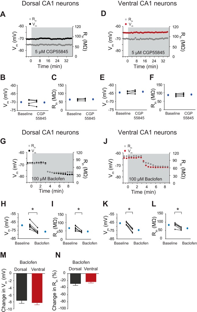
The data presented thus far suggest that there was more basal GIRK activity in dorsal vs. ventral neurons (Figs. 2 and 3). We next examined whether there was more basal activation of neurotransmitter receptors known to couple with GIRK channels in dorsal vs. ventral neurons. In CA1 neurons, the activation of GABAB receptors, A1ARs, and 5-hydroxytryptamine 1A (5-HT1A) serotonin receptors results in hyperpolarization of RMP by GIRK channels (Luscher et al. 1997). Thus it is possible that there is more activity in any one of these receptor systems in dorsal compared with ventral neurons. We first examined RMP and Rin in dorsal and ventral neurons during bath application of a potent and selective GABAB receptor antagonist, CGP52432 (5 μM) (Lanza et al. 1993). There was no significant change in Vm and Rin (at RMP), up to 30–35 min after wash-in of CGP55845 in either dorsal (Fig. 4, B and C, and Table 6) or ventral (Fig. 4, E and F, and Table 6) neurons. Because we did not observe any changes in Vm and Rin after blockade of GABAB receptors in dorsal or ventral neurons, we next applied a GABAB receptor agonist, baclofen, to test for the presence of functional GABAB receptors. Vm and Rin (at RMP) were monitored during 100 μM baclofen application in dorsal (Fig. 4G) and ventral (Fig. 4J) neurons. Baclofen significantly changed Vm (dorsal: Fig. 4H; ventral: Fig. 4K, and Table 6) and Rin (dorsal: Fig. 4I; ventral: Fig. 4L, and Table 6) in both dorsal and ventral neurons. The magnitude of changes in Vm (dorsal: −7.6 ± 0.8 mV vs. ventral: −8.2 ± 0.6 mV; P > 0.05, n = 6; Fig. 4M) and Rin at RMP (dorsal: −28.3 ± 5.2% vs. ventral: −23.2 ± 3.6%; P > 0.05, n = 6; Fig. 4N) was not significantly different after activation of GABAB receptors.
Fig. 4.
Similar changes in Vm and Rin in the presence of GABAB receptor antagonist and agonist in dorsal and ventral CA1 neurons. A and D: time courses of changes in Vm and Rin during CGP55845 (5 μM) wash-in experiments in dorsal (A) and ventral (D) neurons. Dorsal (B and C) and ventral (E and F) neurons showed no changes in Vm and Rin in the presence of CGP55845. G and J: time courses of Vm and Rin in the presence of baclofen in dorsal (G) and ventral (J) neurons. Dorsal (H and I) and ventral (K and L) CA1 neurons showed a significantly hyperpolarized Vm and a decreased Rin following activation of GABAB receptors. M and N: the changes in Vm and Rin were not significantly different in dorsal and ventral CA1 neurons in the presence of baclofen. *P < 0.05.
Table 6.
Subthreshold membrane properties in 5 μM CGP55845 or 100 μM baclofen wash-in experiments (related to Fig. 4)
| Dorsal CA1 | Baseline | 5 μM CGP55845 | Ventral CA1 | Baseline | 5 μM CGP55845 |
|---|---|---|---|---|---|
| Vm, mV | −70.0 ± 0.8 (n = 4) | −70.2 ± 0.9 (n = 4) | Vm, mV | −64.7 ± 0.4 (n = 4) | −64.2 ± 0.5 (n = 4) |
| Rin, MΩ | 62.7 ± 2.7 (n = 4) | 64.5 ± 3.3 (n = 4) | Rin, MΩ | 86.4 ± 4.0 (n = 4) | 89.6 ± 4.0 (n = 4) |
| Dorsal CA1 | Baseline | 100 μM Baclofen | Ventral CA1 | Baseline | 100 μM Baclofen |
|---|---|---|---|---|---|
| Vm, mV | −69.4 ± 1.6 (n = 6) | −76.6 ± 1.0* (n = 6) | Vm, mV | −66.1 ± 0.7 (n = 6) | −74.4 ± 1.0* (n = 6) |
| Rin, MΩ | 66.6 ± 5.7 (n = 6) | 46.5 ± 2.1* (n = 6) | Rin, MΩ | 83.2 ± 5.7 (n = 6) | 63.1 ± 3.4* (n = 6) |
Statistically significant (
P < 0.05, Student's paired t-test compared with baseline).
Effect of A1AR-mediated activation on GIRK conductance in dorsal CA1 neurons.
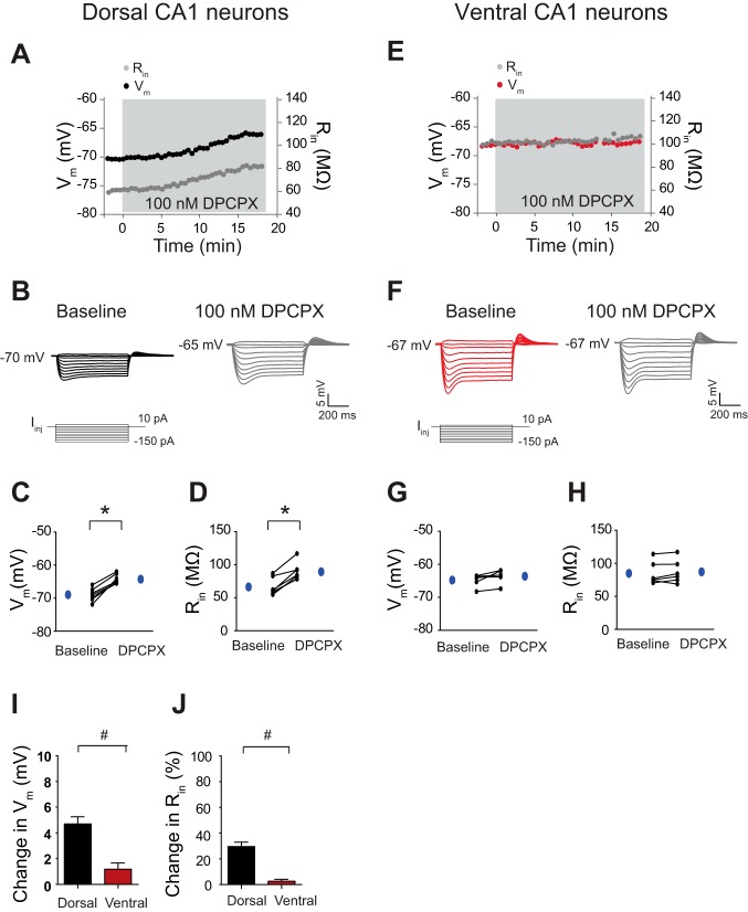
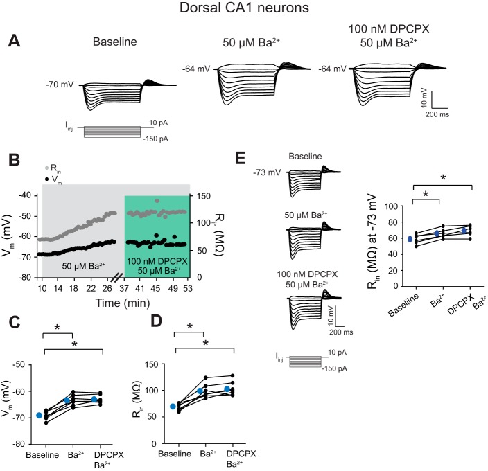
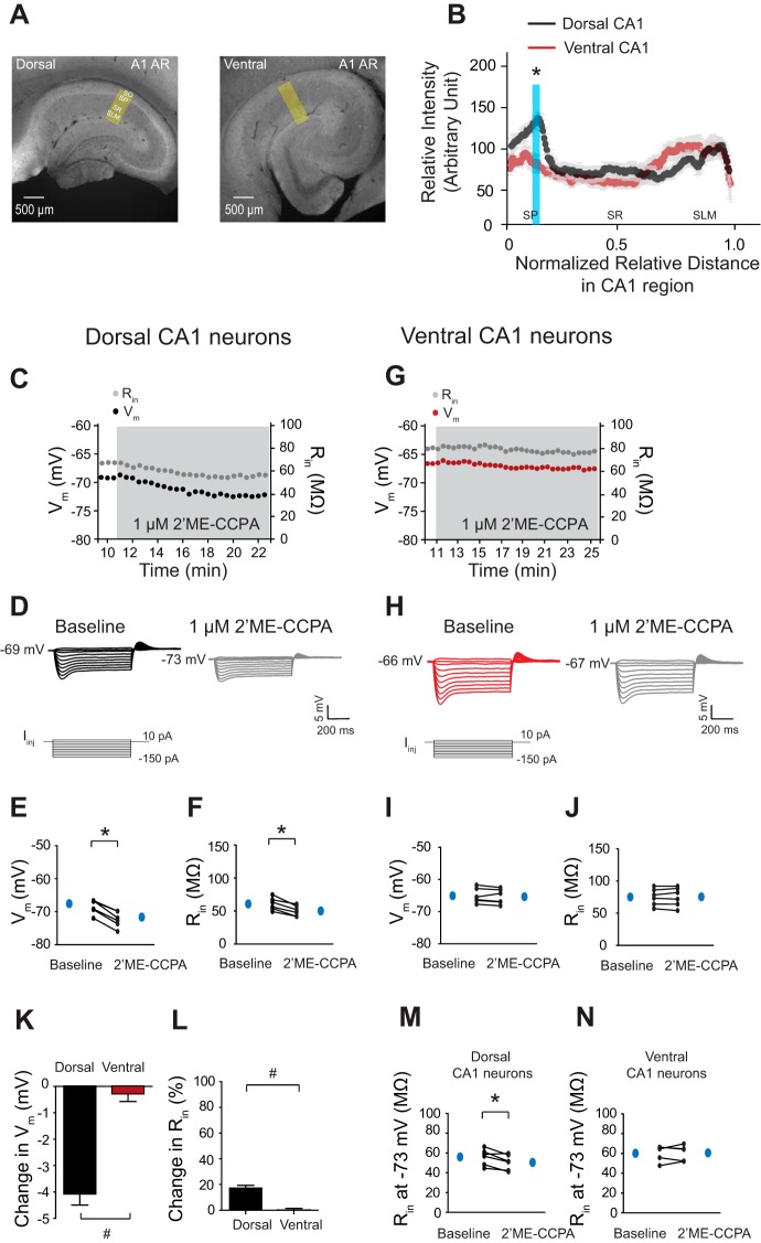
We next examined whether A1ARs are involved in the activation of the resting GIRK conductance in these neurons. DPCPX is a selective A1AR antagonist (Alzheimer et al. 1989). Vm and Rin (at RMP) were monitored during 100 nM DPCPX wash-in in dorsal (Fig. 5A) and ventral (Fig. 5E) neurons. Dorsal neurons had significantly depolarized Vm (Fig. 5, B and C, and Table 7) and an increased Rin at RMP (Fig. 5, B and D, and Table 7) after blockade of A1ARs, whereas no changes were observed in ventral neurons (Fig. 5, G and H, and Table 7). The magnitude of changes in Vm (dorsal: 4.7 ± 0.5 mV vs. ventral: 1.1 ± 0.5 mV; P < 0.05; Fig. 5I) and Rin at RMP (dorsal: 29.5 ± 3.4% vs. ventral: 2.6 ± 1.4%; P < 0.05; Fig. 5J) was significantly greater for dorsal neurons than for ventral neurons after blockade of A1ARs. Because the activation of GIRK conductance is mediated by A1ARs in the hippocampal CA1 neurons (Luscher et al. 1997), we therefore performed successive Ba2+ and DPCPX wash-in experiments to examine whether the effects of DPCPX were occluded by Ba2+ in dorsal neurons. Vm and Rin (at RMP) were monitored during baseline, application of 50 μM Ba2+, and addition of 100 nM DPCPX application in dorsal neurons (Fig. 6B). Ba2+ (50 μM) led to a significant depolarization (Fig. 6, A and C, and Table 8) and an increased Rin (Fig. 6, A and D, and Table 8) in dorsal neurons. Addition of 100 nM DPCPX had no further effects on Vm (Fig. 6, A and C, and Table 8) and Rin (Fig. 6, A and D, and Table 8) in dorsal neurons. At a common Vm (−73 mV), dorsal neurons had a significantly increased Rin after 50 μM Ba2+ application (Fig. 6E and Table 8). When DPCPX was subsequently applied in dorsal neurons, Rin at −73 mV was not further changed (Fig. 6E and Table 8). Given the observation that ventral neurons showed no changes in Vm and Rin (at RMP) after blockade of A1ARs, immunohistochemical staining with antibody against A1ARs was carried out to examine whether ventral CA1 regions express A1ARs. The quantification of A1AR protein expression was again normalized by distance. The A1AR protein expression was highly expressed in the somatic region in the dorsal CA1 region but not in the ventral CA1 region (Fig. 7, A and B; P < 0.05, n = 3). Because there was a significant difference in A1AR protein expression in the somatic region of dorsal and ventral CA1, we thus examined the effects by 2′ME-CCPA, a selective A1 receptor agonist. Vm and Rin (at RMP) were monitored during 1 μM 2′ME-CCPA application in the dorsal (Fig. 7C) and ventral (Fig. 7G) CA1 neurons. 2′ME-CCPA (1 μM) significantly hyperpolarized the Vm (Fig. 7, D and E, and Table 9) and decreased Rin at RMP (Fig. 7, D and F, and Table 9) in dorsal but not ventral neurons (Fig. 7, H–J, and Table 9). The magnitude of changes in Vm (dorsal: −3.9 ± 0.3 mV vs. ventral: −0.3 ± 0.3 mV; P < 0.05, n = 6; Fig. 7K) and Rin at RMP (dorsal: 17.2 ± 2.0% vs. ventral 0.2 ± 1.2%; P < 0.05, n = 6; Fig. 7L) was significantly greater for dorsal neurons than ventral neurons after activation of A1ARs. At a common Vm (−73 mV), Rin for dorsal neurons was decreased significantly after activation of A1ARs (Fig. 7M and Table 9) but not for ventral neurons (Fig. 7N and Table 9).
Fig. 5.
Dorsal CA1 neurons showed greater responses to A1 adenosine receptor (A1AR) antagonist compared with ventral CA1 neurons. A and E: time course of changes in Vm and Rin during 1,3-dipropyl-8-cyclopentylxanthine (DPCPX; 100 nM) wash-in experiments in dorsal (A) and ventral (E) neurons. B and F: representative voltage responses with step current commands at RMP are shown. C and D: DPCPX (100 nM) caused a significant depolarization in dorsal neurons (C) and an increased Rin (D). G and H: ventral neurons showed no changes in Vm (G) and Rin (H) after DPCPX (100 nM) wash-in. I and J: the changes in Vm (I) and Rin (J) after 100 nM DPCPX wash-in were greater for dorsal neurons than ventral neurons. *P < 0.05; #P < 0.05 vs. ventral group.
Table 7.
Subthreshold membrane properties in 100 nM DPCPX wash-in experiments (related to Fig. 5)
| Dorsal CA1 | Baseline | DPCPX | Ventral CA1 | Baseline | DPCPX |
|---|---|---|---|---|---|
| RMP, mV | −69.0 ± 0.7 (n = 7) | −64.2 ± 0.5* (n = 7) | RMP, mV | −64.7 ± 0.7 (n = 6) | −63.6 ± 0.8 (n = 6) |
| Rin, MΩ | 66.4 ± 4.9 (n = 7) | 89.3 ± 5.0* (n = 7) | Rin, MΩ | 84.8 ± 7.0 (n = 6) | 87.2 ± 7.2 (n = 6) |
DPCPX, 1,3-dipropyl-8-cyclopentylxanthine; RMP, resting Vm. Statistically significant (
P < 0.05, Student's paired t-test compared with baseline).
Fig. 6.
Dorsal CA1 neurons have an A1AR-mediated GIRK conductance. A: representative voltage responses with step current commands at RMP. B: time courses of changes in Vm and Rin during successive 50 μM Ba2+ and 100 nM DPCPX in dorsal CA1 neurons. Dorsal CA1 neurons showed a significantly depolarized Vm (C) and an increased Rin (D) at RMP in the presence of Ba2+. Subsequent bath application of DPCPX had no further effects on Vm (C) and Rin (D) at RMP in dorsal neurons. E, left: representative voltage responses with step current commands at −73 mV. Successive Ba2+ and DPCPX application led to a significantly increased Rin at a common Vm (−73 mV). *P < 0.05.
Table 8.
Subthreshold membrane properties in successive Ba2+ and DPCPX wash-in experiments (related to Fig. 6)
| Dorsal CA1 | Baseline | 50 μM Ba2+ wash-in | 100 nM DPCPX 50 μM Ba2+ wash-in |
|---|---|---|---|
| Vm, mV | −68.5 ± 0.6 (n = 7) | −62.8 ± 0.7 (n = 7)* | −62.4 ± 0.6 (n = 7)* |
| Rin, MΩ | 69.8 ± 2.5 (n = 7) | 99.7 ± 4.9 (n = 7) | 103.7 ± 5.0 (n = 7)* |
| Rin, MΩ, at −73 mV | 58.8 ± 2.4 (n = 7) | 65.8 ± 2.2 (n = 7) | 69.1 ± 2.6 (n = 7)* |
Statistically significant (
P < 0.0167, 1-way ANOVA with Bonferroni post hoc test compared with baseline).
Fig. 7.
A1ARs and their physiological responses predominantly exist in dorsal but not in ventral CA1 neurons. A: representative dorsal and ventral hippocampal slices immunolabeled with antibodies against A1ARs. Yellow boxes depict the region of the slice used for quantification of signal intensity. B: quantification of A1AR protein expression from the perisomatic region to the distal dendritic region of CA1 from the dorsal and ventral hippocampus. The protein expression of A1ARs was highly present in the somatic CA1 region of the dorsal hippocampus but not in the ventral CA1 region. The blue shade indicates a significant difference in A1AR protein expression between the dorsal and ventral CA1 region. C and G: time courses of Vm and Rin during 2-chloro-2′-C-methyl-N-6-cyclopentyladenosine (2′ME-CCPA; 1 μM) wash-in experiments in dorsal (C) and ventral (G) CA1 neurons. D and H: representative voltage responses with step current commands at RMP. E and F: dorsal CA1 neurons showed a significant hyperpolarization (E) and a decreased Rin (F) at RMP in the presence of 2′ME-CCPA (1 μM). I and J: ventral CA1 neurons showed no changes in Vm (I) and Rin (J) at RMP after 2′ME-CCPA (1 μM) wash-in. K and L: the changes in Vm (K) and Rin (L) after 2′ME-CCPA (1 μM) application were greater for dorsal CA1 neurons than ventral CA1 neurons. M and N: activation of A1ARs resulted in a significantly decreased Rin at a common Vm (−73 mV) in dorsal (M) but not in ventral (N) CA1 neurons. *P < 0.05; #P < 0.05 vs. ventral group.
Table 9.
Subthreshold membrane properties in 2′ME-CCPA wash-in experiments (related to Fig. 7)
| Dorsal CA1 | Baseline | 2′ME-CCPA | Ventral CA1 | Baseline | 2′ME-CCPA |
|---|---|---|---|---|---|
| Vm, mV | −68.5 ± 0.8 (n = 6) | −72.3 ± 0.9* (n = 6) | Vm, mV | −65.0 ± 0.9 (n = 6) | −65.3 ± 0.94 (n = 6) |
| Rin, MΩ | 60.8 ± 3.8 (n = 6) | 50.2 ± 3.2* (n = 6) | Rin, MΩ | 75.2 ± 5.4 (n = 6) | 75.4 ± 6.0 (n = 6) |
| Rin, MΩ, at −73 mV | 56.0 ± 3.3 (n = 6) | 50.5 ± 3.1* (n = 6) | Rin, MΩ, at −73 mV | 60.2 ± 3.6 (n = 5) | 60.6.±3.1 (n = 5) |
2′ME-CCPA, 2-chloro-2′-C-methyl-N-6-cyclopentyladenosine. Statistically significant (
P < 0.05, Student's paired t-test compared with baseline).
DISCUSSION
In this study, we examined the resting conductances that contribute to the different intrinsic membrane properties in dorsal and ventral CA1 neurons. We found that there was more Ba2+-sensitive conductance in dorsal than in ventral CA1 neurons. Furthermore, Ba2+-dependent changes in Vm and Rin were due to block of GIRK channels. We also found that this resting GIRK conductance was mediated by A1ARs in dorsal CA1 neurons but not in ventral CA1 neurons.
Until recently, the intrinsic electrophysiological properties of CA1 pyramidal neurons along the longitudinal hippocampal axis were assumed to be uniform. However, we and others have recently reported significant differences in the intrinsic electrophysiological properties between dorsal and ventral neurons (Dougherty et al. 2012; Marcelin et al. 2012a). Although differences in the expression of some voltage-gated ion channels between dorsal and ventral CA1 neurons have been reported (Dougherty et al. 2013; Marcelin et al. 2012b), the ionic mechanisms underlying the different intrinsic properties remain unclear. Because dorsal CA1 neurons showed a lower somatic Rin compared with ventral CA1 neurons, this could be due to more dendritic surface area for dorsal than ventral neurons (Dougherty et al. 2013), although Marcelin et al. (2012a, b) reported that somatic and dendritic capacitances were not different between dorsal and ventral neurons. A more hyperpolarized RMP in dorsal neurons compared with ventral neurons, however, could be due to dorsal CA1 neurons possessing a relatively larger resting K+ conductance, which could also contribute to a decrease in Rin. Indeed, low micromolar application of Ba2+ that blocks Kir (Chen and Johnston 2005) had significant effects on Vm and Rin (at RMP and at a common Vm −73 mV) for dorsal neurons but not for ventral neurons. In addition, the changes in V-I curves were greater for dorsal neurons than ventral neurons. It should be noted that higher concentrations of extracellular Ba2+ (>200 μM) block A-type K+ channels (Gasparini et al. 2007), whereas low concentrations of Ba2+ (<200 μM) are known to block Kir (Chen and Johnston 2005; Gerber et al. 1991; Standen and Stanfield 1978; Williams et al. 1988). Furthermore, when 10 μM ZD7288 was applied after blockade of Ba2+-sensitive conductance, we observed that the change in Vm for ventral neurons was larger than dorsal neurons, consistent with more Ih in ventral CA1 neurons (Dougherty et al. 2013). Interestingly, the absolute values of Vm and Rin were similar between dorsal and ventral neurons after blockade of Ba2+-sensitive conductance and Ih, suggesting a larger Ba2+-sensitive conductance in dorsal neurons compared with ventral neurons and more Ih in ventral CA1 neurons compared with dorsal CA1 neurons. Both Ba2+-sensitive conductance and Ih might account for these differences on intrinsic membrane properties between dorsal and ventral CA1 neurons.
Two classes of Kir are highly expressed in hippocampal neurons: IRK channels (Kir2.x) and GIRK channels (Kir3.x) (Chen and Johnston 2005; Karschin et al. 1996; Luscher et al. 1997). Both IRK and GIRK channels undergo voltage-dependent intracellular Mg2+ block, which imparts a rapid hyperpolarization-activated gating mechanism to these channels, and they become blocked and unblocked with depolarization and hyperpolarization, respectively. Because dorsal CA1 neurons showed a larger response to a relatively low concentration of Ba2+ than ventral CA1 neurons, we used a specific GIRK channel blocker, Tertiapin-Q (Jin et al. 1999), in an attempt to identify molecular targets responsible for the resting Ba2+-sensitive conductance. Interestingly, we found that dorsal neurons showed significant changes in Vm and Rin (at RMP and at a common Vm −73 mV) but not ventral neurons after Tertiapin-Q application, indicating that there was more basal GIRK conductance. Furthermore, when action potentials were elicited from a somatic current injection at a common Vm (−73 mV), Tertiapin-Q application significantly increased action-potential firing in dorsal neurons but not in ventral neurons. In agreement with these results, the protein expression of GIRK1 and GIRK2 was enriched significantly in somatic and dendritic CA1 regions of the dorsal hippocampus compared with the ventral hippocampus. The somatic and dendritic protein expression of GIRK subunits in the hippocampus has also been observed by other groups (Liao et al. 1996). Most interestingly, successive Tertiapin-Q and Ba2+ application revealed that a greater Ba2+-sensitive conductance for dorsal CA1 neurons stems from the GIRK conductance.
Hippocampal GIRK channels are activated by a number of neurotransmitter systems, including GABAB receptors, serotonergic 5-HT1A receptors, and A1ARs (Andrade et al. 1986; Luscher et al. 1997; North 1989; North et al. 1987). Given that ventral neurons have a weaker GABAergic fast synaptic inhibition than dorsal CA1 neurons (Papatheodoropoulos et al. 2002), it is possible that there is more GABAB-mediated, slow tonic inhibition, which could activate more GIRK conductance in dorsal neurons compared with ventral neurons. We observed, however, that there are similar changes in Vm and Rin (at RMP) after activation of GABAB receptors in dorsal and ventral neurons, suggesting that the coupling of GABAB receptors to GIRK channels might be similar. Lee et al. (1983) reported that the dorsal CA1 region had a greater A1AR density, resulting in the suppression of extracellular excitatory postsynaptic potentials in the presence of N6-cyclohexyladenosine, a selective A1AR agonist, compared with the ventral CA1 region. In accordance with these results, somatic whole-cell current-clamp recordings revealed that dorsal CA1 neurons showed significant changes in Vm and Rin in the presence of the A1AR agonist or antagonist but not in ventral neurons, suggesting again that A1ARs are more heavily expressed in dorsal CA1 neurons. Consistent with these results, the protein expression of A1AR was highly present in the somatic CA1 region of the dorsal hippocampus compared with the ventral hippocampus. Interestingly, when A1ARs were blocked in dorsal neurons, the absolute values of Vm and Rin at RMP were similar to ventral neurons. Furthermore, successive Ba2+ and DPCPX application revealed that the activation of GIRK conductance is mediated by A1ARs in dorsal CA1 neurons. These results suggest that A1AR-mediated GIRK conductance might contribute to intrinsic membrane properties in dorsal CA1 neurons.
Because GIRK and the adenosinergic system play important roles in the pathophysiology of diseases, such as epilepsy, depression, and anxiety, our results suggest that dorsal CA1 neurons/region (predominantly expressing A1AR-mediated GIRK conductance) might be important for chronic neurological disorders compared with the ventral hippocampus. GIRK channels play an important role in regulating neuronal excitability (Signorini et al. 1997). The deletion of Kir3.2 (GIRK2) channels led to an increase in susceptibility to a convulsant agent and showed sporadic seizures (Signorini et al. 1997). In the present study, we found that there is a more basal GIRK activity (i.e., physiological responses to a specific GIRK channel blocker at RMP) in dorsal CA1 neurons compared with ventral CA1 neurons, which might contribute to different susceptibility to a convulsant agent within the hippocampus. Adenosinergic systems have been implicated in anxiety and depression. In preclinical studies, activation of A1ARs led to antidepressant- and anxiolytic-like behaviors, whereas blockade of A1ARs resulted in an increase in anxiety (Kaster et al. 2004; Maximino et al. 2011; Prediger et al. 2006). In a clinical study, large amounts of caffeine, a nonselective A1AR antagonist, are known to cause anxiety and depression in normal and vulnerable subjects (Broderick and Benjamin 2004). Ligand-stimulated Gi/o protein-coupled receptors, such as A1ARs, induce an inhibition of adenylyl cyclase, which results in a decrease in cAMP concentration (van Calker et al. 1978). Because HCN channels are modulated by cAMP, resulting in an increase in Ih (Wainger et al. 2001), stimulation of an A1AR-induced decrease in cAMP levels might be interacting with HCN channels.
Summary.
In summary, we have investigated the resting conductances that contribute to intrinsic membrane properties of CA1 pyramidal neurons from the dorsal and ventral hippocampus. We found that dorsal CA1 neurons possess more basal activity of GIRK conductance, which was activated by A1ARs. Our results suggest that A1AR-mediated GIRK conductance contributes to the different intrinsic membrane properties in dorsal and ventral CA1 pyramidal neurons.
GRANTS
Support for this study was provided by grants from the National Institute of Neurological Disorders and Stroke (Grants NS084473 and NS077477 to D. Johnston).
DISCLOSURES
The authors declare no competing financial interests.
AUTHOR CONTRIBUTIONS
Author contributions: C.S.K. and D.J. conception and design of research; C.S.K. performed experiments; C.S.K. analyzed data; C.S.K. and D.J. interpreted results of experiments; C.S.K. prepared figures; C.S.K. drafted manuscript; C.S.K. and D.J. edited and revised manuscript; C.S.K. and D.J. approved final version of manuscript.
ACKNOWLEDGMENTS
The authors thank Drs. Rick Gray, Brian Kalmbach, and Darrin Brager for reviewing the manuscript and their comments throughout this study. The authors also thank Drs. Randy Chitwood, Kelly Dougherty, and Ann Clemens for helpful comments.
REFERENCES
- Alzheimer C, Sutor B, ten Bruggencate G. Transient and selective blockade of adenosine A1-receptors by 8-cyclopentyl-1,3-dipropylxanthine (DPCPX) causes sustained epileptiform activity in hippocampal CA3 neurons of guinea pigs. Neurosci Lett 99: 107–112, 1989. [DOI] [PubMed] [Google Scholar]
- Andrade R, Malenka RC, Nicoll RA. A G protein couples serotonin and GABAB receptors to the same channels in hippocampus. Science 234: 1261–1265, 1986. [DOI] [PubMed] [Google Scholar]
- Armstrong CM, Taylor SR. Interaction of barium ions with potassium channels in squid giant axons. Biophys J 30: 473–488, 1980. [DOI] [PMC free article] [PubMed] [Google Scholar]
- Broderick P, Benjamin AB. Caffeine and psychiatric symptoms: a review. J Okla State Med Assoc 97: 538–542, 2004. [PubMed] [Google Scholar]
- Chen X, Johnston D. Constitutively active G-protein-gated inwardly rectifying K+ channels in dendrites of hippocampal CA1 pyramidal neurons. J Neurosci 25: 3787–3792, 2005. [DOI] [PMC free article] [PubMed] [Google Scholar]
- Day M, Carr DB, Ulrich S, Ilijic E, Tkatch T, Surmeier DJ. Dendritic excitability of mouse frontal cortex pyramidal neurons is shaped by the interaction among HCN, Kir2, and Kleak channels. J Neurosci 25: 8776–8787, 2005. [DOI] [PMC free article] [PubMed] [Google Scholar]
- Dong HW, Swanson LW, Chen L, Fanselow MS, Toga AW. Genomic-anatomic evidence for distinct functional domains in hippocampal field CA1. Proc Natl Acad Sci USA 106: 11794–11799, 2009. [DOI] [PMC free article] [PubMed] [Google Scholar]
- Dougherty KA, Islam T, Johnston D. Intrinsic excitability of CA1 pyramidal neurones from the rat dorsal and ventral hippocampus. J Physiol 590: 5707–5722, 2012. [DOI] [PMC free article] [PubMed] [Google Scholar]
- Dougherty KA, Nicholson DA, Diaz L, Buss EW, Neuman KM, Chetkovich DM, Johnston D. Differential expression of HCN subunits alters voltage-dependent gating of h-channels in CA1 pyramidal neurons from dorsal and ventral hippocampus. J Neurophysiol 109: 1940–1953, 2013. [DOI] [PMC free article] [PubMed] [Google Scholar]
- Drake CT, Bausch SB, Milner TA, Chavkin C. GIRK1 immunoreactivity is present predominantly in dendrites, dendritic spines, and somata in the CA1 region of the hippocampus. Proc Natl Acad Sci USA 94: 1007–1012, 1997. [DOI] [PMC free article] [PubMed] [Google Scholar]
- Fanselow MS, Dong HW. Are the dorsal and ventral hippocampus functionally distinct structures? Neuron 65: 7–19, 2010. [DOI] [PMC free article] [PubMed] [Google Scholar]
- Gasparini S, Losonczy A, Chen X, Johnston D, Magee JC. Associative pairing enhances action potential back-propagation in radial oblique branches of CA1 pyramidal neurons. J Physiol 580: 787–800, 2007. [DOI] [PMC free article] [PubMed] [Google Scholar]
- Gerber U, Stevens DR, McCarley RW, Greene RW. Muscarinic agonists activate an inwardly rectifying potassium conductance in medial pontine reticular formation neurons of the rat in vitro. J Neurosci 11: 3861–3867, 1991. [DOI] [PMC free article] [PubMed] [Google Scholar]
- Hoffman DA, Magee JC, Colbert CM, Johnston D. K+ Channel regulation of signal propagation in dendrites of hippocampal pyramidal neurons. Nature 387: 869–875, 1997. [DOI] [PubMed] [Google Scholar]
- Jin W, Klem AM, Lewis JH, Lu Z. Mechanisms of inward-rectifier K+ channel inhibition by Tertiapin-Q. Biochemistry 38: 14294–14301, 1999. [DOI] [PubMed] [Google Scholar]
- Karschin C, Dissmann E, Stuhmer W, Karschin A. IRK(1–3) and GIRK(1–4) inwardly rectifying K+ channel mRNAs are differentially expressed in the adult rat brain. J Neurosci 16: 3559–3570, 1996. [DOI] [PMC free article] [PubMed] [Google Scholar]
- Kaster MP, Rosa AO, Rosso MM, Goulart EC, Santos AR, Rodrigues AL. Adenosine administration produces an antidepressant-like effect in mice: evidence for the involvement of A1 and A2A receptors. Neurosci Lett 355: 21–24, 2004. [DOI] [PubMed] [Google Scholar]
- Kim CS, Chang PY, Johnston D. Enhancement of dorsal hippocampal activity by knockdown of HCN1 channels leads to anxiolytic- and antidepressant-like behaviors. Neuron 75: 503–516, 2012. [DOI] [PMC free article] [PubMed] [Google Scholar]
- Lanza M, Fassio A, Gemignani A, Bonanno G, Raiteri M. CGP 52432: a novel potent and selective GABAB autoreceptor antagonist in rat cerebral cortex. Eur J Pharmacol 237: 191–195, 1993. [DOI] [PubMed] [Google Scholar]
- Lee KS, Schubert P, Reddington M, Kreutzberg GW. Adenosine receptor density and the depression of evoked neuronal activity in the rat hippocampus in vitro. Neurosci Lett 37: 81–85, 1983. [DOI] [PubMed] [Google Scholar]
- Liao YJ, Jan YN, Jan LY. Heteromultimerization of G-protein-gated inwardly rectifying K+ channel proteins GIRK1 and GIRK2 and their altered expression in weaver brain. J Neurosci 16: 7137–7150, 1996. [DOI] [PMC free article] [PubMed] [Google Scholar]
- Luscher C, Jan LY, Stoffel M, Malenka RC, Nicoll RA. G Protein-coupled inwardly rectifying K+ channels (GIRKs) mediate postsynaptic but not presynaptic transmitter actions in hippocampal neurons. Neuron 19: 687–695, 1997. [DOI] [PubMed] [Google Scholar]
- Magee JC. Dendritic hyperpolarization-activated currents modify the integrative properties of hippocampal CA1 pyramidal neurons. J Neurosci 18: 7613–7624, 1998. [DOI] [PMC free article] [PubMed] [Google Scholar]
- Marcelin B, Liu Z, Chen Y, Lewis AS, Becker A, McClelland S, Chetkovich DM, Migliore M, Baram TZ, Esclapez M, Bernard C. Dorsoventral differences in intrinsic properties in developing CA1 pyramidal cells. J Neurosci 32: 3736–3747, 2012a. [DOI] [PMC free article] [PubMed] [Google Scholar]
- Marcelin B, Lugo JN, Brewster AL, Liu Z, Lewis AS, McClelland S, Chetkovich DM, Baram TZ, Anderson AE, Becker A, Esclapez M, Bernard C. Differential dorso-ventral distributions of Kv4.2 and HCN proteins confer distinct integrative properties to hippocampal CA1 pyramidal cell distal dendrites. J Biol Chem 287: 17656–17661, 2012b. [DOI] [PMC free article] [PubMed] [Google Scholar]
- Maximino C, Lima MG, Olivera KR, Picanco-Diniz DL, Herculano AM. Adenosine A1, but not A2, receptor blockade increases anxiety and arousal in zebrafish. Basic Clin Pharmacol Toxicol 109: 203–207, 2011. [DOI] [PubMed] [Google Scholar]
- Mihara S, North RA, Surprenant A. Somatostatin increases an inwardly rectifying potassium conductance in guinea-pig submucous plexus neurones. J Physiol 390: 335–355, 1987. [DOI] [PMC free article] [PubMed] [Google Scholar]
- Moser MB, Moser EI. Functional differentiation in the hippocampus. Hippocampus 8: 608–619, 1998. [DOI] [PubMed] [Google Scholar]
- North RA. Twelfth Gaddum memorial lecture. Drug receptors and the inhibition of nerve cells. Br J Pharmacol 98: 13–28, 1989. [DOI] [PMC free article] [PubMed] [Google Scholar]
- North RA, Williams JT, Surprenant A, Christie MJ. mu and delta Receptors belong to a family of receptors that are coupled to potassium channels. Proc Natl Acad Sci USA 84: 5487–5491, 1987. [DOI] [PMC free article] [PubMed] [Google Scholar]
- Papatheodoropoulos C, Asprodini E, Nikita I, Koutsona C, Kostopoulos G. Weaker synaptic inhibition in CA1 region of ventral compared to dorsal rat hippocampal slices. Brain Res 948: 117–121, 2002. [DOI] [PubMed] [Google Scholar]
- Ponce A, Bueno E, Kentros C, Vega-Saenz de Miera E, Chow A, Hillman D, Chen S, Zhu L, Wu MB, Wu X, Rudy B, Thornhill WB. G-Protein-gated inward rectifier K+ channel proteins (GIRK1) are present in the soma and dendrites as well as in nerve terminals of specific neurons in the brain. J Neurosci 16: 1990–2001, 1996. [DOI] [PMC free article] [PubMed] [Google Scholar]
- Prediger RD, da Silva GE, Batista LC, Bittencourt AL, Takahashi RN. Activation of adenosine A1 receptors reduces anxiety-like behavior during acute ethanol withdrawal (hangover) in mice. Neuropsychopharmacology 31: 2210–2220, 2006. [DOI] [PubMed] [Google Scholar]
- Quayle JM, Standen NB, Stanfield PR. The voltage-dependent block of ATP-sensitive potassium channels of frog skeletal muscle by caesium and barium ions. J Physiol 405: 677–697, 1988. [DOI] [PMC free article] [PubMed] [Google Scholar]
- Sah P. Ca(2+)-Activated K+ currents in neurones: types, physiological roles and modulation. Trends Neurosci 19: 150–154, 1996. [DOI] [PubMed] [Google Scholar]
- Signorini S, Liao YJ, Duncan SA, Jan LY, Stoffel M. Normal cerebellar development but susceptibility to seizures in mice lacking G protein-coupled, inwardly rectifying K+ channel GIRK2. Proc Natl Acad Sci USA 94: 923–927, 1997. [DOI] [PMC free article] [PubMed] [Google Scholar]
- Spruston N. Pyramidal neurons: dendritic structure and synaptic integration. Nat Rev Neurosci 9: 206–221, 2008. [DOI] [PubMed] [Google Scholar]
- Standen NB, Stanfield PR. A potential- and time-dependent blockade of inward rectification in frog skeletal muscle fibres by barium and strontium ions. J Physiol 280: 169–191, 1978. [DOI] [PMC free article] [PubMed] [Google Scholar]
- Stuart GJ, Dodt HU, Sakmann B. Patch-clamp recordings from the soma and dendrites of neurons in brain slices using infrared video microscopy. Pflugers Arch 423: 511–518, 1993. [DOI] [PubMed] [Google Scholar]
- van Calker D, Muller M, Hamprecht B. Adenosine inhibits the accumulation of cyclic AMP in cultured brain cells. Nature 276: 839–841, 1978. [DOI] [PubMed] [Google Scholar]
- van Groen T, Wyss JM. Extrinsic projections from area CA1 of the rat hippocampus: olfactory, cortical, subcortical, and bilateral hippocampal formation projections. J Comp Neurol 302: 515–528, 1990. [DOI] [PubMed] [Google Scholar]
- Vetter P, Roth A, Hausser M. Propagation of action potentials in dendrites depends on dendritic morphology. J Neurophysiol 85: 926–937, 2001. [DOI] [PubMed] [Google Scholar]
- Wainger BJ, DeGennaro M, Santoro B, Siegelbaum SA, Tibbs GR. Molecular mechanism of cAMP modulation of HCN pacemaker channels. Nature 411: 805–810, 2001. [DOI] [PubMed] [Google Scholar]
- Wang HS, Pan Z, Shi W, Brown BS, Wymore RS, Cohen IS, Dixon JE, McKinnon D. KCNQ2 and KCNQ3 potassium channel subunits: molecular correlates of the M-channel. Science 282: 1890–1893, 1998. [DOI] [PubMed] [Google Scholar]
- Williams JT, North RA, Tokimasa T. Inward rectification of resting and opiate-activated potassium currents in rat locus coeruleus neurons. J Neurosci 8: 4299–4306, 1988. [DOI] [PMC free article] [PubMed] [Google Scholar]


