Abstract
The expression of testicular genes following acute heat stress has been reported in layer-type roosters, but few similar studies have been conducted on broilers. This study investigated the effect of acute heat stress on the gene expression in the testes of a broiler-type strain of Taiwan country chickens. Roosters were subjected to acute heat stress (38°C) for 4 h, and then exposed to 25°C, with testes collected 0, 2, and 6 h after the cessation of heat stress, using non-heat-stressed roosters as controls (n = 3 roosters per group). The body temperature and respiratory rate increased significantly (p<0.05) during the heat stress. The numbers of apoptotic cells increased 2 h after the acute heat stress (79 ± 7 vs. 322 ± 192, control vs. heat stress; p<0.05), which was earlier than the time of increase in layer-type roosters. Based on a chicken 44 K oligo microarray, 163 genes were found to be expressed significantly different in the testes of the heat-stressed chickens from those of the controls, including genes involved in the response to stimulus, protein metabolism, signal transduction, cell adhesion, transcription, and apoptosis. The mRNA expressions of upregulated genes, including HSP25, HSP90AA1, HSPA2, and LPAR2, and of downregulated genes, including CDH5, CTNNA3, EHF, CIRBP, SLA, and NTF3, were confirmed through quantitative real-time polymerase chain reaction (qRT-PCR). Moreover, numerous transcripts in the testes exhibited distinct expressions between the heat-stressed broiler-type and layer-type chickens. We concluded that the transcriptional responses of testes to acute heat stress may differ between the broiler-type and layer-type roosters. Whether the differential expression patterns associate with the heat-tolerance in the strains require a further exploration.
Introduction
A high ambient temperature impairs spermatogenesis and leads to low fertility through a decline of the sperm count, motility, and fertilization rate, as well as an elevation of abnormal cells in domestic animals [1–3]. To understand the adverse effect of high temperature on spermatogenesis, cellular and molecular mechanisms in response to heat stress have been studied [4–8]. Overproduced radical oxygen species that is induced through heat stress has been shown to cause oxidative stress and lead to apoptosis in spermatogenic cells, particularly in spermatocytes [4,7]. Moreover, large amounts of radical oxygen species have been shown to interfere with the integrity of sperm DNA and thereby influence embryo development [7]. A microarray was used to explore the expressions of testicular genes in mouse testes, and the expression levels of genes associated with DNA repair and recombination, protein synthesis, protein folding, and cell cycle reduced after heat stress [5]. Conversely, the expressions of heat-shock protein family genes were augmented against heat stress in mouse testes. Furthermore, a number of genes in testes were shown to be differentially expressed between heat-resistant and heat-sensitive murine strains after heat exposure [6]. Kim et al. [8] argued that multiple mechanisms were involved in heat-shock-induced subfertility or infertility.
In poultry, an environmental temperature of 32 to 35°C has been shown to cause poor fertility through impairing sperm penetration, uterovaginal sperm storage, seminal plasma, and intracellular ion concentrations [9–11]. Because of the subtropical climate of Taiwan, summers are typically characterized by high temperature (up to 38°C) and high humidity [12]. Various local breeds of chicken preserved at National Chung Hsing University exhibit a heat resistance superior to that of foreign breeds [13]. The rectal temperature and blood pH value of Taiwan country chickens (TCCs) were lower than those of broilers when they were subjected to 38°C acute heat stress [14]. A previous study showed that genes participating in response to stress, transport, signal transduction, and metabolism in the testes of L2-strain chickens, a layer-type TCCs, changed after acute heat stress [15]. However, no research has explored the transcriptional responses of genes to acute heat stress in the testes of broilers. In this study, broiler-type chickens of the local breed B-strain TCCs were used to investigate the alteration of global gene expression after exposure to acute heat stress in their testes. The result showed that genes related to heat shock protein family, cell adhesion, transcription, development, and apoptosis were differentially expressed in the testes of heat-stressed B-strain TCCs.
Materials and Methods
Experimental animals and ethics statements
Twelve 45-week-old roosters of the B-strain TCC, bred for meat production by National Chung Hsing University [16], were randomly selected and used in this study. The care and use of all animals were complied with the guidelines approved by the Institutional Animal Care and Use Committee (IACUC) of National Chung Hsing University, Taiwan, ROC (IACUC Permit NO. 99–02). Before treatment, roosters were stored in a climate chamber for 2 weeks as an adaptation period under the following conditions: a 14/10-h light/dark photoperiod, 25°C, and 55% relative humidity (RH). Feed and water were provided ad libitum.
Conditions of acute heat stress
The heat treatment was performed according to the procedure of a previous study [15]. After the adaptation period, 9 roosters received acute heat stress (38°C, 55% RH, 4 h) followed by recovery (25°C). Subsequently, the testes of the heat-stressed roosters were collected after 0, 2, and 6 h of recovery and treated as 3 independent groups (H4R0, H4R2, and H4R6, respectively; n = 3 in each group). Controls (n = 3) were maintained at 25°C and 55% RH. Body temperature and respiration rate were recorded during the treatment. For microarray analysis, testis samples were immediately collected and cut into small pieces. The samples were frozen in liquid nitrogen and stored at—80°C until RNA isolation. To prepare tissue sections for apoptosis analysis, the samples were sliced into small pieces, fixed in Bouin’s solution for 24 h, placed into 70% ethanol, and then dehydrated and embedded in paraffin wax.
Sample collection and preparation
A total of 5 μg of RNA was reverse transcribed using moloney murine leukemia virus reverse transcriptase (Promega, Madison, WI, USA). A total of 0.2 μg of RNA from each sample was used to synthesize second-strand complementary DNA (cDNA), which was amplified using a Quick-Amp Labeling kit (Agilent Technologies, Santa Clara, CA, USA). The cDNA was stored at −80°C until further microarray and quantitative real-time polymerase chain reaction (qRT-PCR) analyses.
Gene expressions through microarray analysis
RNA samples from 3 animals in each group were separately used for microarray hybridization as a biological triplicate in each treatment. Briefly, cDNA was used as a target for cRNA labeling Cy3-CTP (CyDye, Agilent Technologies) and hybridized with a chicken 44 K oligo microarray (Agilent Technologies) [17]. Using the cDNA as a template enabled labeling the target cRNA. A total of 1.65 μg of Cy3-labled cRNA was fragmented to an average size of approximately 50 to 100 nucleotides through incubation in a fragmentation buffer at 60°C for 30 min. Correspondingly fragmented cRNA was hybridized in a microarray at 65°C for 17 h. Subsequently, the microarray was washed, dried with a nitrogen gun, and scanned using an Agilent microarray scanner (Agilent Technologies) at 535 nm. The scanned images were analyzed using Feature Extraction 10.5.1.1 software (Agilent Technologies) and GeneSpring software (Agilent Technologies); image-analysis and normalization software were used to quantify the signal and background intensity for each feature. Data acquisition was conducted using the following criteria: (1) a false discovery rate of <0.05; (2) p<0.05 for the difference in gene expression; and (3) a distinct signal from the microarray image that was marked with a flag using GeneSpring software. The differentially expressed genes were considered significant if they exhibited a fold-change cut-off that was twice as low or twice as high as that in the control group. The dataset of microarray analysis were submitted to Gene Expression Omnibus in the National Center for Biotechnology Information under an accession number of GSE65279.
Validation of gene expression through quantitative real-time polymerase chain reaction
The mRNA expressions of 10 differentially expressed genes after heat stress were validated through qRT-PCR. The genes were selected based on that they were commonly differed in both broiler-type (in this study) and layer-type TCCs [15] after acute heat stress, including HSP25, HSPA2 (HPS70), HSP90AA1, and CIRBP. The other genes were selected because of their higher (over 3) fold changes and their relationship to blood-testis barrier, signal transduction, and transcription, including SLA, EHF, NTF3, CDH5, CTNNA3, and LPAR2. Specific oligonucleotide primer pairs were selected from Roche Universal ProbeLibrary for RT-PCR assays (S1 Table). The specificity of each primer pair was validated by performing a RT-PCR reaction using common reference RNA (Stratagene, La Jolla, CA, USA) as a DNA template; the size of the PCR product was checked using a DNA 1000 chip (Agilent Technologies) operated on a Bioanalyzer 2100 (Agilent Technologies). Primer pairs for generating a predicted product size and no other side product were used to conduct the following real-time RT-PCR reaction. Primers for the qRT-PCR and product sizes are listed in S1 Table. The GAPDH gene was used as an internal control to normalize the relative expressions of the target genes.
Real-time PCR reactions were performed on the Roche LightCycler Instrument 1.5, using LightCycler FastStart DNA MasterPLUS SYBR Green I kit (Roche Cat. 03 515 885 001, Castle Hill, Australia). Subsequently, 10 μl reactions containing 2 μl of Master Mix, 2 μl of 0.75 μM forward and reverse primers each, and 6 μl of cDNA sample were performed in triplicate. The RT-PCR protocol specified 95°C for 10 min, 40 cycles of 95°C for 10 sec, 60°C for 15 sec, and 72°C for 10 sec. At the end of the program, a melt curve analysis was performed, the data were automatically analyzed by the system, and an amplification plot was generated for each cDNA sample. Based on each of these plots, the LightCycler3 data analysis software automatically calculated a crossing point (CP) value; the turning point corresponded to the first maximum of the second derivative curve, which functioned as the beginning of the exponential amplification. The fold expression or repression of the target gene relative to the internal control gene GAPDH in each sample was then calculated using the following formula:
To ensure consistency with the microarray analysis, the cut-off value for differentially expressed genes was set to ≥ 2-fold change.
Gene annotation and gene network analysis
The genes with known identities or with homologous sequences and functional definitions were classified according to their cellular components, biological processes, and molecular functions according to annotations in the Gene Ontology (GO) [18] http://www.geneontology.org/]. Differentially expressed genes were mapped to protein interactions of the corresponding genes through functional protein association networks at the String website [19].
Terminal deoxynucleotidyl transferase dUTP nick end labeling assay
The terminal deoxynucleotidyl transferase dUTP nick end labeling (TUNEL) assay for identifying apoptotic cells was performed using the In Situ Cell Death Detection Kit, Peroxidase (Roche, Mannheim, Germany). Paraffin-embedded testis tissues were sectioned, deparaffinized, and rehydrated with EZ-DeWax. The sections were then subjected to a TUNEL assay according to the standard protocol recommended by the manufacturer. The frequencies of apoptotic cells were calculated in 250 seminiferous tubules per section.
Statistical analysis
Fold changes in the microarray and qRT-PCR analysis were presented as the arithmetic mean of 3 replicates in each group. For comparison between the control and heat-stress groups, gene expressions with ≥ 2-fold changes (up or down) were considered to be significantly different. The significant difference of the frequency of apoptotic cells was analyzed using the general linear model procedure with Statistical Analysis System software (SAS Institute, Cary, NC, USA).
Results
Physical response to acute heat stress in broiler-type TCCs
The body temperature in the B-strain TCCs was higher than that in the controls during 0.5 to 4 h of the acute heat stress (p<0.05; S2 Table), and it recovered to the same level as that of the controls 1 h after the acute heat stress. Panting increased at 1 h of heat exposure (p<0.05) and recovered after 1 h of recovery.
The number of apoptotic testicular cells initially increased in heat-stressed broiler-type TCCs (79 ± 7 vs. 253 ± 103, control vs. H4R0; p<0.1) and showed an elevation after 2 h of recovery unlike those of the controls (322 ± 192 vs. 79 ± 7, H4R2 vs. control; p<0.05). However, the numbers of apoptotic cells did not differ after 6 h of recovery (79 ± 7 vs. 117 ± 60, control vs. H4R6; p>0.05).
The alteration of global gene expression in the testes of broiler-type TCCs after acute heat stress
To investigate the gene response to acute heat stress in the testes of broiler-type TCCs, microarray analysis was used to explore whole gene expression patterns. The results of the microarray analysis showed that there were 196 probe sets with significant changes in recovery after the acute heat stress (S1 Fig). These differentially expressed probe sets represented 163 unique genes, including 141 known genes and 22 unknown genes with only expressed sequence tags (S3 Table). A total of 77 genes were upregulated but 86 genes were downregulated in the testes of chickens after heat stress (S2 Fig). Moreover, 45, 38, and 19 genes were upregulated, and 71, 16, and 9 genes were downregulated after 0, 2, and 6 h of recovery, respectively.
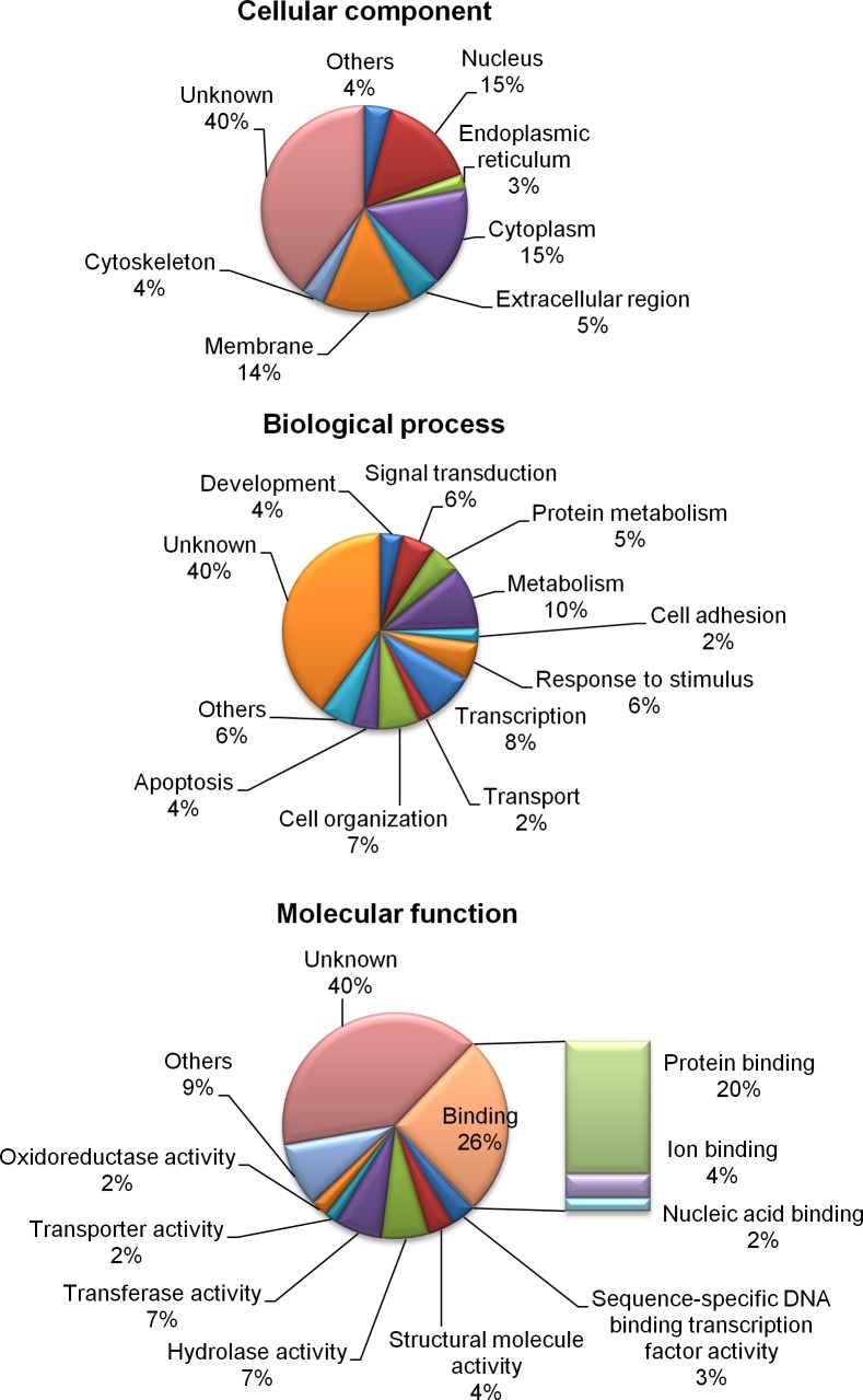
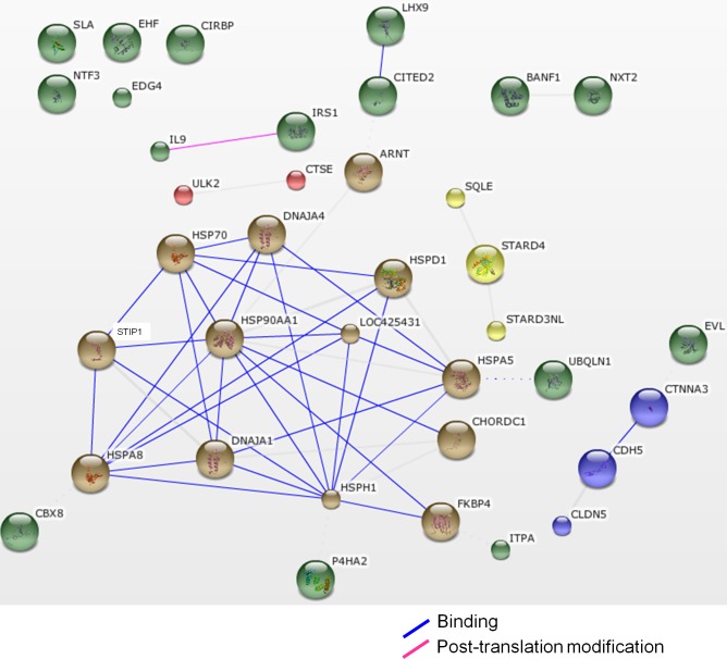
The functions of the differentially expressed genes were defined according to their GO annotations, namely cellular components, biological processes, and molecular functions (Fig 1). The genes were located primarily in the nucleus (15%), cytoplasm (15%), and membrane (14%). Most of the genes participated in the metabolism (10%), transcription (8%), cellular organization (7%), response to stimulus (6%), and signal transduction (6%). The expressions of the genes that were involved in the response to stimulus, such as heat-shock protein 25 (HSP25) and heat-shock 70 kDa protein 2 (HSPA2), increased after heat stress in chicken testes, while the cold inducible RNA binding protein (CIRBP) was downregulated (S3 Table). The results suggested that genes participating in signal transduction were upregulated, including the lysophosphatidic acid receptor 2 (LPAR2), or downregulated, including the interleukin 13 receptor, alpha 1 (IL13RA1), in the testes of B-strain TCCs after heat stress. Genes involved in transcription also changed in the heat-stressed chicken testes. For example, the Ets homologous factor (EHF) was downregulated after heat stress. Genes involved in cell adhesion were also downregulated after heat exposure, including cadherin 5 (CDH5), catenin, alpha 3 (CTNNA3), claudin 5 (CLDN5), and protocadherin-17-like (LOC100551402). In the biological process of apoptotic process, the heat-shock 70 kDa protein 5 (HSPA5), solute carrier family 5, member 11 (SLC5A11), gasdermin A (GSDMA), and heat-shock 60 kDa protein 1 (HSPD1) increased after heat stress. Regarding molecular functions, the differentially expressed genes were primarily sorted into protein binding (20%), hydrolase activity (7%), transferase activity (7%), and structural molecule activity types (4%; Fig 1). A gene network analysis showed that genes of heat-shock proteins (HSPs) and cochaperones exhibited strong relationships (Fig 2).
Fig 1. Pie chart showing the classification of differentially expressed genes in the testes of B-strain TCCs after 4 h of acute heat stress and recovery of 0, 2, and 6 h, in terms of cellular components, biological processes, and molecular functions.
Fig 2. The STRING network of protein interaction in various differentially expressed genes in the testes of heat-stressed B-strain TCCs.
The lines between the protein nodes show the actions among the proteins in Gallus gallus.
Validation of differentially expressed genes in the testes of broiler-type TCCs after acute heat stress
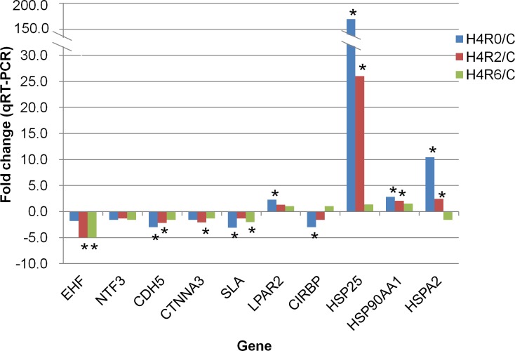
The mRNA expression levels of 10 genes commonly differed in both broiler-type and layer-type TCCs after acute heat stress or with fold changes higher than 3 and relationship to blood-testis barrier, signal transduction, and transcription, namely EHF, NTF3, CDH5, CTNNA3, Src-like-adaptor (SLA), LPAR2, CIRBP, HSP25, HSP90AA1, and HSPA2, were determined through qRT-PCR (Fig 3). The mRNA expressions of HSP25, HSP90AA1, and HSPA2 increased after 0 and 2 h of recovery in heat-stressed chicken testes. The mRNA level of LPAR2 was upregulated after 0 h of recovery, after the acute heat stress. In downregulated genes, CIRBP exhibited a decline after 0 h and CTNNA3 changed after 2 h of recovery. The mRNA level of EHF decreased after 2 and 6 h of recovery, whereas CDH5 was downregulated after 0 and 2 h recovery in chicken testes after the acute heat stress. The mRNA expression level of SLA decreased after 0 and 6 h of recovery after the heat stress. The mRNA expression of NTF3 exhibited no alteration in heat-stressed chicken testes. The mRNA expressions of selected genes determined through the qRT-PCR analysis were consistent with the microarray results. Although the mRNA expressions of NTF3 exhibited no significantly difference, the expression pattern was similar to that identified in the microarray data.
Fig 3. Validation of selected differentially expressed genes through qRT-PCR.
The * indicates the expression of a gene with a significant difference (p<0.05) compared with that of the control (C). H4R0, heat-stressed group without recovery; H4R2, heat-stressed group after 2 h of recovery; H4R6, heat-stressed group after 6 h of recovery. EHF, Ets homologous factor; NTF3, neurotrophin 3; CDH5, cadherin 5; CTNNA3, catenin alpha 3; SLA, Src-like-adaptor; LPAR2, lysophosphatidic acid receptor 2; CIRBP, cold inducible RNA binding protein; HSP25, heat-shock protein 25; HSP90AA1, heat-shock protein 90 kDa alpha, class A member 1; HSPA2, heat-shock 70 kDa protein 2.
Discussion
Heat stress impairs sperm motility and fertility in roosters. Few studies have elaborated on the effect of acute heat stress on gene expression in chicken testes. Our previous study on layer-type TCCs showed that 309 genes were differentially expressed in the testes after acute heat stress [15]. The functional network analysis revealed that genes of HSPs and cochaperones (HSP70, HSP90AA1, HSP25, and DNAJA4) as well as antiapoptotic BAG3 and SERPINB2 were upregulated in heat-stressed chicken testes. The results of this study further showed that broiler-type TCCs exhibited a rapid response to acute heat stress and they recovered immediately after heat stress (S2 Table). The microarray analysis identified 163 genes that were differentially expressed in the testes of broiler-type TCCs after 38°C acute heat stress, most of which changed after 0 h of recovery. The differentially expressed genes participated in transcription, response to stimulus, protein metabolism, cell adhesion, apoptosis, and signal transduction. Moreover, the mRNA levels of the genes were determined through qRT-PCR, and the results were consistent with those of the microarray analysis. In layer-type chickens, genes that expressed differentially were primarily observed after 0 and 2 h of heat stress [15]. We inferred that acute heat stress altered the gene expressions immediately in the testes of broiler-type TCCs.
Alterations of stress-related genes in the testes of broiler-type TCCs after acute heat stress
The mRNA levels of genes belonging to the heat-shock protein family increased in the testes of broiler-type TCCs after 4 h of heat stress, including HSP25, HSPA2, HSP90AA1, HSPA8, HSPD1, HSPH1, and HSPA5 (Fig 2 and S3 Table). Heat stress caused an elevation of HSP expression, attenuating the damage [20]. In heat-stressed mice, HSPA8 expression was shown to increase, protecting the testis from apoptosis and assisting inducible HSP70 (HSPA2) in repairing the stress-induced misfolded proteins [21]. Under acute heat stress, the expression levels of HSPH1 and HSP25 in liver, brain, and leg muscle increased to suppress cell death in boilers [22]. A previous study also indicated that mRNA expressions, including those of HSP25, HSPA2, HSP90AA1, HSPA8, and HSPH1, were upregulated in heat-stressed layer-type roosters [15]. An endoplasmic reticulum stress marker, HSPA5 [23], was induced in human fibroblasts after 30 min of heat stress [24]. The results of this study suggested that the testes responded to acute heat stress through the induction of HSP gene expressions (especially HSP25, HSPA2, HSP90AA1, HSPA8, HSPH1, and HSPA5) in broiler-type TCCs. Whether the overexpressions of the HSP genes attenuate stress in chickens requires further investigation.
Genes related to blood–testis barrier and transcription altered in the testes of broiler-type TCCs after acute heat stress
The genes related to the blood–testis barrier, including CDH5, CATNNA3, CLDN5, and LOC100551402, decreased at recovery in the testes of heat-stressed broiler-type TCCs (S3 Table). A blood–testis barrier is formed by anchoring Sertoli cells through cell junction proteins to prevent system circulation and allow the transit of preleptotene spermatocytes in the testis [25–28]. Various factors, such as cytokines, signal transducers, hormones, and heat stress, can modulate the expression levels or locations of cell junction proteins and thus regulate the integrity of the blood-testis barrier [27,29–32]. A short-term scrotal heat stress disrupts the integrity of the blood–testis barrier by downregulating the expressions of cell junction proteins, including cadherin, catenin, and claudin in mammals [30,32]. The decreased expressions of genes associated with cell junction in the testes of heat-stressed B-strain TCCs might imply that the integrity of the blood-testis barrier was affected in broiler-type chickens.
Our results showed that the transcripts involved in transcription or signal transduction were changed in the testes of broiler-type TCCs after heat stress (S3 Table). For instance, the mRNA expression level of EHF decreased whereas that of LPAR2 increased in heat-stressed chickens. These genes play essential roles in cell proliferation and differentiation [33–36]. Furthermore, the downregulated SLA in the testes of heat-stressed broiler-type TCCs (Fig 3) has been reported to be associated with cell proliferation [37,38]. Previous studies have suggested that SLA is a negative regulator of B-cell development and osteoclastogenesis [37,38]. A lack of SLA was shown to contribute to the proliferation of osteoclast precursors in mice [38]. The E26 transformation-specific (ETS) gene family is one of the largest families of transcriptional regulators regulating a variety of biological processes in normal cell homeostasis [33]. In the ETS gene family, the EHF is also known as epithelial specific ETS-3 and functions in epithelial cell differentiation and proliferation in mammals [34]. In human prostate epithelial cells, EHF can balance cell differentiation and self-renewal [35]. As a member of the G-protein-coupled receptor family, LPAR regulates embryogenesis, development, cell survival, immune cell trafficking, and inflammatory reactions [36]. The deletion of LPAR results in a loss of sperm production in mice [39]. The downregulated EHF and SLA but upregulated LPAR2 in the testes of heat-stressed B-strain TCCs imply that acute heat stress might influence cell proliferation and spermatogenesis of broiler-type chickens.
Expression difference of genes in the testes of heat-stressed broiler-type and layer-type TCCs in response to acute heat stress
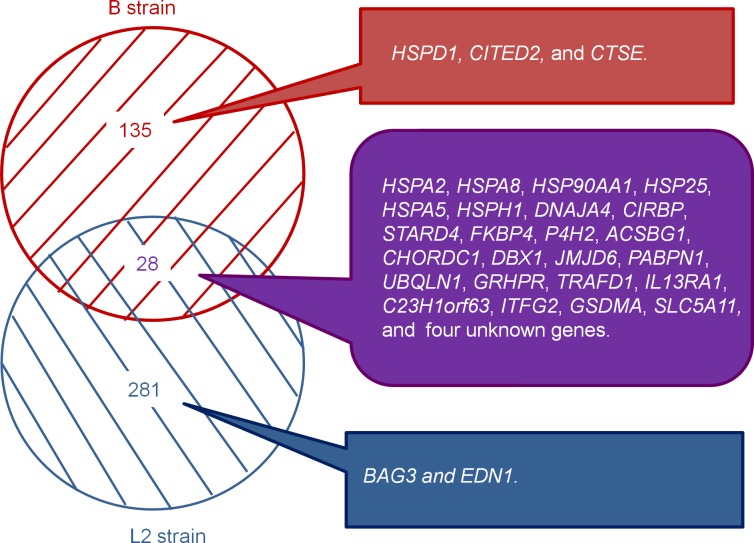
Initial heat-induced apoptosis in the testes of broiler-type TCCs was observed after 0 h of recovery after heat exposure. Furthermore, the apoptotic cells in the testes increased significantly after 2 h of recovery in heat-stressed broiler-type TCCs. In the L2 strain of layer-type TCCs, the number of apoptotic testicular cells was significantly higher than that in the controls after only 2 h of recovery [15]. An increase of apoptotic germ cells in heat-resistant mice occurred later than the increase in controls after exposure to heat stress (43°C, 15 min) [6]. The difference between strains might imply that the testicular cells of broiler-type TCCs were more sensitive than those of layer-type chickens when they were subjected to acute heat stress. Further comparison of the differences of testicular gene expressions between broiler-type and layer-type chickens showed that there were 135 genes exhibited substantial differences, specifically in the testes of heat-stressed broiler-type TCCs, whereas 281 genes showed alterations specifically in layer-type TCCs (Fig 4). A total of 28 genes exhibited substantial changes in both types of chickens and most of the genes were HSP genes. Acute heat stress caused various trends of expressions of apoptosis-related genes between the testes of broiler-type and layer-type TCCs. For example, the Cbp/p300-interacting transactivator together with Glu/Asp-rich carboxy-terminal domain 2 (CITED2) and HSPD1 increased but cathepsin E (CTSE) decreased in the testes of broiler-type TCCs after acute heat stress. Moreover, in layer-type L2-strain TCCs, antiapoptotic genes were upregulated, including BCL2-associated athanogene 3 (BAG3) and endothelin-1 (EDN1; Fig 4). The CTSE, an aspartic protease, plays a role in suppressing cell growth and metastasis in tumor cells [40]. In human prostate-cancer cell lines, a higher expression of CTSE induces growth arrest and apoptosis by catalyzing the proteolytic release of a tumor necrosis factor-related apoptosis-inducing ligand, which induces apoptosis [41]. As a member of the HSP family, HSPD1 plays multiple roles in cellular processes, including apoptosis, anti-apoptosis, and protein folding [42–44]. The mRNA expression of HSPD1 is localized in spermatogonia and Sertoli cells in normal monkey testes and elevated to prevent apoptosis after a short-term heat stress [42]. The BAG3 negatively regulates apoptosis, and it can be induced by heat stress to attenuate apoptosis in chickens [15,45]. The CITED2 acts as an anti-apoptotic gene, and a lack of CITED2 increases apoptosis in the anterior region of testes during early gonad development in mice [46]. Furthermore, hypoxic stress-induced apoptosis can be decreased by stimulating the transcription of CITED2 in fibroblasts [47]. The EDN1 reveals an anti-apoptotic effect in rat uterine leiomyoma ELT3 cells [48]. Moreover, the expression of EDN1 augments apoptosis in cancer cells induced by mild hyperthermia [49]. We inferred that the differential gene expression in response to acute heat stress in the testes of broiler-type and layer-type roosters may thus result in various responses to heat stress (e.g., apoptosis). However, whether the different expression patterns associate with the heat-tolerance as well as the cause-effect relationship in the strains require further exploration.
Fig 4. Comparison of differentially expressed genes between B-strain (broiler-type) and L2-strain (layer-type) TCCs after acute heat stress.
Conclusion
A total of 163 genes changed in the testes of broiler-type B-strain TCCs after acute heat stress. The differentially expressed genes are primarily involved in the protein metabolism, cell adhesion, transcription, development, and apoptosis; this suggests that heat stress may affect the cell survival, differentiation, and development in the testes of broiler-type chickens. Moreover, the partially different expression patterns of genes in the testes of broiler-type and layer-type chickens may suggest that testicular responses to acute heat stress differ between the two types of chickens. As the functions of genes are reflected in their protein levels and post-translational modifications, the results of this study require further validation at translation levels and a follow-up study is now under investigation.
Supporting Information
H4R0, heat stress group after 0 h of recovery; H4R2, heat stress group after 2 h of recovery; H4R6, heat stress group after 6 h of recovery; Control, chickens not subjected to heat stress.
(TIFF)
The numbers in parentheses represent the amount of differentially expressed genes. H4R0, 0 h of recovery after heat stress; H4R2, 2 h of recovery after heat stress; H4R6, 6 h of recovery after heat stress.
(TIFF)
(DOC)
(DOC)
(DOC)
Data Availability
All relevant data are within the paper and its Supporting Information files. All results of microarray dataset files are available from the Gene Expression Omnibus in the National Center for Biotechnology Information database (accession number GSE65279).
Funding Statement
This study was supported in part by grants from National Science Council (NSC# 99-2321-B-005-016-MY3), Executive Yuan and the Ministry of Education, Taiwan, Republic of China, under the ATU plan. The funders had no role in study design, data collection and analysis, decision to publish, or preparation of the manuscript.
References
- 1. Skinner JD, Louw GN. Heat stress and spermatogenesis in Bos indicus and Bos taurus cattle. J Appl Physiol. 1966;21: 1784–1790. [DOI] [PubMed] [Google Scholar]
- 2. Wettemann RP, Wells ME, Omtvedt IT, Pope CE, Turman EJ. Influence of elevated ambient temperature on reproductive performance of boars. J Anim Sci. 1976;42: 664–669. [DOI] [PubMed] [Google Scholar]
- 3. Mieusset R, Quintana Casares P, Sanchez Partida LG, Sowerbutts SF, Zupp JL,Setchell BP. Effects of heating the testes and epididymides of rams by scrotal insulation on fertility and embryonic mortality in ewes inseminated with frozen semen. J Reprod Fertil. 1992;94: 337–343. [DOI] [PubMed] [Google Scholar]
- 4. Ikeda M, Kodama H, Fukuda J, Shimizu Y, Murata M, Kumagai J, et al. Role of radical oxygen species in rat testicular germ cell apoptosis induced by heat stress. Biol Reprod. 1999;61: 393–399. [DOI] [PubMed] [Google Scholar]
- 5. Rockett JC, Mapp FL, Garges JB, Luft JC, Mori C, Dix DJ. Effects of hyperthermia on spermatogenesis, apoptosis, gene expression, and fertility in adult male mice. Biol Reprod. 2001;65: 229–239. [DOI] [PubMed] [Google Scholar]
- 6. Li Y, Zhou Q, Hively R, Yang L, Small C, Griswold MD. Differential gene expression in the testes of different murine strains under normal and hyperthermic conditions. J Androl. 2009;30: 325–337. 10.2164/jandrol.108.005934 [DOI] [PMC free article] [PubMed] [Google Scholar]
- 7. Paul C, Murray AA, Spears N, Saunders PT. A single, mild, transient scrotal heat stress causes DNA damage, subfertility and impairs formation of blastocysts in mice. Reproduction. 2008;136: 73–84. 10.1530/REP-08-0036 [DOI] [PubMed] [Google Scholar]
- 8. Kim B, Park K, Rhee K. Heat stress response of male germ cells. Cell Mol Life Sci. 2013;70: 2623–2636. 10.1007/s00018-012-1165-4 [DOI] [PMC free article] [PubMed] [Google Scholar]
- 9. Karaca AG, Parker HM, Yeatman JB, McDaniel CD. The effects of heat stress and sperm quality classification on broiler breeder male fertility and semen ion concentrations. Br Poult Sci. 2002;43: 621–628. [DOI] [PubMed] [Google Scholar]
- 10. Karaca AG, Parker HM, Yeatman JB, McDaniel CD. Role of seminal plasma in heat stress infertility of broiler breeder males. Poult Sci. 2002;81: 1904–1909. [DOI] [PubMed] [Google Scholar]
- 11. McDaniel CD, Bramwell RK, Wilson JL, Howarth B Jr. Fertility of male and female broiler breeders following exposure to elevated ambient temperatures. Poult Sci. 1995;74: 1029–1038. [DOI] [PubMed] [Google Scholar]
- 12.Monthly data of climate Statistics in Central Weather Bureau website. Taiwan. Available: http://www.cwb.gov.tw/V7/climate/monthlyData/mD.htm. Accessed 2015 Apr 2.
- 13.Lee YP. Taiwan Country Chicken: a slow growth breed for eating quality. Scientific Cooperation in Agriculture between Council of Agriculture (Taiwan, R.O.C.) and Institut National de la Recherche Agronomique (France). 2006.
- 14. Yeh CC. Effect of acute heat stress on the blood characteristics of Taiwan country chickens and broilers. J Chin Soc Anim Sci. 1992;21: 57–66 (in Chinese with English abstract). [Google Scholar]
- 15. Wang SH, Cheng CY, Tang PC, Chen CF, Chen HH, Lee YP, et al. Differential gene expressions in testes of L2 strain Taiwan country chicken in response to acute heat stress. Theriogenology. 2013;79: 374–382. 10.1016/j.theriogenology.2012.10.010 [DOI] [PubMed] [Google Scholar]
- 16.Lee YP. Development and improvement of local chicken in Taiwan. Proceedings of the 5th AAAP Animal Science Congress, Taipei, Taiwan. 1990; pp. 349–353.
- 17. Li X, Chiang HI, Zhu J, Dowd SE, Zhou H. Characterization of a newly developed chicken 44K Agilent microarray. BMC Genomics. 2008;9: 60 10.1186/1471-2164-9-60 [DOI] [PMC free article] [PubMed] [Google Scholar]
- 18.The Gene Ontology website. Available: http://www.geneontology.org/. Accessed 2015 Apr 2.
- 19.Search Tool for the Retrieval of Interacting Genes/Proteins. Available: http://string-db.org/. Accessed 2015 Apr 2.
- 20. Li GC. Elevated levels of 70,000 dalton heat shock protein in transiently thermotolerant Chinese hamster fibroblasts and in their stable heat resistant variants. Int J Radiat Oncol Biol Phys. 1985;11: 165–177. [DOI] [PubMed] [Google Scholar]
- 21. Zaprjanova S, Rashev P, Zasheva D, Martinova Y, Mollova M. Electrophoretic and immunocytochemical analysis of Hsp72 and Hsp73 expression in heat-stressed mouse testis and epididymis. Eur J Obstet Gynecol Reprod Biol. 2013;168: 54–59. 10.1016/j.ejogrb.2012.12.021 [DOI] [PubMed] [Google Scholar]
- 22. Luo QB, Song XY, Ji CL, Zhang XQ, Zhang DX. Exploring the molecular mechanism of acute heat stress exposure in broiler chickens using gene expression profiling. Gene. 2014;546: 200–205. 10.1016/j.gene.2014.06.017 [DOI] [PubMed] [Google Scholar]
- 23. Schröder M, Kaufman RJ. ER stress and the unfolded protein response. Mutat Res. 2005;569: 29–63. [DOI] [PubMed] [Google Scholar]
- 24. Tabuchi Y, Furusawa Y, Kariya A, Wada S, Ohtsuka K, Kondo T. Common gene expression patterns responsive to mild temperature hyperthermia in normal human fibroblastic cells. Int J Hyperthermia. 2003;29: 38–50. [DOI] [PubMed] [Google Scholar]
- 25. Setchell BP, Voglmayr JK, Waites GM. A blood-testis barrier restricting passage from blood into rete testis fluid but not into lymph. J Physiol. 1969;200: 73–85. [DOI] [PMC free article] [PubMed] [Google Scholar]
- 26. Lui WY, Lee WM, Cheng CY. Transforming growth factor-β3 perturbs the inter-Sertoli tight junction permeability barrier in vitro possibly mediated via its effects on occluding, zonula occludens-1, and claudin-11. Endocrinology. 2001;142: 1865–1877. [DOI] [PubMed] [Google Scholar]
- 27. Wong CH, Mruk DD, Lui WY, Cheng CY. Regulation of blood-testis barrier dynamics: an in vivo study. J Cell Sci. 2004;117: 783–798. [DOI] [PubMed] [Google Scholar]
- 28. Bergmann M, Schindelmeiser J. Development of the blood-testis barrier in the domestic fowl (Gallus domesticus). Int J Androl. 1987;10: 481–488. [DOI] [PubMed] [Google Scholar]
- 29. Lui WY, Wong CH, Mruk DD, Cheng CY. TGF-β3 regulates the blood-testis barrier dynamics via the p38 mitogen activated protein (MAP) kinase pathway: an in vivo study. Endocrinology. 2003;144: 1139–1142. [DOI] [PubMed] [Google Scholar]
- 30. Chen M, Cai H, Yang JL, Lu CL, Liu T, Yang W, et al. Effect of heat stress on expression of junction-associated molecules and upstream factors androgen receptor and Wilms’ tumor 1 in monkey Sertoli cells. Endocrinology. 2008;149: 4871–4882. 10.1210/en.2007-1093 [DOI] [PubMed] [Google Scholar]
- 31. Lee NP, Cheng CY. Nitric oxide and cyclic nucleotides: their roles in junction dynamics and spermatogenesis. Oxid Med Cell Longev. 2008;1: 25–32. [DOI] [PMC free article] [PubMed] [Google Scholar]
- 32. Cai H, Ren Y, Li XX, Yang JL, Zhang CP, Chen M, et al. Scrotal heat stress causes a transient alteration in tight junctions and induction of TGF-β expression. Int J Androl. 2011;34: 352–362. 10.1111/j.1365-2605.2010.01089.x [DOI] [PubMed] [Google Scholar]
- 33. Wang Z, Zhang Q. Genome-wide identification and evolutionary analysis of the animal specific ETS transcription factor family. Evol Bioinform Online. 2009;5: 119–131. [DOI] [PMC free article] [PubMed] [Google Scholar]
- 34. Kas K, Finger E, Grall F, Gu X, Akbarali Y. ESE-3, a novel member of an epithelium-specific ets transcription factor subfamily, demonstrates different target gene specificity from ESE-1. J Biol Chem. 2000;275: 2986–2998. [DOI] [PubMed] [Google Scholar]
- 35. Albino D, Longoni N, Curti L, Mello-Grand M, Pinton S, Civenni G, et al. ESE3/EHF controls epithelial cell differentiation and its loss leads to prostate tumors with mesenchymal and stem-like features. Cancer Res. 2012;72: 2889–2900. 10.1158/0008-5472.CAN-12-0212 [DOI] [PubMed] [Google Scholar]
- 36. Skoura A, Hla T. Lysophospholipid receptors in vertebrate development, physiology, and pathology. J Lipid Res. 2009;50 Suppl: S293–298. 10.1194/jlr.R800047-JLR200 [DOI] [PMC free article] [PubMed] [Google Scholar]
- 37. Dragone LL, Myers MD, White C, Sosinowski T, Weiss A. SRC-like adaptor protein regulates B cell development and function. J Immunol. 2006;176: 335–345. [DOI] [PubMed] [Google Scholar]
- 38. Kim HJ, Zou W, Ito Y, Kim SY, Chappel J, Ross FP, et al. Src-like adaptor protein regulates osteoclast generation and survival. J Cell Biochem. 2010;110: 201–209. 10.1002/jcb.22527 [DOI] [PMC free article] [PubMed] [Google Scholar]
- 39. Ye X, Skinner MK, Kennedy G, Chun J. Age-dependent loss of sperm production in mice via impaired lysophosphatidic acid signaling. Biol Reprod. 2008;79: 328–336. 10.1095/biolreprod.108.068783 [DOI] [PMC free article] [PubMed] [Google Scholar]
- 40. Kawakubo T, Okamoto K, Iwata J, Shin M, Okamoto Y, Yasukochi A, et al. Cathepsin E prevents tumor growth and metastasis by catalyzing the proteolytic release of soluble TRAIL from tumor cell surface. Cancer Res. 2007;67: 10869–10878. [DOI] [PubMed] [Google Scholar]
- 41. Wiley SR, Schooley K, Smolak PJ, Din WS, Huang CP, Nicholl JK, et al. Identification and characterization of a new member of the TNF family that induces apoptosis. Immunity. 1995;3: 673–682. [DOI] [PubMed] [Google Scholar]
- 42. Zhang XS, Lue YH, Guo SH, Yuan JX, Hu ZY, Han CS, et al. Expression of HSP105 and HSP60 during germ cell apoptosis in the heat-treated testes of adult cynomolgus monkeys (macaca fascicularis). Front Biosci. 2005;10: 3110–3121. [DOI] [PubMed] [Google Scholar]
- 43. Ostermann J, Horwich AL, Neupert W, Hartl FU. Protein folding in mitochondria requires complex formation with hsp60 and ATP hydrolysis. Nature. 1989; 341: 125–130. [DOI] [PubMed] [Google Scholar]
- 44. Samali A, Cai J, Zhivotovsky B, Jones DP, Orrenius S. Presence of a pre-apoptotic complex of pro-caspase-3, Hsp60 and Hsp10 in the mitochondrial fraction of jurkat cells. EMBO J. 1999;18: 2040–2048. [DOI] [PMC free article] [PubMed] [Google Scholar]
- 45. Rosati A, Ammirante M, Gentilella A, Basile A, Festa M, Pascale M, et al. Apoptosis inhibition in cancer cells: a novel molecular pathway that involves BAG3 protein. Int J Biochem Cell Biol. 2007;39: 1337–1342. [DOI] [PubMed] [Google Scholar]
- 46. Buaas FW, Val P, Swain A. The transcription co-factor CITED2 functions during sex determination and early gonad development. Hum Mol Genet. 2009;18: 2989–3001. 10.1093/hmg/ddp237 [DOI] [PubMed] [Google Scholar]
- 47. Bakker WJ, Harris IS, Mak TW. FOXO3a is activated in response to hypoxic stress and inhibits HIF1-induced apoptosis via regulation of CITED2. Mol Cell. 2007;28: 941–953. [DOI] [PubMed] [Google Scholar]
- 48. Raymond MN, Bole-Feysot C, Banno Y, Tanfin Z, Robin P. Endothelin-1 inhibits apoptosis through a sphingosine kinase 1-dependent mechanism in uterine leiomyoma ELT3 cells. Endocrinology. 2006;147: 5873–5882. [DOI] [PubMed] [Google Scholar]
- 49. Kariya A, Tabuchi Y, Yunoki T, Kondo T. Identification of common gene networks responsive to mild hyperthermia in human cancer cells. Int J Mol Med. 2013;32: 195–202. 10.3892/ijmm.2013.1366 [DOI] [PubMed] [Google Scholar]
Associated Data
This section collects any data citations, data availability statements, or supplementary materials included in this article.
Supplementary Materials
H4R0, heat stress group after 0 h of recovery; H4R2, heat stress group after 2 h of recovery; H4R6, heat stress group after 6 h of recovery; Control, chickens not subjected to heat stress.
(TIFF)
The numbers in parentheses represent the amount of differentially expressed genes. H4R0, 0 h of recovery after heat stress; H4R2, 2 h of recovery after heat stress; H4R6, 6 h of recovery after heat stress.
(TIFF)
(DOC)
(DOC)
(DOC)
Data Availability Statement
All relevant data are within the paper and its Supporting Information files. All results of microarray dataset files are available from the Gene Expression Omnibus in the National Center for Biotechnology Information database (accession number GSE65279).


