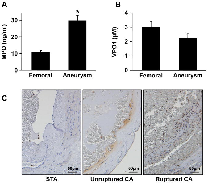
Figure 1.
Plasma concentrations of MPO (A) and VPO1 (B) in femoral artery and cerebral aneurysms of 25 patients with cerebral aneurysms. Patients ranged from 25 to 76 years of age (55±3). Values are mean ± SE, * = p<0.05 vs. femoral artery blood. C. Representative images of immunohistochemistry for MPO in superficial temporal artery (STA), unruptured and ruptured cerebral aneurysm tissue. MPO positive cells were stained brown, with negative cells blue by hematoxylin counterstaining. Scale bars indicate 50 μm.

