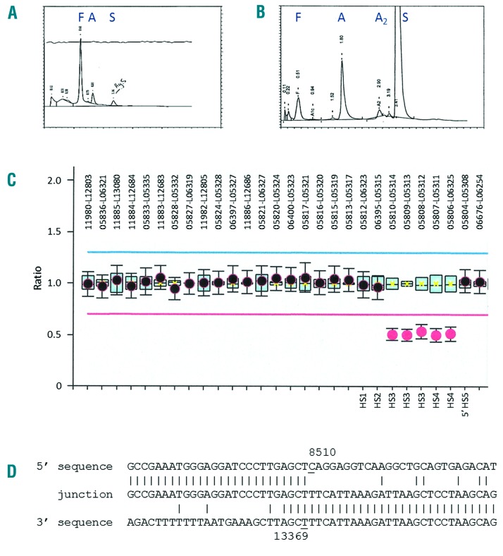
Figure 1.
(A) BioRad HPLC traces of the proband’s hemolysate from newborn screening and (B) at five years of age. Positions of hemoglobins F, A, A2, and S are indicated above the corresponding peaks. (C) MLPA analysis showing the relative probe signals across the β-globin gene cluster (SALSA MLPA probemix P-102-B2 HBB, MRC-Holland, Amsterdam, The Netherlands). The MLPA probes that define the deletion are indicated. (D) Sequence of the deletion junction fragment compared to the normal 5′ and 3′ sequences. The first and last nucleotides of the deleted region are underlined. The deletion junction fragment was amplified as a ~0.6 kb fragment using a pair of flanking primers (forward 5′-ACTTT CAGTC CGGTC CTCA CAGT-3′, NG_000007.3 positions 8111–8133; reverse 5′-GTGGT TTCTA GTCCC TTCAC CATC TTGT-3′, NG_000007.3 positions 135523–13550), which was then sequenced using the reverse primer.

