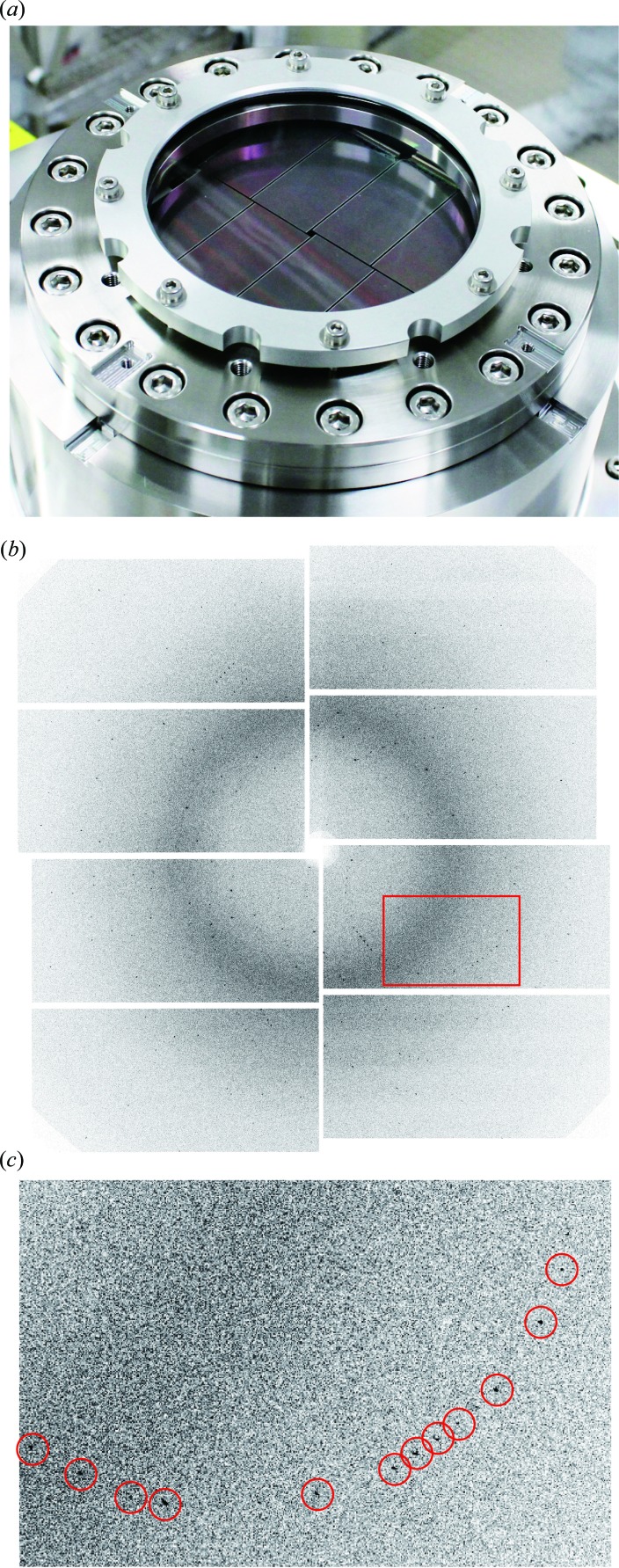
Figure 7.
(a) The camera head of an MPCCD developed for serial femtosecond crystallography (Kameshima et al., 2014 ▶). The detector consists of eight sensors aligned on a flat imaging surface to give a total of 4 Mpixels. The imaging surface can be placed close to the specimen to cover a scattering angle of ±45°. (b) A single-pulse diffraction pattern from a small lysozyme crystal recorded with this detector in combination with an early phase setup at SACLA (Song et al., 2014 ▶). The data were calibrated by the facility’s standard automated procedure. Simple calibration is one advantage of passive pixels (Kameshima et al., 2014 ▶). (c) An enlarged image of the area shown as a red rectangle in (b). Diffraction spots are indicated by red circles.

