Abstract
Turbid white urine ‘albinuria’ is defined as a urine discoloration described as milky or cloudy. One of the most frequent causes of turbid white urine is chyluria complicating filariasis (Table 1). The extant causes of albinuria are non parasitic and rare. Amongst their aetiologies stand excessive mineral sediment excretion such as calciuria and phosphaturia, massive pyuria and fungal infections, and rarely congenital malformations of the lymphatic vessels. Malingering is also possible, in patients adding milk to their urine. We observed a case of albinuria in which the diagnostic work up led to diagnosing an exceptional cause of chyluria in a patient living in a region of Colombia where filariasis is not endemic.
Keywords: albinuria, chyluria, filariasis, lymphatic fistula, turbid white urine
Case
A 40-year-old Colombian man living in central Colombia, Bogota, was referred to local hospital for turbid white urine that had appeared 2 months earlier. The patient worked as an office clerk. He had never lived outside the city of Bogota. The whitish colour of the urine was described as ‘purulent’ or ‘milky’. The phenomenon was intermittent: the patient would pass discoloured urine during 4 or 5 days without complaints of fever, burning or urgency. Thereafter, the urine was clear or pink. His past medical history did not reveal any relevant signs and symptoms. However, he had lost 25 kg over the preceding 6 months. This anxious man had sought medical attention in various facilities, and despite the fact that no urinary tract infection was found by urine cytology and cultures, he had been repeatedly treated by urinary antibiotics that did not produce any effect on his albinuria. Physical examination findings were unremarkable except for a BMI of 30 kg/m2. The patient was not oedematous.
Table 1.
Causes of turbid white urine
| Chyluria |
| Filariasis |
| Schistosomiasis |
| Postsurgery |
| Malignancy |
| Hyperuricosuria |
| Phosphaturia |
| Hyperoxaluria |
| Proteinuria |
| Pyuria |
| Lipiduria |
| Caseous material from renal tuberculosis |
| Congenital malformations of the lymphatic vessels |
At first visit to our nephrology unit, the patient kept describing his urine as ‘milky’. We verified that his description was appropriate by collecting urine in various occasions. Yes, the urine was either white or pink. The patient indignantly denied adding anything into his urine. In fact, analysing the urine revealed a massive proteinuria of 5 g/l, microhaematuria (15–20 red cells per ml) and leucocyturia (10–15 leucocytes/ml). Bacteriologic, fungal and mycobacterial cultures were negative. There was no hypercalciuria or hyperphosphaturia. A closer examination of urinary cytology showed that the white cell population consisted of abundant lymphocytes.
Further investigations revealed a normal renal function, with a creatinine clearance in the order of 105 ml/min. Proteinuria was profuse (6.7 g/24 h), but serum albumin concentration was normal (43 g/l), and so were his serum lipids (total cholesterol: 4.9 mmol/l, triglycerides 1.9 mmol/l). No oedema, no hypoalbuminaemia, no hyperlipidaemia and fasting coincided with a clearing of the urine so it could not be a nephrotic syndrome. So, what was the source of the proteinuria?
The rest of the laboratory work up was also non-contributive: leucocytes 5700/mm3, (neutrophils 64%, lymphocytes 23%), haemoglobin 16.5 g/dl, haematocrit 48.2%, platelets 285.000/mm3. Eosinophilia was not detected. B and C hepatitis tests, HIV and VDRL were all negative. IgE levels were normal. The specific serological tests for the detection of parasites were negative. Abdominal ultrasound examination was normal.
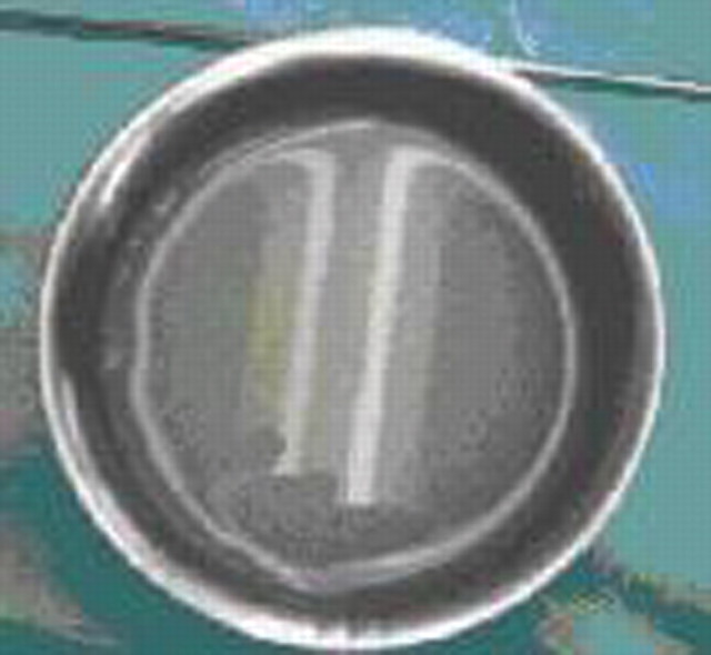
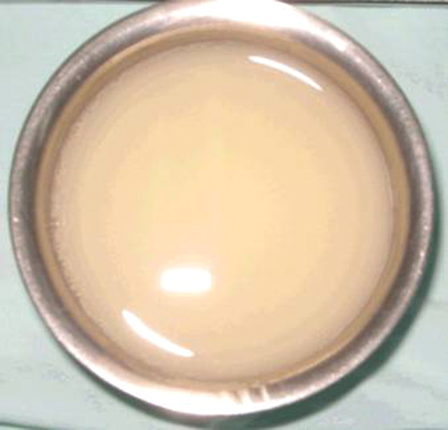
A cystoscopy that found white urine in the bladder (thus excluding malingering) did not disclose tumoural or infectious lesions. These investigations were performed during several hospitalizations. The patient observed that the hospital diet that he found meagre also coincided with a clearing of the urine. This remark prompted us to ask the department of nutrition to give him fat meals. With a lipid-rich diet urine turned white and clotty. With a fat-free diet the urine was found to be clear (Figures 1 and 2). It was found that milky urine was demulsified with chloroform and Sudan dye made it turn red.
Fig. 1.
Clear urine with a fat-free diet.
Fig. 2.
White urine with a fat-rich diet.
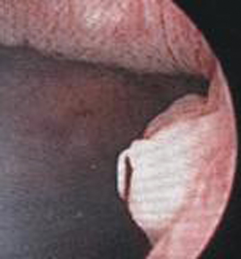
Chyluria it was, but what was the source of lymph? A new cystoscopy was made with previous administration of a fat-rich diet and disclosed a punctiform hole in the posterior wall of the bladder with a leak of white fluid (Figure 3). Iodopovidone closed the fistula and the patient was discharged with instructions to avoid fat foods. The final diagnosis was that of an idiopathic lymphatic fistula.
Fig. 3.

Cystoscopic view of white urine flowing from a hole in the bladder wall (arrow).
Discussion
Albinuria is defined as white urine in relation to any foreign substance. Its differential diagnosis includes phosphates, calcium and urates that in excess give the urine a turbid, white or cloudy appearance, although the amounts of these minerals are rarely high enough to produce albinuria. The urine pH plays a role in provoking a turbid discolouration of the urine: Claude Bernard in his doctoral thesis presented in Paris in 1843 explained that rabbits, a species feeding on vegetal, excrete alkaline turbid urine that turns clear by adding acid to it. Urinalysis and sediment examination may give the missing pieces for diagnosis. When thinking of mineral crystal deposits, some clues are given by the urine pH: phosphaturia is related to alkaline urine [1] and sediment analysis shows identifiable crystals [1]. When chyluria is centrifuged, it remains white, which permits to differentiate it from mineral deposits. Other substances that can induce albinuria are pus in severe urinary tract infections, or caseous material in urinary tuberculosis (TB).
Once such causes have been ruled out, the diagnosis of chyluria is likely to be appropriate, but this has no reason to be in non-endemic regions of filariasis infection, which was the case in our patient who lives in central Colombia.
Chyluria implies an abnormal communication between lymphatics and the urinary tract, mediated by any obstruction of intestinal lymph drainage resulting in lymphatic vessel dilatation and rupture into the urinary tract [2,3]. The most prevalent cause of chyluria is Wuchereria bancrofti filariasis. This parasitic disease is frequent in geographic areas between latitudes 40° North and 30° South. About 10% of the population in these countries suffer from filariasis, and one-tenth of the affected patients present with albinuria caused by urinary-lymphatic fistulae [1,2]. Nonetheless, chyluria can be related to other rare non-parasitic diseases [3,4] such as fungal infections, congenital abnormalities of the lymphatic vessels, malignancy, trauma and pregnancy [1,2].
White urine emission alternates with macrohaematuria and is sometimes accompanied with renal colic produced by chyle clots [2]. Dipstick analysis shows variable proteinuria and haematuria and persistent sterile leucocyturia. Proteinuria is explained by the high concentration of albumin and fibrinogen in chyle (30–60 g/l) [2], and leucocyturia consists of lymphocytes [2,4,5]. A lipid-rich meal is rapidly followed by albinuria, whereas a fat-free diet allows the urine to turn clear.
Our little case has a teaching value. Firstly, it shows that chyluria may occur in any part of the world, and that European readers may come across a case of albinuria in a patient who never lived in an endemic parasitic region where W. bancrofti infection is common. Secondly, diagnosing chyluria may simply be achieved by dietary manoeuvres, alternating a lipid-rich diet followed by a fat-free diet. This is simpler and cheaper than performing a lymphography or a lymphoscintigraphy. However, albinuria and chyluria may be caused by congenital [6] or acquired aetiologies of lymphatic fistulae. When fortune smiles, as it was in our case, the urologist may observe white urine running from a punctiform hole and treat albinuria by sclerotherapy or povidone injection into the fistula. However, a fat-poor diet must be pursued: the fistula might reopen, and besides, a patient whose BMI is 30 cannot but benefit from it.
The patient remained well during a follow-up period of 12 months. When last seen, he had managed to lose 12 kg of weight. There was no new episode of white urine.
Conflict of interest statement. None declared.
References
- 1.Gulati S, Gupta N, Singh NP, et al. Chyluria with proteinuria or filarial nephropathy? An enigma. Parasitol Int. 2007;56:251–254. doi: 10.1016/j.parint.2007.03.002. [DOI] [PubMed] [Google Scholar]
- 2.Cheng J-T, Mohan S, Nasr SH, et al. Chyluria presenting as milky urine and nephrotic-range proteinuria. Kidney Int. 2006;70:1518–1522. doi: 10.1038/sj.ki.5001703. [DOI] [PubMed] [Google Scholar]
- 3.Akisada M, Tani S. Filarial chyluria in Japan. Lymphography, etiology and treatment in 30 cases. Radiology. 1978;90:311–317. [Google Scholar]
- 4.Buck AA. Filariasis. In: Strickland GT, editor. Hunters Tropical Medicine. 7th edn. Philadelphia: WB Saunders; 2002. pp. 713–727. [Google Scholar]
- 5.Zimmer JG, Dewey R, Waterhouse C, et al. The origin and nature of anisotropic urinary lipids in the nephrotic syndrome. Ann Intern Med. 1961;54:205–214. doi: 10.7326/0003-4819-54-2-205. [DOI] [PubMed] [Google Scholar]
- 6.Stalens JP, Falk M, Howmann-Giles R, et al. ‘Milky urine’—a child with chyluria. Eur J Pediatr. 1992;151:61–62. doi: 10.1007/BF02073895. [DOI] [PubMed] [Google Scholar]


