Introduction
The association between secondary syphilis and nephrotic syndrome has been documented in the distant past but is particularly rare in modern times. We report the diagnostic dilemma that was the case of a 41-year-old gentleman who presented to hospital with a profound nephrotic syndrome, rash and simultaneous hepatitis. After extensive investigation, a unifying diagnosis of secondary syphilis was made. Treatment with penicillin resulted in complete resolution of his multi-systemic illness. A summary of the patient’s presentation and progress is provided as well as a concise review of the relevant literature and important teaching points from this challenging case.
Case summary
A 41-year-old male truck mechanic presented to the emergency department with a 3-week history of drenching night sweats and a 1-week history of widespread non-pruritic rash. His work colleagues had commented to him that his face appeared swollen, particularly around the eyes. The patient himself reported 5 days of worsening bilateral leg swelling and production of frothy urine. He had no significant past medical history and did not take any regular medications. His sexual history was noteworthy for occasional unprotected sexual intercourse with both men and women. His most recent sexual contact was unprotected insertive oral intercourse with a female partner 5 weeks prior to presentation. Several days after this encounter, he developed irregular urinary stream associated with penile tip discomfort. These symptoms were transient and resolved spontaneously 5 days later.
At presentation, his blood pressure was 112/68 mm Hg with a heart rate of 93 bpm. His temperature was 37.2°C and oxygen saturation was recorded at 99% on room air. Clinical examination revealed pitting lower limb and sacral oedema. There was a maculo-papular eruption with some superficial scaling over the trunk and limbs with relative sparing of the palms and soles. There were no oral or genital lesions. Multiple small mildly tender inguinal lymph nodes were palpable bilaterally. The liver was enlarged and non-tender with no evidence of splenomegaly. A subsequent abdominal ultrasound confirmed hepatomegaly with a liver span of 20 cm.
Laboratory investigations on admission revealed abnormal liver enymes with alkaline phosphatase (ALP) of 441 U/L (38–126), gamma-glutamyl transferase (GGT) of 328 U/L (0–50), aspartate aminotransferase (AST) of 63 U/L (0–45) and alanine aminotransferase (ALT) of 105 U/L (0–45). Serum bilirubin was normal. Serum albumin reached a nadir of 11 g/L. Urinary protein:creatinine ratio was significantly elevated at 1087 mg/mmol, and 21.96 g of protein was measured in a subsequent 24-h urine collection. Serum total cholesterol was elevated at 9.3 mmol/L (3–5.5), and there was a deterioration in renal function with a serum creatinine of 124 μmol/L (60–110). Urine microscopy showed hyaline casts but no haematuria or red cell casts. Both kidneys appeared structurally normal on ultrasound.
Multiple investigations were performed to look for a secondary cause of his dramatic presentation with nephrotic syndrome. Streptococcal serology, human immunodeficiency virus antibody, hepatitis B surface antigen and hepatitis C antibody were all negative. Urinary polymerase chain reaction for Neisseria gonorrhoeae and Chlamydia trachomatis did not reveal any sexually acquired co-infections. Treponema pallidum antibody, fluorescent treponemal antibodies and rapid plasma reagin (RPR) were all reactive, the latter with a titre of 1:128. Interestingly, anti-cardiolipin IgM and IgG were also detected. Antinuclear antibodies were detected with a titre of 1:80 for both speckled and cytoplasmic antibodies. Double-stranded deoxyribonuclease antibody was negative. An anti-neutrophil cytoplasmic antibody, screened by enzyme-linked immunosorbent assay, was negative. Serum complement levels were within normal limits.
The patient subsequently had a renal biopsy on the fourth day of his admission. The appearances were consistent with early stage membranous glomerulonephritis. The report is as follows:
Light microscopy
Of 13 glomeruli seen, one showed periglomerular fibrosis. The other glomeruli showed rigid appearing, but not thickened, capillary loops with no spikes. No proliferative lesions or areas of segmental sclerosis were seen. There was mild patchy and non-specific chronic interstitial inflammation with minimal focal tubular atrophy. Interlobular arteries and arterioles appeared normal.
Immunofluorescence
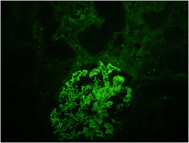
IgG, C3, C1q, kappa and lambda were all strongly positive in a fine granular pattern along the capillary loops (Figure 1). IgM was weakly positive in a granular mesangial distribution. IgA was negative.
Fig. 1.
Immunofluoresence of renal biopsy showing membranous glomerulonephritis.
Electron microscopy
There was marked focal effacement of foot processes over scattered subepithelial dense deposits. The lamina densa of the glomerular basement membranes was normal. No spikes were seen. The tubules, interstitium and vessels were all unremarkable.
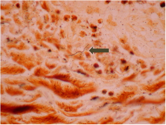
The patient also had a punch biopsy of a skin lesion performed. The appearances were those of a lichenoid reaction pattern. A Warthin–Starry stain showed a single slender spirochaete within the dermis (Figure 2).
Fig. 2.
High power view of skin biopsy showing a single slender spirochaete in the dermis.
He was commenced on intravenous benzylpenicillin (1.8 g four hourly) and the rash improved over the following 2 days. His oedema slowly improved on frusemide, and he was also commenced on an angiotensin-II receptor blocker for its anti-proteinuric effect. At the time of discharge from hospital, his renal function had returned to normal and there was interval improvement of his hepatitis with ALP of 298 U/L, GGT of 296 U/L, ALT of 54 U/L and normal AST of 40 U/L. As an outpatient, he received three doses of weekly benzathine penicillin via intramuscular injection. At clinic follow-up 6 weeks later, his liver function tests were all within normal limits and serum creatinine had returned to 57 μmol/L. The proteinuria had completely resolved as evidenced by a urine protein:creatinine ratio of 8 mg/mmol. Serum cholesterol also returned to normal at 5.5 mmol/L. His RPR titre had fallen to1:64.
Discussion
Our patient presented with night sweats, rash, lymphadenopathy, acute hepatitis and nephrotic syndrome. The subsequent laboratory investigations and skin biopsy findings supported secondary syphilis as the most likely unifying diagnosis. Although there have been a number of previous case reports of nephrotic syndrome in the setting of secondary syphilis, it is a rare presentation. Many of these reports date back to the 1940s and 1950s, and relatively little has been written about syphilis-related nephropathy in more recent times. We felt it was important to highlight this particular case because it represented a diagnostic dilemma and we will most likely see more cases due to the re-emergence of syphilis worldwide.
The association between secondary syphilis and development of the nephrotic syndrome has been recognized for a long time. In a series of over 1000 patients with syphilis in 1935, Hermann and Marr [1] found an incidence of proteinuria in 7.1% and nephrotic syndrome in 0.28%. Historically, there has been a great deal of confusion concerning the pathogenesis of renal injury, partly due to the self-limiting nature of the disease and also because many of these cases predated the advent of more sophisticated renal biopsy techniques. The hypothesized mechanisms of renal injury associated with syphilis included the presence of T. pallidum in the renal parenchyma [2] and the toxic effects of treatment with heavy metals [3] or penicillin [4].
In more recent times, analysis of renal biopsies using immunofluorescence has provided strong evidence that the cause of syphilis-induced nephropathy is an immune complex-mediated glomerulopathy. This would certainly be in keeping with our patient’s biopsy findings with deposition of immunoglobulin and complement (primarily IgG and C3) along the glomerular basement membrane. In their studies of a patient with membranous glomerulonephritis, Gamble and Reardan [5] managed to demonstrate the presence of anti-treponemal antibody within the immune complex deposits in the glomerulus. O’Regan and colleagues demonstrated treponemal antigenic material in the glomeruli of two patients with nephrotic syndrome using immunofluorescence studies [6]. It was around that time in the late 1970s that efforts were being intensified to ascertain the aetiology of membranous nephropathy in experimental animal models, a process that began with Heymann and colleagues in 1957 [7]. The scientific landmarks in the quest to understand the pathogenesis of idiopathic membranous nephropathy have recently been chronicled by Glassock [8]. The fact remains that in the absence of isolating the antigen that triggers the immune cascade leading to glomerular capillary permeability, therapeutic options are very limited. Fortunately, our patient recovered to the point of complete resolution of proteinuria because we found a putative antigen in the form of treponemal infection, and consequently, we were able to direct treatment towards eliminating that antigen from the circulation. Few patients who initially presented with nephrotic syndrome have had repeat renal biopsies performed after treatment of syphilis. In one such patient [9], the glomerular changes found on the first biopsy had almost completely subsided as evidenced by a follow-up biopsy 1 month after presentation and treatment. The glomeruli in the repeat specimen appeared histologically normal and, by electron microscopy, only a few electron-dense deposits were seen along the basement membrane without fusion of foot processes.
Simultaneous acute hepatitis and nephrotic syndrome in secondary syphilis is a very unusual finding and one that has been reported in the literature on only five previous occasions [10–14]. The majority of reported cases of hepatitis in the setting of acquired syphilis comment on a disproportionately elevated alkaline phosphatase, a feature of our patient’s initial presentation. Although there seems to be a consensus that the renal manifestations are immune complex mediated and follow a particular histological pattern, there does not appear to be such a pattern in the case of liver injury and its pathogenesis remains unclear. Feher et al. [15] discovered evidence of liver injury in 17 out of 175 cases of early syphilis. The most common biopsy finding was that of foci of necrotic inflammation around the central vein. They also detected the presence of treponemes in the liver prior to but not after penicillin therapy, suggesting that treponemes were directly responsible for the liver damage. Other biopsies have documented a granulomatous hepatitis [12,13]. Reassuringly, as in the case of the renal manifestations of acquired syphilis, the acute liver injury also appears to resolve after penicillin therapy alone with normalization of liver enzymes [10,12,13].
Conclusion
The case described provides an excellent example of the potentially rare and dramatic manifestations of secondary syphilis. The finding of an early membranous glomerulonephritis on renal biopsy with strongly positive immunofluorescence supports the hypothesis that the renal injury caused by syphilis is driven by immune complex formation or deposition along the glomerular basement membrane. This patient also had an acute hepatitis which resolved after commencing penicillin therapy along a similar time-frame to his rash and proteinuria. Although he made a full recovery, his initial presentation caused much confusion and concern, and his case thus provides a number of important teaching points.
In a patient with confirmed membranous nephropathy, an exhaustive search should be carried out to identify a secondary cause. In recent times, the incidence of syphilis has increased and it has thus re-emerged as a significant cause of morbidity. With that in mind, syphilis must be considered as a potential cause of proteinuria or nephrotic syndrome. It is especially important to consider syphilis because many patients may be reluctant to disclose information regarding their risk factors for sexually transmitted infection. Other patients may not be aware that they are at risk and those who are infected may be relatively asymptomatic in the early stages.
Diagnosis and treatment are quite straightforward. Serological testing has a quick turn-around time enabling prompt diagnosis. The majority of case reports suggest that the nephrosis associated with syphilis infection is readily reversible with penicillin therapy alone, thereby potentially avoiding the need for immunosuppression or even renal replacement therapy.
There are implications for the diagnosis of syphilis. Patients need to be screened and treated for co-transmitted sexually acquired infections and their sexual contacts need to be traced.
Although rarely reported, the simultaneous finding of an acute hepatitis and nephrotic syndrome should prompt one to consider syphilitic infection as a unifying diagnosis.
Conflict of interest statement. None declared.
References
- 1.Hermann G, Marr WL. Clinical syphilitis nephropathies. A study of new cases and a survey of reported cases. Amer J Syph Neurol. 1935;19:1–29. [Google Scholar]
- 2.Warthin AS. The excretion of Spirochaeta pallida through the kidneys. J Infect Dis. 1922;30:569–591. [Google Scholar]
- 3.Patton EW, Corlette MB. Three cases of acute syphilitic nephrosis in adults. Ann Intern Med. 1941;14:1975–1980. [Google Scholar]
- 4.Scott V, Clarke EG. Syphilitic nephrosis as a manifestation of a renal Herxheimer reaction following penicillin therapy for early syphilis. A case report. Am J Syph Gonorrhea Vener Dis. 1946;30:463–467. [PubMed] [Google Scholar]
- 5.Gamble CN, Reardan JB. Immunopathogenesis of syphilitic glomerulonephritis. Elution of antitreponemal antibody from glomerular immune-complex deposits. N Engl J Med. 1975;292:449–454. doi: 10.1056/NEJM197502272920903. [DOI] [PubMed] [Google Scholar]
- 6.O’Regan S, Fong JS, de Chadarévian JP, et al. Treponemal antigens in congenital and acquired syphilitic nephritis. Demonstration by immunofluorescence studies. Ann Intern Med. 1976;85:325–327. doi: 10.7326/0003-4819-85-3-325. [DOI] [PubMed] [Google Scholar]
- 7.Heymann W, Hackel DB, Harwood S, et al. Production of nephrotic syndrome in rats by Freund’s adjuvants and rat kidney suspensions. Proc Soc Exp Biol Med. 1959;100:660–664. doi: 10.3181/00379727-100-24736. [DOI] [PubMed] [Google Scholar]
- 8.Glassock RJ. The pathogenesis of idiopathic membranous nephropathy: a 50-year odyssey. Am J Kidney Dis. 2010;56:157–167. doi: 10.1053/j.ajkd.2010.01.008. [DOI] [PubMed] [Google Scholar]
- 9.Falls WF, Ford KL, Ashworth CT, et al. The nephrotic syndrome in secondary syphilis: report of a case with renal biopsy findings. Ann Intern Med. 1965;63:1047–1058. doi: 10.7326/0003-4819-63-6-1047. [DOI] [PubMed] [Google Scholar]
- 10.Tsai YC, Chen LI, Chen HC. Simultaneous acute nephrosis and hepatitis in secondary syphilis. Clin Nephrol. 2008;70:532–536. doi: 10.5414/cnp70532. [DOI] [PubMed] [Google Scholar]
- 11.Tang AL, Thin RNT, Croft DN. Nephrotic syndrome and hepatitis in early syphilis. Postgrad Med J. 1988;65:14–15. doi: 10.1136/pgmj.65.759.14. [DOI] [PMC free article] [PubMed] [Google Scholar]
- 12.Morrison EB, Norman DA, Wingo CS, et al. Simultaneous hepatic and renal involvement in acute syphilis. Case report and review of the literature. Dig Dis Sci. 1980;25:875–878. doi: 10.1007/BF01338531. [DOI] [PubMed] [Google Scholar]
- 13.Bansal RC, Cohn H, Fani K, et al. Nephrotic syndrome and granulomatous hepatitis in secondary syphilis. Arch Dermatol. 1978;114:1228–1229. [PubMed] [Google Scholar]
- 14.McCracken JD, Hall WH, Pierce HI. Nephrotic syndrome and acute hepatitis in secondary syphilis. Mil Med. 1969;134:682–686. [PubMed] [Google Scholar]
- 15.Feher J, Somogyi T, Timmer M, et al. Early syphilitic hepatitis. Lancet. 1975;2:896–899. doi: 10.1016/s0140-6736(75)92129-7. [DOI] [PubMed] [Google Scholar]


