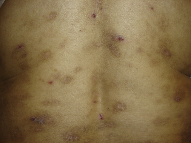
A 50-year-old gypsy woman presented with intense pruritus and skin lesions. She had controlled hypertension and Stage-4 chronic kidney disease with no hyperparathyroidism. No cholestatic disorder was observed. Physical examination revealed a papulonodular eruption on the limbs and the trunk and important lesions due to scratching. We consulted the dermatologist who prescribed sulpiride and a betamethasone and gentamicin cream in 10-day cycles. The lesions disappeared after 2 months of treatment. Sometimes new lesions appeared after a period of rubbing and scratching.
Prurigo nodularis is a dermatosis originating from pruritus and the persistence and progression of skin lesions are strictly correlated to scratching and rubbing. The skin between the lesions is usually normal but can be xerotic or lichenified. It is more frequent in middle aged women. The clinical feature is a papulonodular eruption with a few to hundreds of lesions that range from several millimetres to 2 cm in diameter. There is a tendency for symmetrical distribution with a predilection for extensor surfaces of the limbs and the trunk. The face and the palms are seldom affected. Prominent features include crusting and excoriations with postinflammatory hyperpigmented and hypopigmented macules (Figure 1).
A variety of systemic conditions have been reported to be associated with prurigo nodularis: chronic kidney disease, cholestatic disorders, Hodgkin’s lymphoma, hyperthyroidism, polycythaemia rubra vera and gluten-sensitive enteropathy. The clinical features are usually sufficient for diagnosing the disorder. The first step is to exclude any underlying disease and then to address the causes of general pruritus, which can be monitored by laboratory tests. Computerized tomography scan and chest X-rays are performed in the case of suspicion of lymphoma. Once the general causes have been excluded, the most common dermatologic differential diagnoses for prurigo nodularis are hypertrofic lichen planus, multiple keratoacanthoma and pemphigoid nodularis. If there are few lesions also nodular scabies can be considered.
It is the repetitive rubbing, scratching and picking of the skin that perpetuates the disease. In the absence of any clear underlying association, prurigo nodularis is highly resistant to therapy. Potent topical glucocorticoids such as triamcinolone acetonide are often successfully employed. Topical capsaicin can be effective in the very early manifestations. Ultraviolet-based therapy is useful in diffuse and/or resistant forms of prurigo nodularis. In resistant forms, cyclosporine reduces the severity of pruritus. Thalidomide and naltrexone can also be effective in recalcitrant diffuse forms. Psychosomatic interventions can be suggested.
Fig. 1.
Patient’s back. Excoriations with postinflammatory hyperpigmented and hypopigmented macules.
Acknowledgments
Conflict of interest statement. None declared.


