



Abstract
We present the case of “CL,” a 65 year-old, right-handed, Caucasian female referred for a neuropsychological evaluation of memory difficulties and depression with the rule out of pseudodementia. Mood measures showed elevated depression and apathy symptoms. The neuropsychological profile showed variable effort, intact comprehension but compromised confrontation naming and verbal memory deficits. A brain magnetic resonance imaging (MRI) scan was conducted within 24 hours of the neuropsychology exam. Using normative references from twenty female age and education matched healthy control peers, we identified marked differences in temporal cortex thickness and bilateral hippocampal, right amygdala, and right caudate volumes, supportive of a diagnosis of Semantic dementia. We discuss the neuroanatomical substrates involved in her neurocognitive and mood presentation and how standard measures of effort and malingering have questionable validity and rationale for dementia and pseudodementia referrals. This case highlights the importance of interdisciplinary collaboration and the value for combining normative neuropsychological and neuroradiological measures for clinical purposes.
Keywords: effort testing, pseudodementia, dementia, neuroradiology, MRI
Introduction
Pseudodementia is a cognitive and functional impairment mimicking a neurodegenerative disease but believed to be caused by neuropsychiatric symptoms. Pseudodementia is, in contrast to primary neurodegenerative disease, potentially reversible with management of psychiatric symptoms (Caine, 1981; Nussbaum, 1994). Psychiatric symptoms and depression, however, are risk factors for dementia and associated brain structural changes (Lamberty & Bieliauskas, 1993). Detecting and treating neuropsychiatric and/ or neurodegenerative disorders can thus be challenging. Clinically, red flags for pseudodementia include severe mood disturbance, questionable effort on measures of performance validity, and significant variability within cognitive domains on neuropsychological evaluation.
For neuropsychological assessment, our field has normative values available for comparison and classification of test performance. Normative neuroimaging data for clinical use is not widely available. In this paper we present the interesting case of an individual referred for clarification regarding dementia versus pseudodementia. We combined standardized neuropsychological with standardized norms from neuroimaging data to demonstrate the value of normative neuroimaging for differential diagnosis and clinical interpretation.
CASE PRESENTATION
“CL,” is a 65-year old, Caucasian, right-hand dominant, former reading teacher with 16 years of education and a self-reported ten month history of memory difficulties and depression. Her neurologist referred her to neuropsychology to rule out a possible pseudodementia. Background information was acquired from medical records, patient interview, and a separate interview with CL’s accompanying friend. Medical history from outside providers, including a prior neuropsychological evaluation, was not available at the time of the evaluation.
According to CL, her difficulties began approximately 10 months ago. At that time she began struggling to remember the names of her students and colleagues. Five months prior to our evaluation, she became lost driving to her primary care physician’s (PCP) appointment. The PCP reportedly called the department of motor vehicles and required her to take a driving test. According to CL, she completed two road tests: she failed the first but then passed the second. Her driver’s license had not been reinstated at the time of our evaluation. She reported feeling consequently “very sad” and “depressed”. She stated that her memory problems were attributable to her distress over these recent events in her life. CL stated that she currently feels “depressed and worried” most of her day, every day. At the time of our evaluation she denied suicidal/homicidal ideation and was not receiving psychotherapy services.
By contrast, CL’s accompanying friend and occasional caretaker reported that CL’s memory difficulties began 2-3 years earlier (corroborated by CL’s medical record), with a significant decline observed 6 months prior to her evaluation. Within the year before our assessment, CL’s friend noted increased difficulties with word-finding, which gradually progressed to severe confusion and conversations “devoid of content.” CL now frequently forgets the name of household objects and their purpose (what they are used for). According to her friend, CL’s depression has been ongoing for at least 20 years; the referring neurologist indicated that CL’s first major depressive episode occurred in her 20s. In addition to depression, her neurologist reported that CL’s family was concerned about “personality changes”, with inappropriate laughter, disinhibition, increased irritability, and affective flattening all noted. CL also developed difficulty in understanding humor and sarcasm and associated social strain with her siblings.
Medical system review was significant for sleep difficulties that began over the last year; specifically she wakes frequently during the night with an average of 1-3 hours nightly (per her friend). She reported only four and a half hours of sleep the night prior to our examination; she did not take her sleep medication for fear she would oversleep and miss the neuropsychological appointment. CL denied difficulties with olfaction, hearing, taste, or appetite (weight is stable). Her eyesight is corrected with eyeglasses. There is no past or current use of alcohol, tobacco, or illicit drugs.
Other medical history was remarkable for multiple fall-related head injuries and brief loss of consciousness caused by episodes of syncope (cause unknown), interstitial cystitis, depression, gastro esophageal reflux disease (GERD), and severe unregulated hyperlipidemia. Her medications at the time of the evaluation included Mirtazapine (nightly) for sleep, Prevacid (OTC), and Metamucil (OTC). She had recently discontinued use of Pravastatin (25mg, twice daily) for cholesterol because it reportedly made her body ache. She also recently ceased her antidepressant (type unknown) due to resulting drowsiness. Of note, she reported that her cholesterol level was well over 300 when last checked and stated she is overdue for her updated lab work as well as a brain MRI requested by her neurologist. Family medical history was significant for cancer and heart disease.
Psychosocially, CL lives alone and spends most of her time walking up the road to take care of her family members. She has a bachelor’s degree and denied any history of cognitive or learning disabilities.
Previous neuropsychological testing
An outside provider conducted a neuropsychological evaluation three years prior to our assessment. At that time she had performance scores less than verbal abilities with this considered partially due to visuospatial impairment. Memory testing showed mild encoding difficulties with impaired recall. Semantic and lexical fluencies were mildly impaired and she showed a reduced motor control in her right hand. The report from this evaluation was acquired approximately one week after the current assessment.
Previous brain MRI/EEG
A brain MRI conducted two years prior was interpreted by the neuroradiologist as indicating right greater than left mild temporal lobe atrophy but minimal overall global volume loss. No diagnosis was suggested. She also had a normal EEG at the time. The radiology report was not available at the time of our neuropsychological evaluation.
At the time of our assessment CL had yet to complete a repeat brain MRI. Due to her cognitive performance pattern we arranged for a brain MRI on the same day as our evaluation. For non-clinical purposes and for this case report, her MRI was segmented for gray and white matter analyses. The resulting regions were compared to brain structures from female age normative peers.
For part one of our report, we present her overall cognitive profile. For part two, we separately present the neuroanatomical comparison relative to matched peers.
Part 1: Neuropsychology Findings
Behavioral Observations
CL was oriented to person, place, time, and situation. Gait speed and balance appeared intact. Spontaneous speech was characterized by word-finding difficulties, some circumlocutions, and tangentiality. She frequently returned to the revocation of her driver’s license as the trigger to her mood and current difficulties. She required frequent redirection and clarification of task instructions from the examiner. In the first hours of testing she was fidgety and inappropriately jocular. She talked through tests and made sound effects to accompany her hand gestures (e.g., when instructed to show what people do with scissors she made a cutting sound). Following lunch, she appeared to have very little energy and motivation. Her affect was flat and slightly confrontational in her interactions with examiner. She also began to display odd behaviors such as looking around the room and naming things aloud (e.g., “wall,” “ceiling,” “floor”). Overall, she demonstrated variability between and within cognitive domains. She was unable to complete many tasks due to appearing confused and requiring redirection. Table 1 shows her current neuropsychological assessment scores and percentiles that overlap with those from a previous evaluation conducted in 2010.
Table 1.
Presentation of CL’s Performance On a Comprehensive Neuropsychological Assessment
| 2010 Assessment | Current Assessment | ||||
|---|---|---|---|---|---|
|
| |||||
| Cognitive Domain/Test | Raw Score | Percentile | Raw/Index Score |
T Score |
Percentile |
| Mood | |||||
| Geriatric Depression Scale | -- | -- | 22/30 | -- | -- |
| Apathy Scale | -- | -- | 15 | -- | -- |
| Effort Testing | |||||
| TOMM | |||||
| Trial 1 | -- | -- | 39/50 | -- | -- |
| Trial 2 | -- | -- | 45/50 | -- | -- |
| Rey 15 | 15/15 | -- | 3/15 | -- | -- |
| Reliable Digit Span | -- | -- | 8 | -- | -- |
| Attention, working memory, & processing speed | |||||
| Digit Span Total* | 16 | 50 | 14 | 47 | 37 |
| Forward span | 7 | -- | 6 | 48 | 42 |
| Backward span | 6 | -- | 4 | 47 | 37 |
| Symbol Search* | 29 | 63 | 11 | 33 | 5 |
| Language & language-related skills | |||||
| Boston Naming Test | 29/60 | -- | 6/60 | ||
| Controlled Oral Word Association Test | |||||
| Letter Fluency (FAS) | 27 | 16 | 13 | 27 | 1 |
| Semantic Fluency (Animals) | 15 | 20 | 7 | 23 | <1 |
| Multi-lingual Aphasia Examination | |||||
| Token Test | -- | -- | 41 | 46 | 33 |
| Finger Gnosis | |||||
| Visual Right | -- | -- | 0/5 | -- | -- |
| Visual Left | -- | -- | 2/5 | -- | -- |
| Tactile Right | -- | -- | 5/5 | -- | -- |
| Tactile Left | -- | -- | 5/5 | -- | -- |
| Left-right discrimination | |||||
| Self | -- | -- | 0 errors | -- | -- |
| Other | -- | -- | 0 errors | -- | -- |
| Reading sample | -- | -- | 6 errors | -- | -- |
| Repetition | -- | -- | 6 errors | -- | -- |
| Praxis | |||||
| Right | -- | -- | 2/6 | -- | -- |
| Left | -- | -- | 1/6 | -- | -- |
| Trail Making Test | |||||
| Part A | 41" | -- | 25" | 50 | 50 |
| Part B | 96" | -- | D/C | -- | -- |
| Matrix Reasoning^ | 16 | 75 | 11 | 40 | 16 |
| Visuoperceptual, Spatial, & Construction | |||||
| Clock Drawing Test | |||||
| Command | -- | -- | 58" / 2 errors | -- | -- |
| Copy | -- | -- | 77" / 3 errors | -- | -- |
| Benton Facial Recognition Test | -- | -- | 47 (corrected) | 55 | 71 |
| Learning & Memory | |||||
| Logical Memory# | |||||
| Immediate recall | -- | -- | 8 | 23 | <1 |
| Delayed recall | -- | -- | 2 | 27 | 1 |
| Dementia Screening | |||||
| Dementia Rating Scale-2 | |||||
| Attention | -- | -- | 33 | 40 | 16 |
| Initiation/Perseveration | -- | -- | 23 | 23 | 1 |
| Construction | -- | -- | 5 | 40 | 16 |
| Conceptualization | -- | -- | 37 | 50 | 50 |
| Memory | -- | -- | 21 | 37 | 9 |
| Total | -- | -- | 119 | 27 | 1 |
| Mini Mental State Examination | 29 | -- | 26& | -- | -- |
Comparison to 2010 neuropsychological evaluation is provided where test scores overlap.
Wechsler Adult Intelligence Scale-III.
Wechsler Abbreviated Scale of Intelligence.
Wechsler Memory Scale-III.
Mini Mental State Examination was given by the referring neurologist.
EXAMINATION RESULTS
Previous intellectual estimates
Medical reports indicate that CL was able to complete the full WAIS-III, demonstrating low average verbal and non-verbal intellectual abilities and average processing speed and working memory skills.
Current Mood
CL reported significant symptoms of depression (Yesavage et al., 1982) indicating feeling dissatisfied with her life, having dropped activities, feeling her life is empty, feeling bored, helpless, feeling hopeless, and that most people are better than she is. She endorsed mild levels of apathy (Marin, 1991) such as feeling indifferent to things and needing a push to get started.
Effort
CL completed two free-standing measures of effort and one embedded test of effort to evaluate the extent to which effort and variability might alter test performance (TOMM: Tombaugh, 1996; Reliable Digit Span: Greiffenstein et al., 1994; Rey-15: Rey, 1958). On the TOMM, CL’s performance was suggestive of questionable effort; however, she exhibited difficulty identifying several objects. CL reported being unable to identify “spoon” from a set of two unrelated pictures. When asked to describe what the spoon was used for, she could not provide an answer, nor could she use gesturing to demonstrate its function. On the reliable digit span (longest forward span correct on both trials plus longest backwards span correct on both trials) she scored 8, indicating acceptable performance (Greiffenstein et al., 1994).
On the Rey 15-Item Visual Memory Test, which requires the participant to reproduce a series of objects that are related in some way after a 10 second exposure, CL reported confusion. When prompted, she wrote the word “numbers” on the page. Upon further clarification, she drew three correct objects and one incorrect object, yielding a total score of 3/15 correct. By comparison, she scored 15/15 during her evaluation in 2010.
General Cognitive Screening
CL was administered the Dementia Rating Scale – 2 (DRS-2; Jurica, Leitten, & Mattis, 2001) and a clock drawing task to assess general ‘global’ cognitive functioning and dementia severity. Her DRS-2 performance was impaired relative to age and education-matched peers. Subtests scored at average levels for conceptualization, but low average for attention, memory, and visuoconstruction. She was impaired on initial/perseveration. She struggled more with language-based tasks relative to the movement and graphomotor items. On a clock drawing task CL correctly recalled the clock face and drew the clock proportionally correct but produced executive hand and number errors on the command and copy condition (Price et al., 2011). Figure 1.
Figure 1.

Clock Command/Copy
Command Condition shows executive errors involving incorrect hand placement (she was instructed to set the time to “10 after 11”) and misaligned numbers (particularly 9, 10, and 11) around the circle of the clock face. Copy Condition has the 12 misplaced, slight misalignment in numbers, and despite seeing a clock model with the numbers inside of the face - CL initially perseverates on her original command condition behavior of putting the numbers outside the clock face.
Attention and Processing Speed
Auditory attention span (Wechsler, 1997a) scored average for both forward and backward digit span, with low average for DRS-2 Attention subtest. Trail Making Test Part A was average relative to peers, while WAIS-III Symbol Search was below average.
Language
CL’s expressive and receptive language abilities were markedly compromised. Comprehension assessment via the Token Test (Florida Token Test; adapted from De Renzi & Vignolo, 1962) was intact for simple sentences but requiring additional trials on more syntactically complex sentences. Repetition was intact for simple sentences but impaired for grammatical complex sentences (i.e., “The phantom soared across the foggy heath” resulted in “The phanny heat”; “No ifs, ands, or buts” resulted in “no ifs or no buts”). Visual confrontational naming was strikingly impaired (Boston Naming Test; Kaplan, Goodglass, & Weintrab, 1983) with numerous semantic and circumlocutious errors but no perceptual deficits. Verbal fluency was impaired across letter and semantic conditions (Controlled Oral Word Association; Tombaugh, Kozak, & Rees, 1999); poor performance resulted from few responses rather than loss of set or rule violations. A written response of the Cookie Theft picture was in cursive and largely grammatically intact but with minimal content and limited appreciation for the gestalt (Figure 2). During this task she laughed throughout and required multiple reminders of the instructions.
Figure 2.
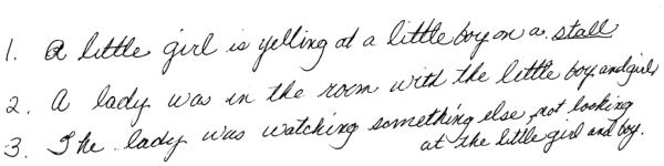
Cookie Theft Writing
Handwriting appears fluid and well-formed. Of note is spelling "stall" instead of "stool". Written answers reveal that only basic visual information about the picture was perceived rather than indicating that CL understood the gestalt of the depicted event.
Regarding language-related skills, ideomotor praxis was bilaterally impaired (i.e., “Pound a nail into a board with a hammer” resulted in poking her finger into the table; “Use a hand saw to cut wood” resulted in a hammer motion). Finger gnosis was impaired bilaterally; she was unable to visually name her right hand fingers and two fingers on her left hand. Right left discrimination was intact for both self and examiner. An assessment for acalculia was significant for spatial misalignment and misusing addition for multiplication although this was later self-corrected.
Visuospatial
Her ability to identify and match a series of faces presented at alternating angles scored average relative to age and education-matched peers (Benton Facial Recognition; Benton, Sivan, Hamsher, Varney, & Spreen, 1994). CL was able to copy a circle, but made errors with a cube. She spontaneously drew a well-formed horse on the back of a piece of paper. Her copy of a clock included misplaced numbers.
Memory
With regard to memory, CL reported difficulty remembering both past and recent events. DRS-2 Memory subtest was low average. However, on a measure of verbal memory requiring recall of simple prose (Wechsler Memory Scale-III Logical Memory; Wechsler, 1997b) CL’s performance was impaired across immediate and delayed recall trials with minor benefit from repetition on the second story. Recognition of the material was within the average range. On a nine-word list-learning task (Philadelphia (repeatable) Verbal Learning Test), CL produced a flat learning curve with five intrusion errors some of which were linguistic blends (i.e., ‘strinapples’). The interference trial resulted in two recall items and one intrusion from the original list. Immediate and delay recall were impoverished and involved intrusions. She did not benefit from category cues. Recognition discriminability was significant for endorsement across all foil types (semantic, prototypical, interference, unrelated).
Abstraction and reasoning
Abstract reasoning of visual matrices scored at low average levels (Wechsler Abbreviated Scale of Intelligence Matrix Reasoning; Wechsler, 1999) and her visual conceptualization on the DRS-2 subtest was average.
Motor
Fine motor manual dexterity was within the average range bilaterally.
Part 1 Discussion
At our evaluation, limited outside medical information was available. In spite of variability on performance validity testing and reduced effort, based on neuropsychology alone, we considered her profile characteristic of an atypical cortical dementia. CL showed marked depression and questionable response consistency secondary to cognitive deficits. Language assessment was most striking with impoverished confrontation naming, letter and semantic fluency, followed secondarily by impaired ideomotor praxis, finger gnosis, and reduced repetition and comprehension for grammatically complex sentences. Learning and memory were significant for impoverished free recall on prose and list-learning, with intrusions suggestive of source error impairment. Recognition performance ranged from average to impaired. Basic attention and basic visual perception were a relative strength, although higher-level appreciation for the gestalt and complex figural integration were qualitatively poor. She also exhibited a range of processing speed (average to impaired), emotional incontinence ranging from jocularity/joyful to flat affect, and variable performance on effort measures. Overall, these findings suggested cortical atrophy of lateral/ medial temporal and possibly parietal lobes, with questionable orbitofrontal and medial frontal lobe integrity. Given the relative sparing of faces and her construction ability, her posterior inferior temporal lobes were considered less compromised.
Due to CL’s overall cognitive pattern, we pushed for an MRI within the next 24 hours. These findings are provided in Part II below.
Part II: Neuroimaging
Methodology
A brain MRI was conducted within 24 hours of our evaluation. CL’s MRI protocol involved clinical 2D T1, T2, T2 fluid attenuated inversion recovery (FLAIR; 320×196 matrix, 28 slices, 5mm gap, 0.625×0.625×4mm voxels, TR/TE=9000/90ms), and diffusion scans. She completed two 3D T1 MP-RAGE sequences (Siemens Avanto 1.5T; 384×384 matrix, 160 contiguous slices, 0.64×0.64×1.2mm voxels, TR/TE=1490/3.45ms) acquired axially without contrast. Her 3D T1 scans were of sufficient quality for automated segmentation and volumetric analysis.
Normative Comparison group
Structural MRI data were from a set of 20 non-demented age (mean = 65.20±2.44; range = 60–69) and education (mean = 15.90±2.84; range = 12–20) matched females who participated as part of separate federally funded investigations that included MRI and neuropsychological testing. Each individual completed a brain MRI from a Siemens 3T Verio scanner using an 8-channel head coil. The protocol included an MP-RAGE T1-weighted scan with the following parameters: 176 contiguous slices, 1×1×1mm voxel, TR/TE = 2500/3.77ms, sagittal acquisition. These data were used to provide a standardized reference point for CL’s brain volumes.
MRI Segmentation
For both CL and the normative peers, cortical reconstruction and volumetric segmentation was completed with FreeSurfer image analysis suite version 5.3, which is documented and freely available for download online (http://surfer.nmr.mgh.harvard.edu/; Fischl, 2012). The technical details of these procedures are described in prior publications (Dale, Fischl, & Sereno, 1999; Fischl & Dale, 2000; Fischl et al., 2002; Fischl, Salat, et al., 2004a; Fischl, Sereno, & Dale, 1999; Fischl, van der Kouwe, et al., 2004b; Han et al., 2006). FreeSurfer morphometric procedures have been demonstrated to show good test-retest reliability across field strengths (Han et al., 2006), which is important given the different field strengths used for CL’s MRI and the normative group’s MRI. Both CL and the normative group had adequate white matter signal to noise and gray/white contrast.
Primary neuroimaging variables of interest included cortical thickness by brain lobe, hippocampus, amygdala, caudate, thalamus, and white matter volume by hemisphere, as well as leukoaraiosis (LA) volume. Hippocampal volumes from the normative group match reported volumes for healthy older adults (Sánchez-Benavides et al., 2010). Procedures for the measurement of cortical thickness have been validated against histological analysis (Rosas et al., 2002) and manual measurements (Kuperberg et al., 2003; Salat et al., 2004). FreeSurfer segmentations were checked for accuracy. CL’s segmentation required manual ‘control points’ placed in fusiform gyral white matter and reprocessing to fix suboptimal temporal lobe segmentation. All volumes were calculated as a ratio to total intracranial volume (TICV). TICV was derived from FreeSurfer’s brainmask volume, which has high correlation with manual TICV measurements (r = 0.93; ICC = 0.92; p < 0.01) and high spatial overlap (Dice Similarity Coefficient = 0.95). T1-weighted volumes and thickness values are reported in FreeSurfer conformed space (1mm3 voxels, 256×256×256 matrix, coronal reslice). Leukoaraiosis (LA) volumes for controls and CL were created using in-house tools using methods previously reported (Price et al., 2014).
Results
Table 2. Supplemental Table 1. Figure 4. CL’s total intracranial volume and total brain volume were below the mean of the peer sample (CL’s TICV z = −1.00; brain volume correcting for TICV = −2.24).
Table 2.
Raw (uncorrected) Neuroimaging Cortical Gray, Subcortical Gray, and White Matter Metrics for Peers (n=20) and CL
| Peers (n=20) | CL | |||||
|---|---|---|---|---|---|---|
| Mean | S.D. | Min | Max | Measure | ||
|
|
||||||
| TICV | 1471042.35 | 106449.65 | 1304282 | 1698745 | 1364140 | |
| Brain Vol | 981789.95 | 77591.12 | 847652 | 1175235 | 843661 | |
|
|
||||||
| Cortical Gray Thickness | L Frontal | 2.40 | 0.11 | 2.23 | 2.60 | 2.48 |
| R Frontal | 2.35 | 0.09 | 2.21 | 2.50 | 2.35 | |
| L Temporal | 2.59 | 0.11 | 2.41 | 2.80 | 2.25 | |
| R Temporal | 2.64 | 0.12 | 2.42 | 2.82 | 1.79 | |
| L Parietal | 2.24 | 0.11 | 2.05 | 2.39 | 2.22 | |
| R Parietal | 2.25 | 0.10 | 2.05 | 2.37 | 2.07 | |
|
|
||||||
| Gray Structures | L Hipp | 3658.77 | 408.57 | 3077 | 4883 | 2376 |
| R Hipp | 3826.82 | 383.08 | 3176 | 4604 | 2368 | |
| L Amyg | 1395.92 | 205.65 | 885 | 1875 | 1179 | |
| R Amyg | 1465.72 | 201.29 | 950 | 1769 | 697 | |
| L Caudate | 3381.16 | 506.90 | 2481 | 4349 | 2766 | |
| R Caudate | 3550.29 | 495.68 | 2722 | 4641 | 2113 | |
| L Thalamus | 6649.56 | 732.70 | 5412 | 8208 | 5835 | |
| R Thalamus | 6240.18 | 640.64 | 5256 | 7394 | 5367 | |
|
|
||||||
| White Matter |
L WM Vol | 202932.24 | 21974.24 | 162892 | 246205 | 174754 |
| R WM Vol | 205193.42 | 22851.20 | 164359 | 254182 | 165313 | |
| LA* | 2973.51 | 2710.31 | 742 | 73967 | 2080 | |
|
|
||||||
Results demonstrate CL’s thinner temporal lobes, small hippocampi bilaterally and small right caudate, amygdala, and white matter volumes.
TICV = Total Intracranial Volume in mm3. Brain Vol = Total parenchyma volume without ventricles. Hipp = Hippocampus. Amyg = Amygdala. WM Vol = Volume of parenchyma white matter by hemisphere.
LA = Leukoaraiosis (n=14). Structural volumes are in mm3 in FreeSurfer interpolated space. Thickness is in mm.
Figure 4.

Representative Images from CL’s MRI
Cortical gray matter thickness metrics show CL had thinner temporal cortices with thinning more pronounced on the right side (temporal left z = −3.15; right z = −7.04); temporal atrophy was primarily anterior (Figure 4). The right parietal lobe was below the peer sample mean (parietal left z = −0.13; right = −1.80). Frontal metrics were within the average range (frontal left z = 0.74; right z = −0.04).
Hippocampus, amygdala, caudate, and thalamus volumetrics (TICV corrected) were asymmetric (R < L). Hippocampal volumes were bilaterally smaller than peers (left z = −3.08; right z = −3.25). Amygdala volumes were strikingly asymmetric (left z = −0.60; right z = −3.42). Caudate volumes were also dramatically asymmetric (left z = −0.94; right z = −3.33). Thalamus volumes were asymmetric but within reasonable ranges relative to peers (left z = −0.59, right z = −1.19).
White matter volumetric (TICV corrected) comparisons showed asymmetry relative to the peers (white matter left z = −1.38; right z = −2.39). Leukoaraiosis load was minimal and consistent with peer ranges (z = −.27).
Part 2 Discussion
Neuroimaging comparisons showed a remarkable brain volume difference relative to peers involving an asymmetric pattern (right < left). There was clear temporal lobe disruption as evidenced by bilateral thinness and severely reduced bilateral hippocampi. Although asymmetric, there was evidence of disease in both hemispheres. The subcortical regions revealing the most striking asymmetry included the amygdala, caudate, and general white matter metrics. Asymmetry was noted in parietal cortex and, to a lesser extent, in the thalamic hemispheres; these structures were in the low average range relative to peers. The overall volume loss corresponds to the larger ventricles seen in Figure 4. Of all regions, the frontal lobe appeared the most preserved with thickness values well within the average range relative to peers. Based on leukoaraiosis metrics, there was also no convincing evidence that small vessel vascular disease contributed to her profile.
For her current MRI, the neuroradiologist commented on global cerebral volume loss, severe bilateral temporal lobe atrophy, moderate parietal lobe atrophy, mild symmetric hippocampal loss, and “normal” basal ganglia. The neuroradiologist interpreted the MRI as “suggestive of Alzheimer’s type dementia.” Our data add to this reading. First, we found strong asymmetry of atrophy in the amygdala, caudate, and white matter as well as temporal and parietal cortex. Second, moderate bilateral hippocampal atrophy was also apparent relative to the normative sample.
DISCUSSION
In this case report we presented data from a 65-year old woman, “CL,” who was referred for neuropsychological evaluation to assist with differential diagnosis and rule out pseudodementia. CL had variability in her scores, mood disturbance, and poor performance on measures of effort. Using information from just effort and mood measures one could surmise pseudodementia. With full neuropsychological assessment, however, there are clear strengths and weaknesses that demonstrate a marked pattern. Data from assessment revealed global cognitive impairment as well as language and memory impairment. CL exhibited heterogeneity of performance across and within domains but generally struggled on language and memory tasks. While there was evidence of language and memory decline compared to her 2010 assessment, other domains, visuospatial in particular, did not show dramatic declines. Her overall neuropsychological pattern strongly suggested a neurodegenerative process although some could consider her profile suggestive of pseudodementia due to the extent of her mood disturbance, her behavioral presentation, and her variable effort.
Neuroimaging results demonstrated marked anterior temporal atrophy with reduced bilateral hippocampal and right amygdala and caudate volumes (Figure 4). The extent and asymmetry of her atrophy is clear relative to peers. Neuroimaging thus confirmed the impairment, ruling out pseudodementia.
CL was first diagnosed with depression in her 20s but struggled with it more particularly within the past 2-3 years. Early depressive and emotional signs can herald developing dementia (Cummings, 1986) and are associated with limbic system changes (Catani, Dell'Acqua, & Thiebaut de Schotten, 2013). CL’s emotional variability observed throughout the exam and stated in medical record reports is consistent with right hemisphere disruption. Although CL did not have striking evidence of reduced global frontal thickness, she had significantly impoverished temporal thickness (right > left) and asymmetric atrophy of amygdala, caudate, and white matter. CL’s right hemisphere changes possibly explain her performance variability and likely explain her emotional variability (Bickart et al., 2014). Additionally, right hemisphere damage is likely one of the causes of her reduced humor appreciation (W. Liu et al., 2004; Rosen et al., 2002; Shammi & Stuss, 1999).
CL’s language difficulties are supported by her bilateral temporal atrophy. It is important to note that although CL had strikingly impoverished confrontation naming, she was fluent in writing and wrote in cursive. Consequently, she had adequate use of her right hand for accessing graphemic systems (e.g., her description of the ‘cookie theft’ was fluently written in script). CL’s written language was significant only for the paucity of nouns and an integration of the overall picture gestalt. This is a possible symptom of right parietal thinning.
CL’s memory deficits are supported by her bilateral atrophy in temporal cortex and medial temporal structures. CL’s observed memory impairment includes encoding and retrieval deficits with significant recognition intrusions. In general, increasing entorhinal cortex volume or blood flow associates with aspects of verbal memory (Cabeza, Dolcos, Graham, & Nyberg, 2002; Goto et al., 2011; Price et al., 2010; Rodrigue & Raz, 2004). The cingulum and entorhinal cortices are components of the limbic system, which supports both memory and emotion; CL’s MRI demonstrates the extent of her limbic system disruption. In addition, the temporo-amygdala-orbitofrontal network is certainly affected given CL’s extensive amygdala and temporal atrophy; this network is important for integrating cognition and emotion (Catani, de Schotten, & Slater, 2013).
CL’s right caudate was much smaller than peers. The caudate plays a role in integrating attentional systems and mediating communication to the frontal lobes. CL’s right frontal lobe, while within the normal range, was approximately one standard deviation below the left frontal lobe with regard to thickness, likely indicative of asymmetric atrophy. Additionally, both the caudate and medial temporal lobe connect directly to the frontal lobes. CL’s overall frontal cortex is largely intact but closely connected network regions are impaired, likely reducing the integrity of the white matter connections to frontal regions. Specifically, right caudate input and output could be disrupted thereby resulting in understimulation and reduced communication to the right frontal lobe.
CL’s relative sparing of frontal lobes and thalamus is consistent with intact basic attention; there is, however, a suggestion of frontal disease progression due to CL’s reduced repetition and comprehension of complex syntax (Grossman & Moore, 2005). CL’s thalamic volumes were within normal ranges, indicating that disease progression/ pathology, while likely affecting the caudate, has not spread measurably to the thalamus. It is not known if white matter pathways to and from the thalamus are affected, although it is likely given the extent of brain regions directly connected to the thalamus. CL’s clinical diffusion-weighted neuroimaging data were not of sufficient quality for such analyses.
Other contributing factors to her degeneration include reported unregulated hyperlipidemia and previously reported head injuries. Blood lab work conducted the day following her neuropsychological evaluation revealed severely elevated cholesterol levels, with the exception of HDL, which was within normal limits. Remarkably, however, the T2 FLAIR brain image revealed only one small spot of very intense signal, indicating minimal white matter disease. This indicates that her depression is not driven by cerebrovascular changes. Elevated cholesterol and history of head injury (she experienced minor but repeated head injuries due to falls) have been linked with fronto-temporal changes and so perhaps played a role in her disease progression (Deutsch, Mendez, & Teng, 2014).
Diagnosis
Considering only her behavioral profile, differential diagnoses could include Semantic dementia and Alzheimer’s disease given the extent of her memory and language impairment. Her first-reported symptoms were “memory problems”, although it is possible that word-finding difficulty was reported as a “memory” problem. However, when combining neuropsychology with neuroimaging, her pattern of atrophy on MRI, particularly when compared to normative imaging data, revealed marked temporal cortex loss that exceeded her hippocampal volume loss (right temporal thickness was 7.04 standard deviations below peers whereas right hippocampus was 3.25 standard deviations below peers); her results thus indicate a primary temporal cortex process with pronounced but less hippocampal atrophy. The neuroradiologist commented on bilateral temporal, parietal, and hippocampus atrophy but interpreted the pattern as suggestive of Alzheimer’s disease, rather than frontotemporal dementia. The results from our analyses, in contrast, indicated a pattern that is not typical of Alzheimer’s disease, though it is possible she has comorbid Alzheimer’s disease given her moderate bilateral hippocampal atrophy.
Medial temporal lobe atrophy is a hallmark of early Alzheimer’s disease (Fox et al., 1996; Jack et al., 1997; Jack et al., 1998). Atrophy in Alzheimer’s disease is usually bilateral and does not typically occur in anterior temporal lobes early in the disease process, initially affecting medial temporal lobes (Chan et al., 2001). In contrast, Semantic dementia commonly presents with asymmetric anterior temporal atrophy (Chan et al., 2001; Galton et al., 2001) with left prominent atrophy more common than right (Thompson, Patterson, & Hodges, 2003).
Semantic dementia is a subtype of frontotemporal dementia (FTD; Neary et al., 1998), which is a term encapsulating a broad array of deficits and symptoms and thus has limited utility for deficit specificity. CL’s age is typical age of onset for Semantic dementia (Hodges & Patterson, 2007). Her pattern of extensive anterior temporal atrophy matched what is commonly seen in Semantic dementia. While left hemisphere atrophy in Semantic dementia is more common than right, right atrophy is well-documented in the literature (Thompson et al., 2003); CL’s right>left atrophy was only surprising given the extent of her language difficulties and her mild to unimpaired visuospatial abilities (Hodges & Patterson, 2007). CL exhibited visuospatial difficulties on her 2010 neuropsychological evaluation but our assessment yielded only mild deficits, indicating little or no decline in abilities over time. She did have striking isolated instances of object recognition impairment (such as on the TOMM) but visuoperceptual and visuospatial deficits were not remarkable relative to her overall profile, suggesting that object recognition deficits were likely a result of decline in the semantic system.
Irritability, impulsiveness, and bizarre changes in behaviors are common in individuals with Semantic dementia with greater right side atrophy (Edwards-Lee et al., 1997). CL exhibited increased negativity, depression, and apathy but her friend and occasional caretaker did not report increased irritability or impulsiveness; she was overly jocular at times during the evaluation but external report places more emphasis on her word finding and language than on her behavioral changes. In general, however, CL’s behavioral and cognitive profile is expected given the extent of her right and left temporal atrophy. It is also possible that language functions for CL are right hemisphere lateralized or are bilaterally distributed, although this would be unexpected given her right-handedness.
While CL’s neuropsychological and neuroimaging data are supportive of a diagnosis of Semantic dementia the heterogeneity of behavior, atrophy, and pathology within Semantic dementia limit the utility of the label. Applying specific labels, while concise and useful for clinical billing, does not adequately summarize CL’s behavioral and brain deficits. The argument for describing deficits rather than or in addition to diagnosis has been made previously and is worth repeating (Cosentino et al., 2004; Lamberty & Bieliauskas, 1993).
Effort
The case of CL is also useful to demonstrate the value of performance validity measures on neuropsychological testing. She ‘failed’ one stand-alone measure (Rey-15), had marginal performance on another (TOMM), but performed adequately on an embedded measure (reliable digit span). While symptom and performance validity testing play an important role in neuropsychological evaluations, some cautions are in order. The utility of measures of effort or performance validity tests within dementia populations has been called into question (Rudman, Oyebode, Jones, & Bentham, 2011; Dean, Victor, Boone, Philpott, & Hess, 2009). Specifically, it has been shown that the relationship between emotional function and effort is poor and common measures of performance validity might not be valid in dementia (Rudman et al., 2011).
Where a diagnosis of dementia is not immediately clear, such as with CL who had variable performance on performance validity testing and neuropsychological measures and who presented with mood disturbance and reduced effort throughout testing, performance validity test findings should be interpreted with caution (Van Dyke, Millis, Axelrod, & Hanks, 2013). For a case like CL’s, it is conceivable that a neuropsychologist with limited outside background medical information (such as was the case with CL) who starts with mood and performance validity testing, might terminate the examination due to the patient’s questionable effort. While such situations might occur infrequently, they could negatively affect patient care. Thus in general, the use of performance validity testing in any population at increased risk of dementia (older adults) should be applied and interpreted with caution without neuroimaging to support one interpretation or another. Interpreting the results of CL’s performance validity testing is thus of limited utility. It is possible but speculative that CL’s variability in performance on performance validity testing and within cognitive domains is tied to her severe right hemisphere atrophy as well as her severe anterior temporal lobe atrophy.
Limitations
We recognize limitations with our study. MRI for CL was acquired at 1.5T while the normative group was acquired at 3T. Different sequence parameters were used, which could potentially introduce error into analyses; however, FreeSurfer is reliable across field strengths (Han et al., 2006). Further, CL’s clinical scan had comparable gray-white contrast and signal to noise to the control group (data not reported but derived in part from a FreeSurfer QA tool: https://surfer.nmr.mgh.harvard.edu/fswiki/QATools). All images were checked for segmentation quality; only CL had suboptimal segmentations due to significant temporal atrophy. Segmentation errors were fixed manually and then reprocessing occurred. Final segmentation was of excellent quality. For MRI with atypical atrophy or damage, automated segmentation tools will likely require manual intervention to yield valid results, a significant limitation for clinical use.
Manual segmentation or manual cleaning of automated segmentations would improve accuracy of hippocampal segmentations (FreeSurfer overestimates hippocampal volumes, for example; refer to Tae, Kim, Lee, Nam, & Kim, 2008; Sánchez-Benavides et al., 2010) but manual segmentation is time-intensive and can require months of training to gain expertise. Time required for segmenting multiple brain regions, including portions of the cortex and selected subcortical regions, would be prohibitive for clinical use. Small, well-defined regions (e.g., caudate nucleus) can take 15-30 minutes for an expert rater to measure but larger or more complex brain regions require multiple hours per region. Manual methods also introduce human rater error. We recognize that we had a limited normative sample size (n = 20) but they were matched closely for sex, age, and education. The normative values we used, however, are similar to values recently reported in the literature (Jiang et al., 2014; Long et al., 2012), although direct comparisons in thickness and volume can be limited by demographic differences between studies. We also did not investigate specific sub-regions of lobar cortex, which might clarify the pattern of atrophy further (e.g., anterior more than posterior).
Clinical Recommendations
Given the extent of CL’s cognitive difficulties, it was recommended that her daily activities be supervised more closely by family or friends and then, as her abilities continue to decline, CL receive formal assistance. Closer monitoring of her home environment, medications, eating habits, and other self-care would benefit her overall health, especially in light of her unregulated hyperlipidemia. Given the extent of her deficits and brain atrophy, repeat neuropsychological assessment would not benefit her care.
Strengths and future directions
The strength of the current case report is our use of age and sex matched normative neuroimaging data to document the extent of CL’s brain disease and asymmetrical atrophy and argue against pseudodementia and poor effort. Previous reports on poor effort in dementia have limited integration with neuroimaging. Normative imaging variables clarified the extent of her atrophy and helped support her diagnosis with dementia. At least one FDA-approved tool exists for quantifying brain volumes for clinical use (i.e., NeuroQuant, which is based on FreeSurfer segmentation algorithms and thus comparable to our methods; www.cortechslabs.com), but it is not used widely. There are, in contrast, many research tools available to reliably quantify brain volumes and lesion load but they have not yet transitioned to clinical applications. Marketed software for clinical requires FDA approval given the definitions of “medical devices” in Section 201(h) of the Federal Food, Drug & Cosmetic Act (FD&C; 21 U.S.C. 301). Some argue that this will also be the case for computerized neuropsychological assessment systems in the future (Bauer et al., 2012).
With this case report, we offer a glimpse of the possibilities and challenges for integrating neuropsychology and standardized neuroimaging for differential diagnosis classification. Integration, however, requires careful consideration to age, sex, education, comorbidities, MRI field strength and acquisition parameters, as well as other potentially salient factors that might alter brain-behavioral inferences. The same rigor used to derive psychological assessment tools and normative values must be applied to creating neuroimaging normative values. Neuropsychologists with sub-specialty interests in neuroimaging appear uniquely positioned to initiate the integration of normative-based assessment and normative-based neuroimaging for clinical care.
Figure 3.

Raw and standardized z-score values for cortical and subcortical regions.
Charts in the left column display raw thickness and TICV-corrected gray and white volumetrics by group. Charts in the right column display normative-based (n = 20) z scores for thickness and gray and white volumes corrected for TICV. Charts in the left column display standard deviations for the control group data whereas those in the right column (z scores) display standard error bars.
Acknowledgements
Supported in part by NINDS K23 NS60660; NINDS R01 NS082386; NINR R01 NR014181
References
- Bauer RM, Iverson GL, Cernich AN, Binder LM, Ruff RM, Naugle RI. Computerized neuropsychological assessment devices: Joint position paper of the American Academy of Clinical Neuropsychology and the National Academy of Neuropsychology. The Clinical Neuropsychologist. 2012;26(2):177–196. doi: 10.1080/13854046.2012.663001. [DOI] [PMC free article] [PubMed] [Google Scholar]
- Benton AL, Sivan AB, Hamsher K, Varney NR, Spreen O. Contributions to Neuropsychological Assessment: A Clinical Manual. 2nd Oxford University Press; New York: 1994. [Google Scholar]
- Bickart KC, Brickhouse M, Negreira A, Sapolsky D, Barrett LF, Dickerson BC. Atrophy in distinct corticolimbic networks in frontotemporal dementia relates to social impairments measured using the Social Impairment Rating Scale. Journal of Neurology, Neurosurgery, and Psychiatry. 2014;85(4):438–448. doi: 10.1136/jnnp-2012-304656. doi:10.1136/jnnp-2012-304656. [DOI] [PMC free article] [PubMed] [Google Scholar]
- Cabeza R, Dolcos F, Graham R, Nyberg L. Similarities and differences in the neural correlates of episodic memory retrieval and working memory. NeuroImage. 2002;16(2):317–330. doi: 10.1006/nimg.2002.1063. doi:10.1006/nimg.2002.1063. [DOI] [PubMed] [Google Scholar]
- Caine ED. Pseudodementia. Archives of General Psychiatry. 1981;38(12):1359–1364. doi: 10.1001/archpsyc.1981.01780370061008. doi:10.1001/archpsyc.1981.01780370061008. [DOI] [PubMed] [Google Scholar]
- Catani M, de Schotten MT, Slater D. Connectomic approaches before the connectome. NeuroImage. 2013 doi: 10.1016/j.neuroimage.2013.05.109. doi:10.1016/j.neuroimage.2013.05.109. [DOI] [PubMed] [Google Scholar]
- Catani M, Dell'Acqua F, Thiebaut de Schotten M. A revised limbic system model for memory, emotion and behaviour. Neuroscience & Biobehavioral Reviews. 2013;37(8):1724–1737. doi: 10.1016/j.neubiorev.2013.07.001. doi:10.1016/j.neubiorev.2013.07.001. [DOI] [PubMed] [Google Scholar]
- Chan D, Fox NC, Scahill RI, Crum WR, Whitwell JL, Leschziner G, Rossor MN. Patterns of temporal lobe atrophy in semantic dementia and Alzheimer's disease. Annals of neurology. 2001;49(4):433–442. doi:10.1002/ana.92. [PubMed] [Google Scholar]
- Cosentino SA, Jefferson AL, Carey M, Price CC, Davis-Garrett K, Swenson R, Libon DJ. The clinical diagnosis of vascular dementia: A comparison among four classification systems and a proposal for a new paradigm. The Clinical Neuropsychologist. 2004;18(1):6–21. doi: 10.1080/13854040490507118. [DOI] [PubMed] [Google Scholar]
- Cummings J. Subcortical dementia. Neuropsychology, neuropsychiatry, and pathophysiology. The British Journal of Psychiatry. 1986;149(6):682. doi: 10.1192/bjp.149.6.682. doi:10.1192/bjp.149.6.682. [DOI] [PubMed] [Google Scholar]
- Dale AM, Fischl B, Sereno MI. Cortical surface-based analysis. I. Segmentation and surface reconstruction. NeuroImage. 1999;9(2):179–194. doi: 10.1006/nimg.1998.0395. doi:10.1006/nimg.1998.0395. [DOI] [PubMed] [Google Scholar]
- De Renzi E, Vignolo LA. The token test: A sensitive test to detect receptive disturbances in aphasics. Brain : a Journal of Neurology. 1962;85(4):665–678. doi: 10.1093/brain/85.4.665. doi:10.1093/brain/85.4.665. [DOI] [PubMed] [Google Scholar]
- Dean AC, Victor TL, Boone KB, Philpott LM, Hess RA. Dementia and effort test performance. The Clinical Neuropsychologist. 2009;23(1):133–152. doi: 10.1080/13854040701819050. doi:10.1080/13854040701819050. [DOI] [PubMed] [Google Scholar]
- Deutsch M, Mendez M, Teng E. Medical Risk Factors For Frontotemporal Degeneration: Analysis of the NACC Uniform Dataset (S58. 002) Neurology. 2014 [Google Scholar]
- Edwards-Lee T, Miller BL, Benson DF, Cummings JL, Russell GL, Boone K, Mena I. The temporal variant of frontotemporal dementia. Brain: A Journal of Neurology. 1997;120(6):1027–1040. doi: 10.1093/brain/120.6.1027. doi:10.1093/brain/120.6.1027. [DOI] [PubMed] [Google Scholar]
- Fischl B. FreeSurfer. NeuroImage. 2012:1–15. doi: 10.1016/j.neuroimage.2012.01.021. doi:10.1016/j.neuroimage.2012.01.021. [DOI] [PMC free article] [PubMed] [Google Scholar]
- Fischl B, Dale AM. Measuring the thickness of the human cerebral cortex from magnetic resonance images. Proceedings of the National Academy of Sciences of the United States of America. 2000;97(20):11050–11055. doi: 10.1073/pnas.200033797. doi:10.1073/pnas.200033797. [DOI] [PMC free article] [PubMed] [Google Scholar]
- Fischl B, Salat DH, Busa E, Albert M, Dieterich M, Haselgrove C, Dale AM. Whole brain segmentation: automated labeling of neuroanatomical structures in the human brain. Neuron. 2002;33(3):341–355. doi: 10.1016/s0896-6273(02)00569-x. [DOI] [PubMed] [Google Scholar]
- Fischl B, Salat DH, van der Kouwe AJW, Makris N, Ségonne F, Quinn BT, Dale AM. Sequence-independent segmentation of magnetic resonance images. NeuroImage. 2004a;23:S69–S84. doi: 10.1016/j.neuroimage.2004.07.016. doi:10.1016/j.neuroimage.2004.07.016. [DOI] [PubMed] [Google Scholar]
- Fischl B, Sereno MI, Dale AM. Cortical surface-based analysis. II: Inflation, flattening, and a surface-based coordinate system. NeuroImage. 1999;9(2):195–207. doi: 10.1006/nimg.1998.0396. doi:10.1006/nimg.1998.0396. [DOI] [PubMed] [Google Scholar]
- Fischl B, van der Kouwe A, Destrieux C, Halgren E, Ségonne F, Salat DH, Dale AM. Automatically parcellating the human cerebral cortex. Cerebral cortex. 2004;14(1):11–22. doi: 10.1093/cercor/bhg087. doi:10.1093/cercor/bhg087. [DOI] [PubMed] [Google Scholar]
- Fox NC, Warrington EK, Freeborough PA, Hartikainen P, Kennedy AM, Stevens JM, Rossor MN. Presymptomatic hippocampal atrophy in Alzheimer's disease: A longitudinal MRI study. Brain: A Journal of Neurology. 1996 doi: 10.1093/brain/119.6.2001. doi:10.1093/brain/119.6.2001. [DOI] [PubMed] [Google Scholar]
- Galton CJ, Patterson K, Graham K, Lambon-Ralph MA, Williams G, Antoun N, Hodges JR. Differing patterns of temporal atrophy in Alzheimer’s disease and semantic dementia. Neurology. 2001;57(2):216–225. doi: 10.1212/wnl.57.2.216. doi:10.1212/WNL.57.2.216. [DOI] [PubMed] [Google Scholar]
- Goto M, Abe O, Miyati T, Yoshikawa T, Hayashi N, Takao H, Ohtomo K. Entorhinal cortex volume measured with 3T MRI is positively correlated with the Wechsler Memory Scale-Revised logical/verbal memory score for healthy subjects. Neuroradiology. 2011;53(8):617–622. doi: 10.1007/s00234-011-0863-1. doi:10.1007/s00234-011-0863-1. [DOI] [PubMed] [Google Scholar]
- Greiffenstein MF, Baker WJ, Gola T. Validation of malingered amnesia measures with a large clinical sample. Psychological Assessment. 1994;6(3):218. [Google Scholar]
- Grossman M, Moore P. A longitudinal study of sentence comprehension difficulty in primary progressive aphasia. Journal of Neurology, Neurosurgery, and Psychiatry. 2005;76(5):644–649. doi: 10.1136/jnnp.2004.039966. doi:10.1136/jnnp.2004.039966. [DOI] [PMC free article] [PubMed] [Google Scholar]
- Han X, Jovicich J, Salat D, van der Kouwe A, Quinn B, Czanner S, Fischl B. Reliability of MRI-derived measurements of human cerebral cortical thickness: the effects of field strength, scanner upgrade and manufacturer. Neuroimage. 2006;32(1):180–194. doi: 10.1016/j.neuroimage.2006.02.051. doi:10.1016/j.neuroimage.2006.02.051. [DOI] [PubMed] [Google Scholar]
- Hodges JR, Patterson K. Semantic dementia: A unique clinicopathological syndrome. The Lancet Neurology. 2007;6(11):1004–1014. doi: 10.1016/S1474-4422(07)70266-1. doi:10.1016/S1474-4422(07)70266-1. [DOI] [PubMed] [Google Scholar]
- Jack CR, Petersen RC, Xu YC, Waring SC, O'Brien PC, Tangalos EG, Kokmen E. Medial temporal atrophy on MRI in normal aging and very mild Alzheimer's disease. Neurology. 1997;49(3):786–794. doi: 10.1212/wnl.49.3.786. doi:10.1212/WNL.49.3.786. [DOI] [PMC free article] [PubMed] [Google Scholar]
- Jack CR, Petersen RC, Xu Y, O'Brien PC, Smith GE, Ivnik RJ, Kokmen E. Rate of medial temporal lobe atrophy in typical aging and Alzheimer's disease. Neurology. 1998;51(4):993–999. doi: 10.1212/wnl.51.4.993. [DOI] [PMC free article] [PubMed] [Google Scholar]
- Jiang J, Sachdev P, Lipnicki DM, Zhang H, Liu T, Zhu W, Wen W. A longitudinal study of brain atrophy over two years in community-dwelling older individuals. NeuroImage. 2014;86:203–211. doi: 10.1016/j.neuroimage.2013.08.022. doi:10.1016/j.neuroimage.2013.08.022. [DOI] [PubMed] [Google Scholar]
- Jurica PJ, Leitten CL, Mattis S. DRS-2: Dementia Rating Scale-2. 2001 [Google Scholar]
- Kaplan E, Goodglass H, Weintrab S. The Boston Naming Test. Lea & Febiger; Philadelphia: 1983. [Google Scholar]
- Kuperberg GR, Broome MR, McGuire PK, David AS, Eddy M, Ozawa F, Fischl B. Regionally localized thinning of the cerebral cortex in schizophrenia. Archives of general psychiatry. 2003;60(9):878–888. doi: 10.1001/archpsyc.60.9.878. doi:10.1001/archpsyc.60.9.878. [DOI] [PubMed] [Google Scholar]
- Lamberty GJ, Bieliauskas LA. Distinguishing between depression and dementia in the elderly: a review of neuropsychological findings. Archives of Clinical Neuropsychology: The Official Journal of the National Academy of Neuropsychologists. 1993;8(2):149–170. doi:10.1016/0887-6177(93)90032-V. [PubMed] [Google Scholar]
- Long X, Liao W, Jiang C, Liang D, Qiu B, Zhang L. Healthy aging: an automatic analysis of global and regional morphological alterations of human brain. Academic Radiology. 2012;19(7):785–793. doi: 10.1016/j.acra.2012.03.006. doi:10.1016/j.acra.2012.03.006. [DOI] [PubMed] [Google Scholar]
- Liu W, Miller BL, Kramer JH, Rankin K, Wyss-Coray C, Gearhart R, Rosen HJ. Behavioral disorders in the frontal and temporal variants of frontotemporal dementia. Neurology. 2004;62(5):742–748. doi: 10.1212/01.wnl.0000113729.77161.c9. doi:10.1212/01.WNL.0000113729.77161.C9. [DOI] [PMC free article] [PubMed] [Google Scholar]
- Marin RS. Apathy: A neuropsychiatric syndrome. The Journal of Neuropsychiatry and Clinical Neurosciences. 1991;3(3):243–254. doi: 10.1176/jnp.3.3.243. [DOI] [PubMed] [Google Scholar]
- Neary D, Snowden JS, Gustafson L, Passant U, Stuss D, Black SA, Benson DF. Frontotemporal lobar degeneration: A consensus on clinical diagnostic criteria. Neurology. 1998;51(6):1546–1554. doi: 10.1212/wnl.51.6.1546. [DOI] [PubMed] [Google Scholar]
- Nussbaum PD. Pseudodementia: A slow death. Neuropsychology Review. 1994;4(2):71–90. doi: 10.1007/BF01874829. [DOI] [PubMed] [Google Scholar]
- Price CC, Cunningham H, Coronado N, Freedland A, Cosentino S, Penney DL, Libon DJ. Clock drawing in the Montreal Cognitive Assessment: Recommendations for dementia assessment. Dementia and geriatric cognitive disorders. 2011;31(3):179–187. doi: 10.1159/000324639. doi:10.1159/000324639. [DOI] [PMC free article] [PubMed] [Google Scholar]
- Price CC, Tanner JJ, Schmalfuss I, Garvan CW, Gearen P, Dickey D, Monk TG. A pilot study evaluating presurgery neuroanatomical biomarkers for postoperative cognitive decline after total knee arthroplasty in older adults. Anesthesiology. 2014;120(3):601–613. doi: 10.1097/ALN.0000000000000080. doi:10.1097/ALN.0000000000000080. [DOI] [PMC free article] [PubMed] [Google Scholar]
- Price CC, Wood MF, Leonard CM, Towler S, Ward J, Montijo H, Schmalfuss I. Entorhinal cortex volume in older adults: Reliability and validity considerations for three published measurement protocols. Journal of the International Neuropsychological Society. 2010;16(05):846–855. doi: 10.1017/S135561771000072X. doi:10.1017/S135561771000072X. [DOI] [PMC free article] [PubMed] [Google Scholar]
- Rey A. L'examen clinique en psychologie. Presses Universitaries De France; Oxford, England: 1958. The clinical examination in psychology. [Google Scholar]
- Rodrigue KM, Raz N. Shrinkage of the entorhinal cortex over five years predicts memory performance in healthy adults. The Journal of Neuroscience: The Official Journal of the Society for Neuroscience. 2004;24(4):956–963. doi: 10.1523/JNEUROSCI.4166-03.2004. doi:10.1523/JNEUROSCI.4166-03.2004. [DOI] [PMC free article] [PubMed] [Google Scholar]
- Rosas HD, Liu AK, Hersch S, Glessner M, Ferrante RJ, Salat DH, Fischl B. Regional and progressive thinning of the cortical ribbon in Huntington’s disease. Neurology. 2002;58(5):695–701. doi: 10.1212/wnl.58.5.695. [DOI] [PubMed] [Google Scholar]
- Rosen HJ, Perry RJ, Murphy J, Kramer JH, Mychack P, Schuff N, Miller BL. Emotion comprehension in the temporal variant of frontotemporal dementia. Brain. 2002;125(10):2286–2295. doi: 10.1093/brain/awf225. doi:10.1093/brain/awf225. [DOI] [PubMed] [Google Scholar]
- Rudman N, Oyebode JR, Jones CA, Bentham P. An investigation into the validity of effort tests in a working age dementia population. Aging & Mental Health. 2011;15(1):47–57. doi: 10.1080/13607863.2010.508770. doi:10.1080/13607863.2010.508770. [DOI] [PubMed] [Google Scholar]
- Salat DH, Buckner RL, Snyder AZ, Greve DN, Desikan RS, Busa E, Fischl B. Thinning of the cerebral cortex in aging. Cerebral cortex. 2004;14(7):721–730. doi: 10.1093/cercor/bhh032. doi:10.1093/cercor/bhh032. [DOI] [PubMed] [Google Scholar]
- Sánchez-Benavides G, Gómez-Ansón B, Sainz A, Vives Y, Delfino M, Peña-Casanova J. Manual validation of FreeSurfer's automated hippocampal segmentation in normal aging, mild cognitive impairment, and Alzheimer Disease subjects. Psychiatry Research: Neuroimaging. 2010;181(3):219–225. doi: 10.1016/j.pscychresns.2009.10.011. [DOI] [PubMed] [Google Scholar]
- Shammi P, Stuss DT. Humour appreciation: A role of the right frontal lobe. Brain: A Journal of Neurology. 1999;122(4):657–666. doi: 10.1093/brain/122.4.657. Pt 4. doi:10.1093/brain/122.4.657. [DOI] [PubMed] [Google Scholar]
- Tae WS, Kim SS, Lee KU, Nam EC, Kim KW. Validation of hippocampal volumes measured using a manual method and two automated methods (FreeSurfer and IBASPM) in chronic major depressive disorder. Neuroradiology. 2008;50(7):569–581. doi: 10.1007/s00234-008-0383-9. [DOI] [PubMed] [Google Scholar]
- Thompson SA, Patterson K, Hodges JR. Left/right asymmetry of atrophy in semantic dementia: Behavioral-cognitive implications. Neurology. 2003;61(9):1196–1203. doi: 10.1212/01.wnl.0000091868.28557.b8. doi:10.1212/01.WNL.0000091868.28557.B8. [DOI] [PubMed] [Google Scholar]
- Tombaugh TN. Test of memory malingering: TOMM. Multi-Health Systems; North Tonawanda, NY: 1996. [Google Scholar]
- Tombaugh TN, Kozak J, Rees L. Normative data stratified by age and education for two measures of verbal fluency: FAS and animal naming. Archives of Clinical Neuropsychology: The Official Journal of the National Academy of Neuropsychologists. 1999;14(2):167–177. doi:10.1016/S0887-6177(97)00095-4. [PubMed] [Google Scholar]
- Van Dyke SA, Millis SR, Axelrod BN, Hanks RA. Assessing effort: Differentiating performance and symptom validity. The Clinical Neuropsychologist. 2013;27(8):1234–1246. doi: 10.1080/13854046.2013.835447. doi:10.1080/13854046.2013.835447. [DOI] [PubMed] [Google Scholar]
- Wechsler D. Wechsler Adult Intelligence Scale, Third Edition (WAIS-III) Psychological Corporation; 1997a. [Google Scholar]
- Wechsler D. Wechsler Memory Scale - Third Edition (WMS-III) 3rd Psychological Corporation; 1997b. [Google Scholar]
- Wechsler D. Wechsler Abbreviated Scale of Intelligence. Harcourt Assessment; San Antonio, TX: 1999. [Google Scholar]
- Yesavage JA, Brink TL, Rose TL, Lum O, Huang V, Adey M, Leirer VO. Development and validation of a geriatric depression screening scale: A preliminary report. Journal of Psychiatric Research. 1982;17(1):37–49. doi: 10.1016/0022-3956(82)90033-4. [DOI] [PubMed] [Google Scholar]