Abstract
Intra-abdominal neoplastic testicular torsion is a very rare clinical condition, which is normally not considered during differential diagnosis of acute abdomen. In any male patient with acute abdominal symptoms and absence of scrotal testis, a high index of suspicion for intra-abdominal testicular torsion should be maintained. An increased diagnostic yield is dependent on an expedient and comprehensive preoperative evaluation. An illustrated case report is presented.
Keywords: Cryptorchidism, Testicular torsion, Seminoma, Intra-abdominal testis
Introduction
An undescended testis (cryptorchidism) is at a 3.5–5 times higher risk for development of testicular germ cell tumour, most commonly seminoma, compared with scrotal testis [1]. Such a testis is also vulnerable to torsion due to rapid increase in tumour size and free mobility, which is a rare and difficult preoperative diagnosis. The clinical presentation suggests an acute abdomen, and the majority of patients undergo exploratory laparotomy only to reveal this elusive pathologic entity [2]. There are a few case reports in the literature depicting such a rare complication.
Case Report
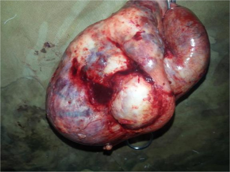
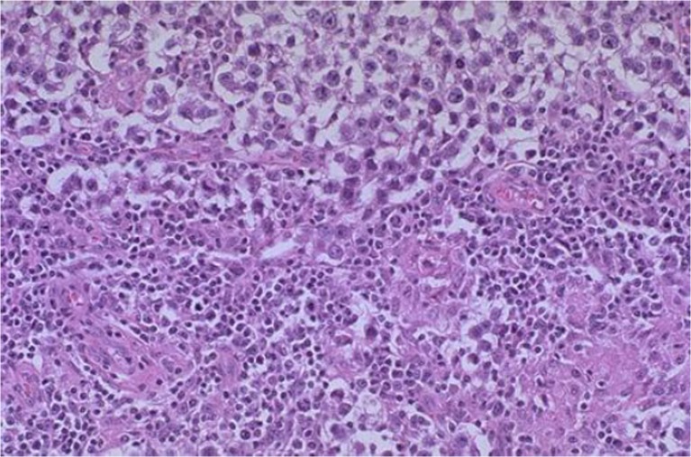
A 38-year-old male presented to our hospital with complaints of acute right lower abdominal pain accompanied by recurrent vomiting and fever for the last 48 h. The patient was febrile with pulse rate of 86/min and blood pressure of 130/70 mmHg. His abdominal examination revealed tenderness and lump in the right iliac fossa. His blood tests were within normal limits except for leucocytosis (TLC = 13,000/cumm). Emergency sonography was performed which showed a tender, mixed echogenic mass in the right iliac fossa and an appendix mass was presumed. The patient was put on conservative Ochsner-Sherren regimen. Regular re-examination revealed an increasing abdominal pain and increasing size of appendix mass. A contrast-enhanced CT examination of the abdomen was then performed which revealed a mass 12 × 7.4 × 7.4 cm in size arising from right side of the pelvis, extending superior and anterior to the bladder, suggestive of a solid mass/bowel mass. Exploratory laparotomy was performed through a right paramedian incision which revealed a large 12 × 8 × 8 cm well-capsulated mass, torted 360° on its pedicle. The pedicle was clamped, and the mass was removed (Fig. 1). No lymphadenopathy was appreciated. The mass weighed 560 g. A cut section revealed cysto-solid mass with foci of haemorrhage and necrosis. Postoperative histopathology report was classical seminoma with features of round to polyhedral cells arranged in sheets and trabeculae separated by septae traversed by lymphocytes (Fig. 2). The stroma showed extensive areas of haemorrhage and necrosis with no peri-capsular infiltration. Subsequent clinical examination revealed the absence of testis in the right scrotal sac, which was missed clinically and in the imaging studies. Postoperative human beta subunit chorionic gonadotropin and alpha-fetoprotein levels were normal. Later, the patient received adjuvant chemotherapy in the form of 4 cycles of bleomycin, etoposide and cisplatin (BEP) regimen.
Fig. 1.
The right testicular tumour after excision
Fig. 2.
Histopathology showing classical seminoma
Discussion
Torsion of an intra-abdominal testicular tumour is a rare event. The first case reported in the literature was by Gerster in 1897 [2]. Subsequently, sporadic small clusters of cases have appeared in the literature [3].
One in every 500 men has an undescended testis that may be associated with complications such as cancer, ischaemia, infertility and torsion. The cancer risk of an ectopic testis is 40 times higher than in a normal testis, and an abdominal testis is 4 times more likely to undergo malignant degeneration than an inguinal testis [4].
The cancer of undescended testis peaks in the third or fourth decade of life. Pure seminoma is the most frequent germ cell neoplasm in the undescended testis. However, 5–10 % of testicular tumours occur in the contralateral, normally descended testis [5].
By 12 months of age, about 1 % of all boys have cryptorchidism. Orchiopexy is now recommended for patients younger than 2 years old and even as young as 6 months old. It was found that the risk of testicular tumour among those treated at 13 years of age or older was twice the risk among those who were treated at a younger age [6].
Because torsion of the intra-abdominal testis is more common on the right [7], confusion with the diagnosis of acute appendicitis or appendix mass may arise because of a failure to examine the external genitalia as part of the abdominal examination [8], as in this case. Clinical presentations of malignant intra-abdominal testis can range from an asymptomatic mass to symptoms simulating appendicitis or retroperitoneal mass, incarcerated hernia, urinary frequency or dysuria from mass effect on bladder or acute abdominal pain due to torsion or haemorrhage [1]. Often, radiologic and preoperative diagnosis of torsion of intra-abdominal testicular tumour is rare and difficult because, usually, the history of cryptorchidism is not provided and the imaging features can be non-specific. However, a detailed history taking, thorough physical examination of the abdomen and genitalia, meticulous inspection of radiological films and, above all, the awareness of such complications involving maldescended testis can enhance the diagnostic yield.
Conclusion
Although a rare event, the diagnosis of torsion of an intra-abdominal testicular tumour should be considered in any patient presenting with an acute abdomen and a history of cryptorchidism.
References
- 1.Manisha M, Satyanarayana S, Anindita M, Rao KV, Rao GB. Seminoma of undescended testis presenting as acute abdomen. Indian J Pathol Microbiol. 2009;52(2):278–280. doi: 10.4103/0377-4929.48948. [DOI] [PubMed] [Google Scholar]
- 2.Ronald GF, Perry SG, Jude TB, Lindsay K, Wise GJ. Torsion of an intraabdominal testis tumour presenting as an acute abdomen. Urol Radiol. 1990;12:50–52. doi: 10.1007/BF02923966. [DOI] [PubMed] [Google Scholar]
- 3.Hutcheson JC, Khorasani R, Capelouto CC, Moore FD, Silverman SG, Loughlin KR. Torsion of intraabdominal testicular tumour: a case report. Cancer. 1996;77(2):339–343. doi: 10.1002/(SICI)1097-0142(19960115)77:2<339::AID-CNCR17>3.0.CO;2-2. [DOI] [PubMed] [Google Scholar]
- 4.Mishra HA. A giant intraabdominal testicular seminoma. Biomed Res. 2010;21(3):227–229. [Google Scholar]
- 5.Imaz AY, Bayraktar B, Sagiroglu J, Gucluer B. Giant seminoma in an undescended testis presenting as an abdominal wall mass. J Surg Case Rep. 2011;12:9–12. doi: 10.1093/jscr/2011.12.9. [DOI] [PMC free article] [PubMed] [Google Scholar]
- 6.Andreas P, Lorenzo R, Agneta N, Kaijser M, Akre O. Age at surgery for undescended testis and risks of testicular cancer. N Engl J Med. 2007;356:1835–1841. doi: 10.1056/NEJMoa067588. [DOI] [PubMed] [Google Scholar]
- 7.Osime OC, Momoh MI, Elusoji SO. Torsed intraabdominal testis: a rarely considered diagnosis. Calif J Emerg Med. 2006;7(2):31–33. [PMC free article] [PubMed] [Google Scholar]
- 8.Tomasz S, Lukasz S, Miroslaw T. Torsed intraabdominal testicular tumour diagnosed during surgery performed due to suspicion of acute appendicitis. Pol J Surg. 2012;84(10):526–530. doi: 10.2478/v10035-012-0088-y. [DOI] [PubMed] [Google Scholar]


