Figure 2.
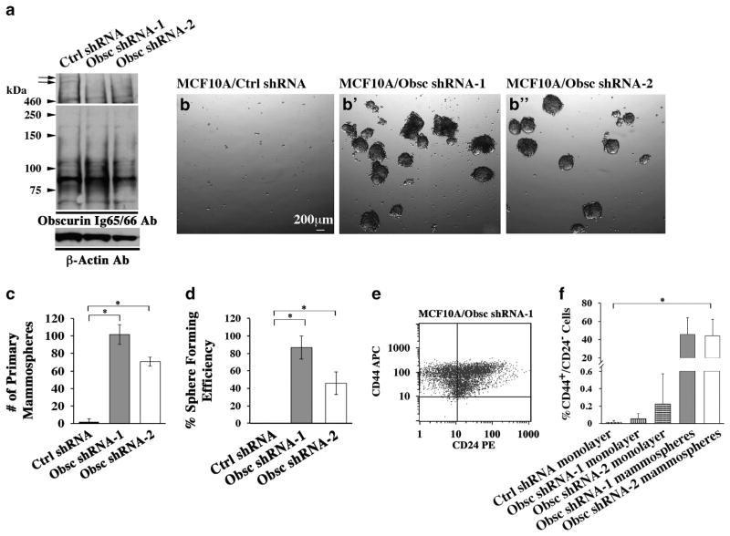
Obscurin-KD MCF10A breast epithelial cells form mammospheres with enriched markers for cancer-initiating cells. (a) Giant obscurins (arrows) are downregulated in MCF10A cells stably transduced with obscurin shRNA-1 or obscurin shRNA-2 compared with control cells, as indicated by immunoblotting using the Ig65/66 antibody. Notably, the expression of small obscurins remains unchanged in MCF10A cells expressing obscurin shRNA-1 or obscurin shRNA-2 compared with control cells. Equal loading of protein homogenates was ensured by measuring protein concentration and probing for β-actin. (b, b′) MCF10A cells stably transduced with obscurin shRNA-1 (b′) or obscurin shRNA-2 (b″) are able to form primary mammospheres, while control cells are not (b). (c, d) Quantification of primary (c) and secondary (d) mammospheres formed by MCF10A cells stably expressing obscurin shRNA-1 or obscurin shRNA-2, compared with MCF10A cells expressing scramble control shRNA; n =3, error bars =s.d., *P<0.03; t-test. (e) Representative plot obtained from fluorescence activated cell sorting (FACS) analysis of primary mammospheres generated from MCF10A obscurin shRNA-1 expressing cells, indicating that they are highly enriched in a cell population with the surface marker signature CD44+/CD24−, which is associated with stem-like properties. (f) Approximately 40% of cells within the MCF10A obscurin shRNA-1 and obscurin shRNA-2 mammospheres are CD44+/CD24−, compared with <1% of adherent MCF10A control shRNA, obscurin shRNA-1 and obscurin shRNA-2 cell monolayers, as measured by FACS; n =3, error bars =s.d., *P<0.03; t-test.

