Abstract
Telomeres, comprised of short repetitive sequences, are essential for genome stability and have been studied in relation to cellular senescence and aging. Telomerase, the enzyme that adds telomeric repeats to chromosome ends, is essential for maintaining the overall telomere length. A lack of telomerase activity in mammalian somatic cells results in progressive shortening of telomeres with each cellular replication event. Mammals exhibit high rates of cell proliferation during embryonic and juvenile stages but very little somatic cell proliferation occurs during adult and senescent stages. The telomere hypothesis of cellular aging states that telomeres serve as an internal mitotic clock and telomere length erosion leads to cellular senescence and eventual cell death. In this report, we have examined telomerase activity, processivity, and telomere length in Daphnia, an organism that grows continuously throughout its life. Similar to insects, Daphnia telomeric repeat sequence was determined to be TTAGG and telomerase products with five-nucleotide periodicity were generated in the telomerase activity assay. We investigated telomerase function and telomere lengths in two closely related ecotypes of Daphnia with divergent lifespans, short-lived D. pulex and long-lived D. pulicaria. Our results indicate that there is no age-dependent decline in telomere length, telomerase activity, or processivity in short-lived D. pulex. On the contrary, a significant age dependent decline in telomere length, telomerase activity and processivity is observed during life span in long-lived D. pulicaria. While providing the first report on characterization of Daphnia telomeres and telomerase activity, our results also indicate that mechanisms other than telomere shortening may be responsible for the strikingly short life span of D. pulex.
Introduction
Telomeres, the ends of linear chromosomes, have been studied extensively in relation to cellular aging and senescence [1,2,3]. Composed of repetitive nucleotide sequences (TTAGGG for mammals) associated with proteins, telomeres protect important genetic information of linear chromosomes from deletion arising due to the “end replication” problem [1,3]. The process of DNA replication leads to progressive shortening of linear chromosomes at the telomeres due to the fact that DNA polymerases can only polymerize in a 5’ to 3’ direction and require a primer with a free 3’-OH group [1,3]. This inability to replicate linear DNA on the lagging strands all the way to ends necessitates the telomerase, an enzyme responsible for de novo addition of telomeric repeats to chromosomal ends [4]. Telomerase is a ribonucleoprotein complex, comprised of a protein catalytic subunit TERT (Telomeric Reverse Transcriptase), and an RNA template termed TERC (telomeric RNA Component) [1,3]. Telomerase activity is essential for maintaining telomere length throughout cellular lifespan. Early in human development, telomerase is constitutively active in cells but after birth it is active predominately in stem cells and germ cells with most somatic tissues having no telomerase activity [5,6]. Each individual DNA replication event of human telomerase-negative somatic cells leads to a loss of 100 bp of telomeric sequence, resulting in a progressive decline in telomere length with each cellular division [7]. Because of this progressive telomere shortening, human somatic cells can only undergo approximately 50 to 80 cellular replication events before becoming senescent [7]. Thus, telomere length is essential for normal cellular function and proliferation as well as chromosome stability. In the absence of proper telomere complex formation, the double-stranded break repair pathway can be initiated resulting in apoptosis or senescence [8,9]. Thus, telomeres serve a protective molecular role by shielding the loss of important genetic information as well as by maintaining chromosome stability throughout the cellular lifespan. Telomerase has also been implicated in nuclear DNA damage repair and plays a protective role for mitochondrial DNA during oxidative stress response during which telomerase shuttles from the nucleus to the mitochondria [10,11,12,13,14,15].
In this study we investigated telomerase activity, the telomeric repeat sequence, and telomere lengths in Daphnia, a freshwater crustacean, and an emerging model in aging liuresearch. Daphnia has been used extensively as a model in ecotoxicology studies [16] and with a fully sequenced genome of D. pulex, it is an emerging model in biomedical research [17,18]. Two ecotypes of Daphnia are of interest in relation to aging, D. pulex and D. pulicaria [19,20,21,22,23,24]. D. pulex is found in small transitory ponds, in which selection favors short longevity due to the limited time the ponds have water. In a laboratory environment D. pulex exhibits a lifespan on average of about 20 days [20,23]. D. pulicaria lives primarily in a stable environment of stratified lakes that are present all year long. In the lab D. pulicaria exhibits lifespans on average of about 70 days [20,23]. Genetically, the two ecotypes are almost identical and are capable of interbreeding with viable offspring in the wild [21]. Daphnia can be easily cultured and undergo cyclic parthenogenduesis [16], thus enabling creation of clonal lineages without the genetic variation normally associated with sexually reproducing organisms. Being crustaceans, Daphnia constantly shed their outer carapace and have regenerative cellular capacities [16]. Due to these unique characteristics Daphnia is a interesting model organism for understanding cellular processes associated with of aging.
We present characterization of the telomere length, telomerase activity and processivity in the two ecotypes, the short-lived D. pulex and the long lived D. pulicaria. In the short-lived D. pulex, telomere length did not decline with age; however, in the long-lived D. pulicaria, telomere length decreased with age. Accordingly, telomerase activity in D. pulex is relatively constant throughout the life span, whereas in D. pulicaria, it declines considerably with age. In addition, the telomerase processivity increased with age in D. pulex, whereas in D. pulicaria it declined with age. This is an important initial study to investigate telomere length and telomerase activity in a newly emerging model system for research on aging.
Materials and Methods
Daphnia Cultures
Daphnia pulex (clone RW 20) and Daphnia pulicaria (clone Lake XVI-11) were isolated from populations in southwest Michigan in 2008 and have since been cultured in the lab. No specific permissions are required to collect zooplankton from these public-access waterbodies in Michigan. D. pulex and D. pulicara are neither endagered nor protected. For further details on the source populations, see Dudycha [21]. Daphnia were maintained at 20°C with a photoperiod of 12:12 L:D (12 hours of light followed by 12 hours of dark) within a Percival growth chamber. Daphnia were maintained at 3 to 5 animals per 250 mL beaker in 200 mL of filtered lake water. Daphnia were cleared of young and transferred to a new beaker with fresh water on alternate days. They were fed every day with vitamin supplemented algae Ankistrodesmus falcatus at a concentration of 20,000 cells/mL. To generate experimental animals, even-aged cohorts were obtained by placing neonates individually in 100 mL of hardwater COMBO, or artificial lake water. Experimental animals were otherwise maintained as in the source cultures.
All Daphnia were maintained clonally in the lab and all Daphnia used for experimentation are diploid females. Once individuals were collected from the field, they were placed in beakers of fresh lake water and allowed to clonally reproduce. In this manner, each isolate, or clone, of Daphnia was produced (for example, individuals from the pond named Rough Wood were placed individually into beakers and designated as clone RW followed by a number which corresponds to the original analysis of the collected Daphnia). After each clone was established, Daphnia were maintained by constant feeding of algae and changing of the lake water to create an isogenic clonal line of Daphnia. Care was taken not to induce stress for the Daphnia such that sexual reproduction would not occur in the clonal lines.
Telomeric Repeat Amplification Protocol (TRAP) Assay
Telomeric repeat amplification protocol (TRAP) Assay was performed using the TRAPeze Telomerase Detection kit (Millipore). D. pulex and D. pulicaria were collected at different ages. For D. pulex (RW20), we used 1, 2, and 3 week old individuals and for D. pulicaria (LakeXVI-11) we used 1, 4, and 8 week old individuals. To prepare cell extracts from D. pulex and D. pulicaria, 25 to 30 individual Daphnia were drained of all residual water, and homogenized in 200 μl of 1X CHAPS Lysis Buffer on ice. The homogenate was incubated on ice for 30 min, centrifuged at 12000 X g for 20 min at 4°C, 160 μl of extract was transferred to a new tube, and the total protein concentration in the extract was determined using a Bradford Assay. Samples were stored at -80°C.
The TRAP Assay was performed as per the instructions in the TRAPeze kit. A 50 μl reaction was performed for all samples and controls. A positive control of human cancer cell line extract was provided with the kit. Each reaction contained 5 μl of 10X TRAP reaction buffer, 1 μl of 50X dNTP mix, 1 μl of TS Primer, 1 μl of TRAP primer mix, 0.4 microliters (2 units) of Taq polymerase, 39.6 μl of deionized water, and 2 μl of extract (either control or sample). Tubes containing each reaction were placed in the thermocycler and heated to 30°C for 30 min. Then 30–33 cycles of 94°C for 30 sec, 59°C for 30 sec, and 72°C for 1 min were performed. The heat-inactivated control samples, were incubated at 85°C for 10 min prior to TRAP assay.
Following PCR amplification, 5 μl of loading dye (with bromophenol blue (0.25% in 50% glycerol/50 mM EDTA) and xylene cyanol (0.25% in 50% glycerol/50 mM EDTA) was mixed with the TRAPeze product. 25 μl was loaded into a 10% non-denaturing polyacrylamide gel and the gel was run at 400 V for 1.5 hours in 0.5 X TBE buffer, stained using Sybr Green for 30 min before scanning in a Typhoon FLA 7000.
Quantification of the TRAP Assay Results
TRAP Assay results were quantified using a formula described in TRAPeze kit manual. The formula takes into account the signal measured in each reaction/lane (x) as well as the heat inactivated control (xO), the no-template control (rO), and the TSR8 quantitation control (r) reactions/lanes. All of these reactions contain an internal standard (36 bp band) engineered into the assay, which was also used in the formula (c for samples, cr for TSR8 control). The resulting quantification was in units of Total Product Generated (TPG), which corresponds to the number of TS primers extended by at least 4 telomeric repeats in 30 min at 30°C.
Sequencing of the TRAP Product
A TRAP Assay reaction that contained a strong positive result from the Daphnia samples was used to determine the telomeric repeat sequence for Daphnia. 5 μl of the reaction products was purified using a Qiagen PCR Purification kit, and ligated into the vector pGEMT Easy (Promega), and sequenced.
Survivorship studies
We compared survivorship of the XVI-11 and RW20 clones using standard life table methods for Daphnia [25]. Experimental conditions were 20°C, a 12:12 L:D photoperiod, with animals kept in individual 150-ml pyrex beakers in 100 ml COMBO hardwater artificial lakewater [26]. Two generations were maintained under experimental conditions prior to initiating the life table to minimize maternal effects variation. Experimental individuals were fed 20,000 cells/ml Ankistrodesmus falcatus daily, a food level that produces normal aging processes in Daphnia [20]. Individuals were transfered to fresh beakers and COMBO every other day, and survivorship was observed daily until all experimental animals died. For each clone, n = 60 females.
Terminal Restriction Fragment (TRF) Assay
We preformed a Terminal Restriction Fragment Assay using a previously established protocol by Herbert et al. [27]. Genomic DNA was extracted from D. pulex and D. pulicaria at various ages using a CTAB (Hexadecyltrimethylammonium Bromide) based protocol optimized for use with Daphnia. For each aged set of Daphnia, 25 individuals were pooled for the extraction of genomic DNA. Each individual Daphnia was cleared of all embryos before the genomic DNA was extracted to ensure the results were reflective of the adult telomere length. One μg of genomic DNA was digested at 37°C overnight with 1 U/μl of the following restriction enzymes: HinfI, RsaI, MspI, CfoI, HaeII and AluI. Digests were run on a 0.7% agarose gel. Samples were run for 4 h at 120 V in 1X TAE Buffer to achieve desirable separation in size range 1 kb- 25 kb. The gel was denatured for 20 min in denaturing solution (0.5 M NaOH, 1.5M NaCl), and rinsed in distilled water for 10 min. The gel was then dried upside down between 2 sheets of Whatman 3MM filter paper under vacuum for 1 hour at 50°C. After removing the gel from the dryer, the gel was neutralized for 15 min in neutralizing solution (1.5 M NaCl, 0.5 M Tris-Cl, pH 8.0) and then rinsed with distilled water for 10 min. The gel was then soaked in 10 ml of prehybridization buffer (5X SSC Buffer, 5X Denhardt solution, 10 mM Na2HPO4, 1 mM Na2H2P2O7) for 10 min. The gel was then transferred to hybridization solution (5X SSC Buffer, 5X Denhardt solution, 10 mM Na2HPO4, 1 mM Na2H2P2O7) containing the radiolabeled telomeric sequence probe (see below). The gel was hybridized overnight at 42°C. After hybridization, the gel was washed once in 2X SSC for 15 min at 22°C, washed four times for 10 min each in 0.1X SSC/0.1% SDS. Following washes, the gel was exposed to a phosphor screen overnight and scanned on a Typhoon FLA 7000 phosphor imager and visualized with ImageQuant software.
Daphnia telomeric probe design and labeling
Based on the telomeric repeat sequence for Daphnia, a telomeric probe was designed as 6 repeats of the telomeric repeat: 5’-TTAGGTTAGGTTAGGTTAGGTTAGGTTAGG-3’. The probe was 5’ end-labeled with γ-P32 ATP using polynucleotide kinase. To label the probe, 2 μl of 20 pmol/μl of oligonucleotide, 24 μl of γ-P32 ATP (3000 Ci/mmol), 10 μl 5X forward reaction buffer, 2 μl 10 U/μl T4 polynucleotide kinase, and 12 μl H20 were added to the reaction mix and incubated at 37°C for 30 min. A QIAquick nucleotide removal kit was used to remove the unincorporated radioactivity. The labeling and specific activity of probe was determined by counting total cpm in a 1 μl aliquot (total 200 μl) using a scintillation counter.
Statistics
To determine statistical significance of results of the TRAP assay, as well as TRF Assay, a two-tailed Student’s T-test was performed, assuming equal variance. Each figure legend denotes p values as denoted by brackets and special characters. Note that our alpha level was p = 0.05. A nonparametric log-rank test was performed for significance of the survival curves.
Results
Telomerase activity is present in Daphnia samples
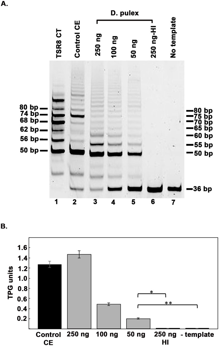
To detect and assay for telomerase activity from adult Daphnia, we performed a telomerase repeat amplification protocol (TRAP) assay, using the TRAPeze (Millipore) kit. The kit is optimized for human telomerase activity, thus we tested it for use with Daphnia extracts. As represented in Fig 1A, we could detect strong telomerase activity in extracts prepared from adult Daphnia (lanes 3–5). Telomerase activity showed dose-dependence with amount of total protein in extracts. Each TRAP reaction mixture contains primers as well as a template for amplification of a 36 bp internal control, which also serves to determine false negative results due to the presence of a telomerase inhibitor in the extracts. Daphnia extracts did not show any inhibition of the telomerase activity compared to the positive control (lane 2) provided with the kit. The dependence of the band ladder on telomerase activity was confirmed by heat treatment of the Daphnia extract (lane 6), which completely eliminated the ladder due to inactivation of telomerase, thus confirming the presence of telomerase activity in Daphnia extract. Absence of bands with no template added (lane 7), indicated no contamination in the PCR step of the assay and further confirmed the presence of telomerase activity in Daphnia extract. The periodicity of the bands generated using Daphnia extract (lanes 3–5) was different than the periodicity obtained with extract from telomerase positive human cell line HEK293 (positive control- lane 1). Fig 1B represents quantification of telomerase activity.
Fig 1. Telomerase activity in Daphnia.
A) Telomeric repeat amplification protocol (TRAP) assay of D. pulex extracts. The amount of cell extract used is indicated above each lane. HI: Heat Inactivated, CE: Cell Extract. TSR8 CT: positive control for PCR step. B) Quantification of the TRAP Assay in 1A. Student T-test was performed, p values are as follows: * = 6.15x10⁻⁶, ** = 6.63x10⁻⁶ (n = 3).
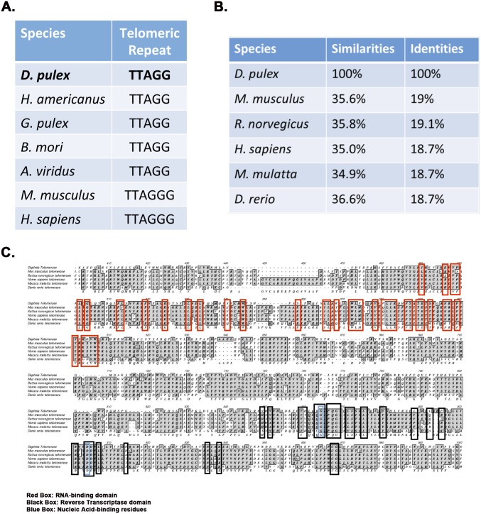
Human telomeres have a six-nucleotide telomeric repeat sequence (TTAGGG). To determine the telomeric repeat length and sequence in Daphnia, we cloned and sequenced the Daphnia TRAP reaction products. As seen in Fig 2A, the sequence analysis of the clones revealed that the telomeric repeat was TTAGG, a sequence identical to crustaceans H. americanus and G. pulex [28]. The five nucleotide repeat sequence corresponds to the observed difference in the periodicity of bands in TRAP assay (Fig 1A, lanes 3–5), as the bands are expected to be shorter by one nucleotide as compared to TRAP assay products obtained with human cell extract (Fig 1A, lane 2). When telomerase (TERT) sequences across various species were compared, the similarity between the D. pulex TERT sequence and those of other organisms is relatively low for the entire protein (Fig 2B). The highest similarity is found in the essential functional domains of the protein: the RNA binding domain and the reverse transcriptase domain (Fig 2C), with high degree of sequence conservation.
Fig 2. Telomeric repeat sequence from Daphnia and sequence alignment of Daphnia telomerase (dTERT) with telomerase proteins from other species.
A) Telomeric repeat sequence of Daphnia and other species. H. americanus: Homarus americanus—Lobster, G. pulex: Gammarus pulex—Freshwater amphipod, B. mori: Bombyx mori—Silkworm/moth, A. viridus: Amaranthus viridus—Beetle, M. musculus: Mus musculus—Mouse, H. sapiens: Homo sapiens—Human. B) Identity and similarity percentages of dTERT with TERT from other species. C) D. pulex telomerase reverse transcriptase (DTERT) sequence alignment for the RNA-binding and reverse transcriptase domains. Green Boxes: conserved residues within the RNA binding domain. Black Boxes: conserved residues within the reverse transcriptase domain. Blue boxes: residues within the reverse transcriptase domain that are essential for nucleic acid binding.
Telomerase in Daphnia embryos is more processive than in adult organisms
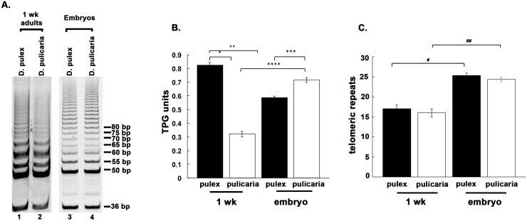
In order to compare the relative telomerase activities in embryos and adult organisms we assayed the extracts from embryos and 1 week-old adults of two ecotypes, D. pulex (RW20) and D. pulicaria (LakeXVI-clone11). As is shown in Figs 3 and 1 week old D. pulex (lane 1) shows more telomerase activity than D. pulicaria of the same age (lane 2). On the other hand, both ecotypes have very comparable levels of telomerase activity at the embryonic stage (lanes 3 and 4). D. pulex adults at 1 week (lane 1) show some increase in telomerase activity (as measured by total intensity of the bands in entire lane) as compared to embryos (lane 3). In contrast to this, D. pulicaria adults at 1 week show a significant decrease in total telomerase activity (lane 2) as compared to embryos (lane 4). Fig 3B shows quantification of total telomerase activity in Fig 3A. In addition to determining total telomerase activity based on band intensities, the TRAP assay can also measure the processivity of telomerase, which is the ability of an enzyme to catalyze multiple reactions without releasing the substrate. The greater the processivity, the greater the number of repeats added by the enzyme during the time interval of the assay. This can be visualized as the ladder reaching to higher molecular weights. As seen in Fig 3C, a decline in telomerase processivity is observed from embryo stage to 1 week-old stage of both ecotypes.
Fig 3. Telomerase from Daphnia embryos shows high processivity.
A) TRAP assay from egg stage 1 embryos and 1 week old adults of D. pulex and D. pulicaria. 250 ng of Daphnia extract was used in each lane. B) Quantification of the TRAP assay displayed in 2 A. C) Quantification of the processivity of telomerase from each displayed sample. The p values calculated from student T-tests are as follows: * = 6.15x10⁻⁶, ** = 5.56x10⁻⁶, *** = 0.0005,**** = 1.5x10⁻⁵; # = 0.0002, ## = 0.0002 (n = 4).
Comparison of telomerase activity from D. pulex and D. pulicaria at different ages
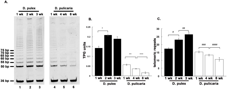
We further investigated the telomerase activity of D. pulex (RW20 ecotype) and D. pulicaria (LakeXVI-clone11) at equivalent points in their respective life spans. We performed the TRAP Assay with extracts from 1, 2, and 3 week-old D. pulex and 1, 4, and 8 week-old D. pulicaria which were previously determined to correspond to equivalent time points in their respective life spans [24]. As seen in Fig 4A and 4B, D. pulex (lanes 1–3) showed an increase in telomerase activity from 1 week to 2 week but showed a marginal decline in 3-week-old organisms. In contrast, D. pulicaria displayed a steady decline of telomerase activity with age, telomerase activity being the highest at 1 week and the lowest at 8 weeks (Fig 4A, lanes 4–6). The telomerase activity was quantified using the Imagequant software and is shown in Fig 4B, which shows a 50% increase in telomerase activity from 1 week-old to 2 week-old organisms in D. pulex. In contrast, D. pulicaria showed an age-dependent decline in telomerase activity with about 30% decrease from ages 1 week to 4 week and another 30% decrease from ages 4 week to 8 week. The processivity of the telomerase from these samples was determined and is represented in Fig 4C. In D. pulex, the processivity of telomerase increased considerably from 1 week to 2 week and showed a small but consistent increase from 2 week to 3 week. In contrast, the processivity of telomerase in D. pulicaria samples showed a steady decline with age from 1 week to 8 week.
Fig 4. Comparison of telomerase activity at different ages in D. pulex and D. pulicaria.
A) TRAP assay was performed using Daphnia extracts prepared at indicated ages. Lanes 1–3: D. pulex, lanes 4–6: D. pulicaria. B) Quantification of the TRAP assay displayed in 4 A. C) Quantification of the processivity of telomerase from each displayed sample. Student T-tests were performed, and p values are as follows * = 0.0005, ** = 0.0006, *** = 0.002, # = 0.001, ## = 0.034, ### = 0.0132, #### = 0.029 (n = 4).
Telomere length in D. pulex and D. pulicaria at various ages
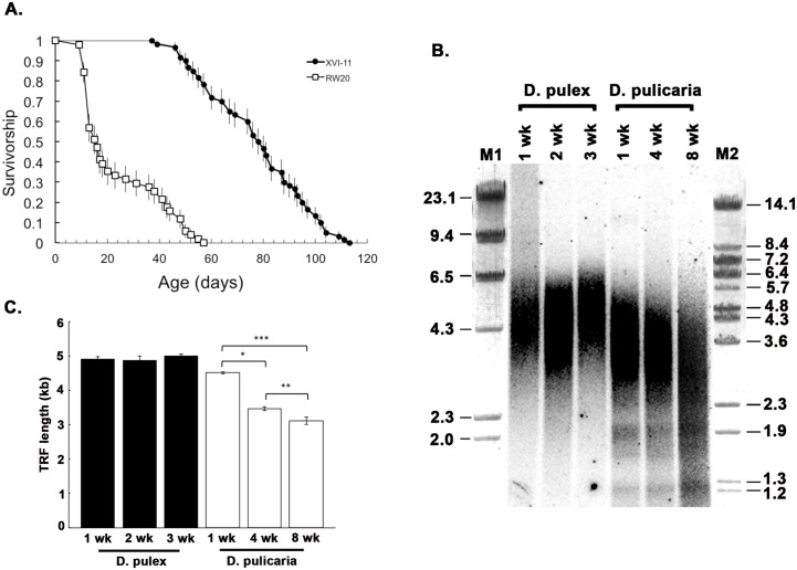
As shown in Fig 5A, survivorship patterns are substantially different between D. pulex (clone RW20) and D. pulicaria (clone Lake XVI-11). RW20 had a median lifespan of 16 d (maximum = 56 d), whereas XVI-11 had a median lifespan of 79 d (maximum = 112 d). A nonparametric log-rank test showed that the survival curves are significantly different (X2 = 120.894, df = 1, p <0.0001). To analyze the average telomere lengths during life span in these two Daphnia clones that display markedly different lifespans as well as significantly different telomerase activities (Fig 4B), we performed a terminal restriction fragment (TRF) assay using genomic DNA isolated from 1, 2, and 3 week old D. pulex and 1, 4, and 8 week old D. pulicaria. As seen in Fig 5B, the telomere-specific probe detected the TRFs at various ages in both ecotypes. The average telomere length was calculated and is represented in Fig 5B. D. pulex and D. pulicaria telomeres at age of 1 week were quite similar in length with D. pulex at 4.9 kb and D. pulicaria at 4.5 kb. In D. pulex, the telomere length did not shorten with age, with the 2 week old and 3 week old telomeres averaging at 4.9 kb and 5.0 kb respectively. In contrast to this, D. pulicaria telomeres exhibited age-dependent shortening with telomere lengths being 3.5 kb and 3.1 kb at 4 weeks and 8 weeks of age respectively. Therefore, at all equivalent ages, D. pulex’s average TRF is longer than D. pulicaria’s average TRF. Thus, the short-lived ecotype D. pulex displayed no shortening of telomere length with age while the long-lived ecotype pulicaria showed an age-dependent decline of telomere length.
Fig 5. Life spans and telomere lengths in D. pulex (RW20) and D. pulicaria (LakeXVI-clone11).
A) Survivorship curves of clones RW20 (open squares) and XVI-11 (black circles). Error bars show age-specific standard errors from Kaplan-Meier survival probability estimates. B) Terminal Restriction Fragment (TRF) assay was performed to estimate the average telomere length of the Daphnia at various ages. The age in weeks is indicated above each lane. Lanes 1–3: D. pulex, lanes 4–6: D. pulicaria. M1 and M2: molecular weight markers; lambda HindIII digest and lambda BstEII digest respectively. C) Quantification of 5 A, error bars indicate standard deviation. Student T-tests were performed and p values are as follows: * = 1.03x10⁻⁵, ** = 0.007, *** = 2.48x10⁻⁵ (n = 3).
Discussion
Aging is associated with a progressive deterioration of cellular functions leading to a functional decline of organs and tissues. Molecular processes that are considered as characteristics of aged organisms include loss of telomere function, epigenetic genomic changes, declining protein homeostasis, increased cellular senescence, depletion of the stem cell pool, and altered intercellular communication [29]. Telomerase deficiency in humans is associated with premature onset of diseases that are typical of old age [30]. There is evidence of a causal link between telomere loss, cellular senescence and organismal aging that emerged from genetically-modified animal models. Mice with shortened or lengthened telomeres exhibit decreased or increased lifespan, respectively [31,32,33] and aging could be reverted by telomerase activation in telomerase deficient mice [34]. In humans, recent meta-analyses have indicated a strong correlation between short telomeres and mortality risk [35]. Along with its telomere-associated function, telomerase is also involved in DNA damage response and shuttles from the nucleus to the mitochrondria upon oxidative stress to protect the mitochondrial DNA from sustaining oxidative damage [10,13,14,15].
Daphnia telomerase protein shows a high degree of homology in the essential functional domains of the protein: the RNA binding domain and the reverse transcriptase domain. Daphnia telomeric repeat sequence is TTAGG, a sequence identical to crustaceans H. americanus and G. pulex [28]. Shelterin, a complex formed by six telomere-specific proteins (TRF1, TRF2, TIN2, Rap1, TPP1, and POT1) binds to the telomeric repeats and protects the chromosome ends in mammalian cells [36]. Without the shelterin complex, telomeres are not protected from being recognized by the DNA damage surveillance and are inappropriately processed by DNA repair pathways [36]. Although the shelterin complex is not found in some organisms, there are functional orthologs of the shelterin component proteins. Using the data and tools in PANTHER (a comprehensive, curated database of protein families, trees, subfamilies, and functions) [37] we could identify a Daphnia homolog of POT1. POT1-like proteins are present in nearly all eukaryotes [38], and thus it may be of interest in future to identify other proteins that complex with Daphnia POT1 since such proteins may be involved in telomere maintenance and chromosome integrity in Daphnia.
In our present study, we characterized the telomerase activity, telomerase processivity, and telomere length during the lifespan in two ecotypes of Daphnia, the short-lived D. pulex and the long-lived D. pulicaria. Our results demonstrate a clear age-associated decline in telomerase activity, telomerase processivity, and telomere length in the long-lived ecotype D. pulicaria. Surprisingly, the short-lived ecotype D. pulex showed no decline in telomerase activity and telomere length but an age-dependent increase in processivity of telomerase. The telomere hypothesis of cellular aging states that the telomeres shorten with each cellular replication event until the telomeres are completely eroded and the resulting genomic instability leads to the cellular death in telomerase negative mammalian somatic cells [39,40,41]. Organismal aging, however, is more complex with several different factors in addition to the telomere length and telomerase activity playing a role in the overall lifespan of an organism. Supporting this multifaceted organismal aging process, our results indicate that in the D. pulex ecotype, telomere length does not decline with age and thus is not the main cause of its short life span.
Overall, our results are in agreement with previous studies in other model organisms with respect to the relationship between the telomerase activity and corresponding telomere length [7,42,43,44]. In D. pulex, which has high telomerase activity throughout lifespan, there is no decline in overall telomere length. For D. pulicaria, a decrease in telomerase activity coincides with a decrease in telomere length with age. Comparing telomere lengths of D. pulex and D. pulicaria at equivalent points in their lifespan, D. pulex always has longer telomeres than D. pulicaria corresponding to higher telomerase activity in D. pulex. The processivity of Daphnia telomerase could be of biological significance in terms its impact on the length of telomeres. Although the telomerase activity may be high in terms of high rate of telomeric repeat addition in TRAP assay, if the enzyme is not processive, the overall telomere length may not be maintained. Thus, in D. pulex with high telomerase activity and processivity, individual cellular replication events do not effectively erode the telomeres since telomerase can add de novo telomeric repeats to the ends of telomeres. However, in D. pulicaria with telomerase activity and processivity decreasing with age, each cellular replication event leads to shortening of telomeres with age. This is particularly relevant in Daphnia as it shows indeterminate overall growth during the entire life span. Although telomerase processivity as a function of aging has not been explored in the past, several different factors are known to contribute to telomerase processivity. These include telomerase RNA template structure, telomere structure, and various proteins that stabilize telomeres [45,46,47]. It’s possible that there is a difference in any of these three factors between the two ecotypes of D. pulex and D. pulicaria, which could be investigated in future studies. While our study provides an initial characterization of telomerase activity, processivity, and telomere length in D. pulex and D. pulicaria, this work utilizes one isolate or clone from each ecotype. Investigating telomerase activity and telomere lengths in other Daphnia species and more isolates or clones of D. pulex and D. pulicaria will be of value.
Although much work has been done investigating cellular aging and telomere length, the connection between telomere length and organismal longevity is not straightforward [7,42,43,44]. Formulated by Harley et al, the telomere hypothesis of cellular aging postulated that telomeres serve as an internal mitotic clock in telomerase negative mammalian somatic cells and when telomere length is exhausted cellular senescence and the eventual death ensues [39,40,41,48]. Multiple studies have found varying results in terms of telomere length and overall organismal longevity [7,42,44]. Studies involving C. elegans, a well-established model for molecular biology of aging, have demonstrated that overall telomere length does not affect or predict the organismal longevity [7]. Danio rerio shows high levels of telomerase activity throughout its lifespan and maintains telomere length even into late stages of life [44]. No telomere shortening with increasing age is seen in wild-derived mouse strains, as well as in the marine bird Oceanodroma leucorhoa [42,49]. The crustacean, Homarus americanus (lobster), was investigated to determine overall telomere length and telomerase activity during aging. These organisms display such extensive lifespans that some predict would be nearly immortal if provided with optimal environment [50,51]. Lobsters display telomerase activity throughout their lifespan with a relatively unchanged telomere length [28]. Although cellular aging may be well defined by the telomere hypothesis of cellular aging, organismal aging is a multifactor process with a more complicated relationship between telomere length and organismal longevity [7,42,44,49].
The above results lead to the question that if telomere erosion and hence genomic instability is not the cause of the characteristic short life span of D. pulex what may be causing this remarkably short life span in D. pulex? Organismal aging is a multifaceted process and the ability to respond to and survive the proteotoxic stress is an important factor in determining the longevity of an organism [52,53]. There are several theories of aging; however, studies have shown that the pathologies and phenotypes associated with aging may stem from damaged proteins and the inability to repair or eliminate these damaged molecules from cells [52,53,54]. Our previous work has established that the induction of chaperone HSP70 in response to heat shock in D. pulex declines rapidly with age making it highly susceptible to proteotoxicity. In contrast, D. pulicaria continues to show a robust heat shock response and chaperone HSP70 induction past the midpoint in its life span, thus enabling it to survive proteotoxicity [24]. Although there are many different aspects of the aging process, it is possible that the ability to respond to proteotoxic stress is a better determinant of organismal life span instead of overall length of the telomeres. In other words, the cellular damage arising from being unable to respond appropriately to proteotoxic stress may lead to death before the telomere length declines. In addition to heat stress, oxidative damage to proteins also causes proteotoxicity and in multiple organisms investigated, better ability to respond to oxidative stress is correlated with longer lifespans [29]. In the marine crustacean, Acartia tonsa, oxidative damage was found to be greater in the shorter lived male individuals of the species indicating that oxidative damage may play a role in the overall lifespan of this crustacean [55]. A study done in honeybees identified vitellogenin as a protein that protects the organism from oxidative stress and contributes to longevity [56]. Daphnia contain a vitellogenin homolog that could be investigated for any differences in D. pulex vs D. pulicaria for its role in protection from oxidative damage [17]. As Daphnia is emerging as a model system in aging studies our results reveal a non-concordance between telomerase activity, telomere length and the overall organismal aging and indicate that factors other than telomere length maintenance contribute to strikingly short life span in D. pulex.
Data Availability
All relevant data are within the paper.
Funding Statement
This work was supported by National Institutes of Health grant 1R01AG037969-01 awarded to JLD and RCP. The funder had no role in study design, data collection and analysis, decision to publish, or preparation of the manuscript.
References
- 1. de Lange T. Protection of mammalian telomeres. Oncogene. 2002;21: 532–540. [DOI] [PubMed] [Google Scholar]
- 2. Hayflick L. The limited in vitro lifetime of human diploid cell strains. Exp Cell Res. 1965;37: 614–636. [DOI] [PubMed] [Google Scholar]
- 3. Shay JW, Wright WE. Hayflick, his limit, and cellular ageing. Nat Rev Mol Cell Biol. 2000;1: 72–76. [DOI] [PubMed] [Google Scholar]
- 4. Greider CW, Blackburn EH. Identification of a specific telomere terminal transferase activity in Tetrahymena extracts. Cell. 1985;43: 405–413. [DOI] [PubMed] [Google Scholar]
- 5. Liu L, Bailey SM, Okuka M, Munoz P, Li C, Zhou L, et al. Telomere lengthening early in development. Nat Cell Biol. 2007;9: 1436–1441. [DOI] [PubMed] [Google Scholar]
- 6. Morgan G. Telomerase Regulation and the Intimate Relationship with Aging. Research and Reports in Biochemistry. 2013;3: 71–78. [Google Scholar]
- 7. Raices M, Maruyama H, Dillin A, Karlseder J. Uncoupling of longevity and telomere length in C. elegans. PLoS Genet. 2005;1: e30 [DOI] [PMC free article] [PubMed] [Google Scholar]
- 8. d'Adda di Fagagna F, Reaper PM, Clay-Farrace L, Fiegler H, Carr P, Von Zglinicki T, et al. A DNA damage checkpoint response in telomere-initiated senescence. Nature. 2003;426: 194–198. [DOI] [PubMed] [Google Scholar]
- 9. Takai H, Smogorzewska A, de Lange T. DNA damage foci at dysfunctional telomeres. Curr Biol. 2003;13: 1549–1556. [DOI] [PubMed] [Google Scholar]
- 10. Ahmed S, Passos JF, Birket MJ, Beckmann T, Brings S, Peters H, et al. Telomerase does not counteract telomere shortening but protects mitochondrial function under oxidative stress. J Cell Sci. 2008;121: 1046–1053. 10.1242/jcs.019372 [DOI] [PubMed] [Google Scholar]
- 11. Bommer UA, Borovjagin AV, Greagg MA, Jeffrey IW, Russell P, Laing KG, et al. The mRNA of the translationally controlled tumor protein P23/TCTP is a highly structured RNA, which activates the dsRNA-dependent protein kinase PKR. Rna. 2002;8: 478–496. [DOI] [PMC free article] [PubMed] [Google Scholar]
- 12. Geserick C, Blasco MA. Novel roles for telomerase in aging. Mech Ageing Dev. 2006;127: 579–583. [DOI] [PubMed] [Google Scholar]
- 13. Haendeler J, Drose S, Buchner N, Jakob S, Altschmied J, Goy C, et al. Mitochondrial telomerase reverse transcriptase binds to and protects mitochondrial DNA and function from damage. Arterioscler Thromb Vasc Biol. 2009;29: 929–935. 10.1161/ATVBAHA.109.185546 [DOI] [PubMed] [Google Scholar]
- 14. Indran IR, Hande MP, Pervaiz S. hTERT overexpression alleviates intracellular ROS production, improves mitochondrial function, and inhibits ROS-mediated apoptosis in cancer cells. Cancer Res. 2011;71: 266–276. 10.1158/0008-5472.CAN-10-1588 [DOI] [PubMed] [Google Scholar]
- 15. Singhapol C, Pal D, Czapiewski R, Porika M, Nelson G, Saretzki GC. Mitochondrial telomerase protects cancer cells from nuclear DNA damage and apoptosis. PLoS One. 2013;8: e52989 10.1371/journal.pone.0052989 [DOI] [PMC free article] [PubMed] [Google Scholar]
- 16. Benzie J (2005) The Genus Daphnia (Including Daphniopsis) (Anomopoda: Daphniidae) Guides to the Identification of the Microinvertebrates of the Continental Waters of the World.; Productions K, editor: Ghent and Backhuys Publishers, Leiden. [Google Scholar]
- 17. Colbourne JK, Pfrender ME, Gilbert D, Thomas WK, Tucker A, Oakley TH, et al. The ecoresponsive genome of Daphnia pulex. Science. 2011;331: 555–561. 10.1126/science.1197761 [DOI] [PMC free article] [PubMed] [Google Scholar]
- 18. Murthy M, Ram J. Invertebrates as a Model Organisms for Research on Aging Biology. Invertebrate Reproduction and Development. 2014;59: 1–4. [DOI] [PMC free article] [PubMed] [Google Scholar]
- 19. Dudycha JL. The senescence of Daphnia from risky and safe habitats. Ecology Letters. 2001;4: 102–105. [Google Scholar]
- 20. Dudycha JL. A multi-environment comparison of senescence between sister species of Daphnia. Oecologia. 2003;135: 555–563. [DOI] [PubMed] [Google Scholar]
- 21. Dudycha JL. Mortality dynamics of Daphnia in contrasting habitats and their role in ecological divergence. Freshwater Biology. 2004;49: 505–514. [Google Scholar]
- 22. Dudycha JL, Hassel C. Aging in sexual and obligately asexual clones of from temporary ponds. J Plankton Res. 2013;35: 253–259. [DOI] [PMC free article] [PubMed] [Google Scholar]
- 23. Dudycha JL, Tessier AJ. Natural Genetic Variation of Life Span, Reproduction, and Juvenile Growth in Daphnia. Evolution 1999;53: 1744–1756. [DOI] [PubMed] [Google Scholar]
- 24. Schumpert C, Handy I, Dudycha JL, Patel RC. Relationship between heat shock protein 70 expression and life span in Daphnia. Mech Ageing Dev. 2014;139: 1–10. 10.1016/j.mad.2014.04.001 [DOI] [PMC free article] [PubMed] [Google Scholar]
- 25. Kim E, Ansell CM, Dudycha JL. Resveratrol and food effects on lifespan and reproduction in the model crustacean Daphnia. J Exp Zool A Ecol Genet Physiol. 2014;321: 48–56. 10.1002/jez.1836 [DOI] [PMC free article] [PubMed] [Google Scholar]
- 26. Kilham SS, Kreeger DA, Lynn SG, Goulden CE, Herrera L. COMBO: a defined freshwater culture medium for algae and zooplankton. Hydrobiologia. 1998;377: 147–159. [Google Scholar]
- 27. Herbert BS, Shay JW, Wright WE. Analysis of telomeres and telomerase. Curr Protoc Cell Biol. 2003;Chapter 18: Unit 18.16. [DOI] [PubMed] [Google Scholar]
- 28. Klapper W, Kuhne K, Singh KK, Heidorn K, Parwaresch R, Krupp G. Longevity of lobsters is linked to ubiquitous telomerase expression. FEBS Lett. 1998;439: 143–146. [DOI] [PubMed] [Google Scholar]
- 29. Lopez-Otin C, Blasco MA, Partridge L, Serrano M, Kroemer G. The hallmarks of aging. Cell. 2013;153: 1194–1217. 10.1016/j.cell.2013.05.039 [DOI] [PMC free article] [PubMed] [Google Scholar]
- 30. Armanios M, Blackburn EH. The telomere syndromes. Nat Rev Genet. 2012;13: 693–704. 10.1038/nrg3246 [DOI] [PMC free article] [PubMed] [Google Scholar]
- 31. Armanios M, Alder JK, Parry EM, Karim B, Strong MA, Greider CW. Short telomeres are sufficient to cause the degenerative defects associated with aging. Am J Hum Genet. 2009;85: 823–832. 10.1016/j.ajhg.2009.10.028 [DOI] [PMC free article] [PubMed] [Google Scholar]
- 32. Rudolph KL, Chang S, Lee HW, Blasco M, Gottlieb GJ, Greider C, et al. Longevity, stress response, and cancer in aging telomerase-deficient mice. Cell. 1999;96: 701–712. [DOI] [PubMed] [Google Scholar]
- 33. Tomas-Loba A, Flores I, Fernandez-Marcos PJ, Cayuela ML, Maraver A, Tejera A, et al. Telomerase reverse transcriptase delays aging in cancer-resistant mice. Cell. 2008;135: 609–622. 10.1016/j.cell.2008.09.034 [DOI] [PubMed] [Google Scholar]
- 34. Jaskelioff M, Muller FL, Paik JH, Thomas E, Jiang S, Adams AC, et al. Telomerase reactivation reverses tissue degeneration in aged telomerase-deficient mice. Nature. 2011;469: 102–106. 10.1038/nature09603 [DOI] [PMC free article] [PubMed] [Google Scholar]
- 35. Boonekamp JJ, Simons MJ, Hemerik L, Verhulst S. Telomere length behaves as biomarker of somatic redundancy rather than biological age. Aging Cell. 2013;12: 330–332. 10.1111/acel.12050 [DOI] [PubMed] [Google Scholar]
- 36. de Lange T. Shelterin: the protein complex that shapes and safeguards human telomeres. Genes Dev. 2005;19: 2100–2110. [DOI] [PubMed] [Google Scholar]
- 37. Mi H, Muruganujan A, Thomas PD. PANTHER in 2013: modeling the evolution of gene function, and other gene attributes, in the context of phylogenetic trees. Nucleic Acids Res. 2013;41: D377–386. 10.1093/nar/gks1118 [DOI] [PMC free article] [PubMed] [Google Scholar]
- 38. Palm W, de Lange T. How shelterin protects mammalian telomeres. Annu Rev Genet. 2008;42: 301–334. 10.1146/annurev.genet.41.110306.130350 [DOI] [PubMed] [Google Scholar]
- 39. Allsopp RC, Vaziri H, Patterson C, Goldstein S, Younglai EV, Futcher AB, et al. Telomere length predicts replicative capacity of human fibroblasts. Proc Natl Acad Sci U S A. 1992;89: 10114–10118. [DOI] [PMC free article] [PubMed] [Google Scholar]
- 40. Harley CB, Futcher AB, Greider CW. Telomeres shorten during ageing of human fibroblasts. Nature. 1990;345: 458–460. [DOI] [PubMed] [Google Scholar]
- 41. Harley CB, Vaziri H, Counter CM, Allsopp RC. The telomere hypothesis of cellular aging. Exp Gerontol. 1992;27: 375–382. [DOI] [PubMed] [Google Scholar]
- 42. Hemann MT, Greider CW. Wild-derived inbred mouse strains have short telomeres. Nucleic Acids Res. 2000;28: 4474–4478. [DOI] [PMC free article] [PubMed] [Google Scholar]
- 43. Joeng KS, Song EJ, Lee KJ, Lee J. Long lifespan in worms with long telomeric DNA. Nat Genet. 2004;36: 607–611. [DOI] [PubMed] [Google Scholar]
- 44. Lund TC, Glass TJ, Tolar J, Blazar BR. Expression of telomerase and telomere length are unaffected by either age or limb regeneration in Danio rerio. PLoS One. 2009;4: e7688 10.1371/journal.pone.0007688 [DOI] [PMC free article] [PubMed] [Google Scholar]
- 45. Evans SK, Lundblad V. Positive and negative regulation of telomerase access to the telomere. J Cell Sci. 2000;113 Pt 19: 3357–3364. [DOI] [PubMed] [Google Scholar]
- 46. Lue NF. Adding to the ends: what makes telomerase processive and how important is it? Bioessays. 2004;26: 955–962. [DOI] [PubMed] [Google Scholar]
- 47. Smogorzewska A, de Lange T. Regulation of telomerase by telomeric proteins. Annu Rev Biochem. 2004;73: 177–208. [DOI] [PubMed] [Google Scholar]
- 48. Morin GB. Telomere control of replicative lifespan. Exp Gerontol. 1997;32: 375–382. [DOI] [PubMed] [Google Scholar]
- 49. Nakagawa S, Gemmell NJ, Burke T. Measuring vertebrate telomeres: applications and limitations. Mol Ecol. 2004;13: 2523–2533. [DOI] [PubMed] [Google Scholar]
- 50. Klapper W, Heidorn K, Kuhne K, Parwaresch R, Krupp G. Telomerase activity in 'immortal' fish. FEBS Lett. 1998;434: 409–412. [DOI] [PubMed] [Google Scholar]
- 51. Krupp G, Klapper W, Parwaresch R. Cell proliferation, carcinogenesis and diverse mechanisms of telomerase regulation. Cell Mol Life Sci. 2000;57: 464–486. [DOI] [PMC free article] [PubMed] [Google Scholar]
- 52. Anckar J, Sistonen L. Regulation of HSF1 function in the heat stress response: implications in aging and disease. Annu Rev Biochem. 2011;80: 1089–1115. 10.1146/annurev-biochem-060809-095203 [DOI] [PubMed] [Google Scholar]
- 53. Morimoto RI, Cuervo AM. Protein homeostasis and aging: taking care of proteins from the cradle to the grave. J Gerontol A Biol Sci Med Sci. 2009;64: 167–170. 10.1093/gerona/gln071 [DOI] [PMC free article] [PubMed] [Google Scholar]
- 54. Clancy D, Birdsall J. Flies, worms and the Free Radical Theory of ageing. Ageing Res Rev. 2013;12: 404–412. 10.1016/j.arr.2012.03.011 [DOI] [PubMed] [Google Scholar]
- 55. Rodriquez-Gana L, Calliari D, Tiselius P, Hansen B, Skold H. Gender specific ageing and non-Mendelian inheritance of oxidative damage in marine copepods. Marine Ecology Progress Series 2010;401: 1–13. [Google Scholar]
- 56. Seehuus SC, Norberg K, Gimsa U, Krekling T, Amdam GV. Reproductive protein protects functionally sterile honey bee workers from oxidative stress. Proc Natl Acad Sci U S A. 2006;103: 962–967. [DOI] [PMC free article] [PubMed] [Google Scholar]
Associated Data
This section collects any data citations, data availability statements, or supplementary materials included in this article.
Data Availability Statement
All relevant data are within the paper.


