Abstract
Background
Gastroschisis (GS) is a congenital abdominal wall defect that results in the development of GS-related intestinal dysfunction (GRID). Transforming growth factor-β, a pro-inflammatory cytokine, has been shown to cause organ dysfunction through alterations in vascular and airway smooth muscle. The purpose of this study was to evaluate the effects of TGF-β3 on intestinal smooth muscle function and contractile gene expression.
Methods
Archived human intestinal tissue was analyzed using immunohistochemistry and RT-PCR for TGF-β isoforms and markers of smooth muscle gene and micro-RNA contractile phenotype. Intestinal motility was measured in neonatal rats ± TGF-β3 (0.2 and 1 mg/kg). Human intestinal smooth muscle cells (hiSMCs) were incubated with fetal bovine serum ±100 ng/ml of TGF-β 3 isoforms for 6, 24 and 72 h. The effects of TGF-β3 on motility, hiSMC contractility and hiSMC contractile phenotype gene and micro-RNA expression were measured using transit, collagen gel contraction assay and RT-PCR analysis. Data are expressed as mean ± SEM, ANOVA (n = 6–7/group).
Results
GS infants had increased immunostaining of TGF-β3 and elevated levels of micro-RNA 143 & 145 in the intestinal smooth muscle. Rats had significantly decreased intestinal transit when exposed to TGF-β3 in a dose-dependent manner compared with Sham animals. TGF-β3 significantly increased hiSMC gel contraction and contractile protein gene and micro-RNA expression.
Conclusion
TGF-β3 contributed to intestinal dysfunction at the organ level, increased contraction at the cellular level and elevated contractile gene expression at the molecular level. A hyper-contractile response may play a role in the persistent intestinal dysfunction seen in GRID.
Keywords: Gastroschisis, Intestinal dysfunction, Smooth muscle, Contraction
Introduction
Gastroschisis (GS) is the leading cause of pediatric intestinal failure and intestinal transplantation. GS intestinal injury results in edema, ileus, failure of intestinal defense mechanisms and severe intestinal dysfunction. GS-related intestinal dysfunction (GRID) delays full enteral autonomy and increases morbidity that is associated with prolonged hospital stays [1–3].
Normal intestinal motility is regulated by active and passive mechanical properties. Active mechanical properties include smooth muscle tone, phasic contractility and luminal fluid flow [4]. Smooth muscle tone is important in the initiation and maintenance of peristalsis and propagation of food content [5]. Smooth muscle hypertrophy, collagen deposition and delayed smooth muscle maturity are characteristics of GS intestinal injury, and some studies suggest that these characteristics alter the active mechanical properties which may result in GRID [1, 6, 7].
Two risk factors for GRID development have been identified in humans and animal models: (1) the mesenteric constriction of GS abdominal wall defect and (2) intestinal exposure to amniotic fluid (AF). Based on these risk factors, we developed a postnatal rat model to study whether the mechanical influence of the abdominal wall defect would cause GRID [8]. We found simulating the abdominal wall defect elevated the mesenteric venous pressure producing non-occlusive mesenteric venous hypertension (NMH). NMH without the presence of AF contributed to intestinal dysmotility, smooth muscle hypertrophy and bowel shortening recreating GRID in our model.
Pro-inflammatory mediators have been shown to impair intestinal motility in a variety of settings in animal models. Our model also provided evidence that NMH produced intestinal inflammation [9–12]. Transforming growth factor-beta (TGF-β), an inflammatory cytokine, has been associated with a number of human smooth muscle diseases such as asthma and atherosclerosis. These disorders exhibit smooth muscle migration, hypertrophy, hyperplasia, extracellular matrix deposition and inflammation as a part of end-organ remodeling and dysfunction, a characteristic shared with GRID. Using cDNA microarray, we found that the intestine in NMH animals had increased gene expression of TGF-β proteins and receptors. We confirmed this finding by staining the intestine for TGF-β proteins and found that the intestinal smooth muscle cells had increased levels of TGF-β3 (unpublished data). Since limited data exist on TGF-β3′s role in the intestine, we hypothesized that TGF-β3 is an important signal for intestinal smooth muscle dysfunction promoting the GRID phenotype.
Materials and Methods
The University of Texas Animal Welfare Committee approved all procedures according to the National Institutes of Health Guide for the Care and Use of Laboratory Animals. The University of Texas Institutional Review Board approved all human studies performed in this study.
Reagents and Devices
The following cells and reagents were used in our experiments: Sprague–Dawley rats (Harlan Labs, Indianapolis, Indiana, USA), human intestinal smooth muscle cells (hiSMC, ScienCell), smooth muscle cell medium (FBS, ScienCell), Dulbecco’s phosphate-buffered saline (DPBS, Thermo Scientific), human TGF beta 1 and TGF beta 2 (TGF-β1 & β2 (100 ng/ml), Biolegend) and TGF-β3 (TGF-β3 (100 ng/ml), Prospec) recombinant proteins, rabbit polyclonal antibody to all TGF-βs (Abcam), mouse monoclonal antibody α-smooth muscle actin (α-SMA, Abcam), cell dissociation solution (Mediatech, 25-056CI), Alexa Fluor® goat anti-mouse IgG (H + L) highly cross-adsorbed secondary antibody (Life technologies), Alexa Fluor® goat anti-rabbit IgG (H + L) highly cross-adsorbed secondary antibody (Life technologies), 4’,6-diamidino-2-phenylindole (DAPI, Life technologies) and rat tail collagen I (BD Biosciences). Western blot signals were detected by using Kodak image station 4000R. Micro-RNA materials included: miRNeasy FFPE and miScript II RT kits (Qiagen) and miRNA assays (SA Biosciences, Valencia, CA).
TGF-β Isoform Immunohistochemistry
Human Intestinal Tissue
We obtained archived intestinal tissue samples from infants with GS that underwent surgical resection secondary intestinal atresia. GS infants that had intestinal volvulus or perforation and/or necrotic segments were excluded from evaluation. We used intestinal tissue from age-matched infants that died without intestinal pathology for our control tissue. We evaluated the pathologist’s autopsy reports to determine whether the tissue sections were taken from healthy intestinal tissue segments. After deparaffinization and antigen recovery, slides were incubated at 4 °C overnight with a primary TGF-β1, 2 or 3 antibody and co-stained with α-actin antibody. The slides were then incubated for 1 h with a PE-conjugated fluorescent secondary antibody. The slides were then stained with DAPI for nuclear visualization, and 2D deconvolution microscopy was utilized to visualize TGF-β isoforms and α-actin localization staining.
Functional Studies
Determination of Smooth Muscle Contractility in Cell Culture
Primary hISMCs were suspended (500,000 cells/mL) in a rat tail collagen I solution (2 mg/mL), and FBS was added to the diluent. 1 N NaOH 0.023 × collagen stock volume was used to adjust pH to neutral immediately after cells were added. TGF-β1, 2 or 3 was added to the mixture. A 250 μl cell suspension was dispensed into a 48-well plate. The collagen gel was allowed to solidify at 37 °C for 30 min. After loosening the gel from each well, we added 250 μl FBS ± TGF-β1, 2 or 3 to the wells and imaged the gels using a Kodak image station 4000R at 6, 24 and 72 h.
Determination of Intestinal Transit in Rats
Male neonatal Sprague–Dawley rats, weighing between 40 and 60 g, were fasted 10–12 h prior intraperitoneal (i.p.) injection of low dose (0.2 mg/kg) or high dose (1 mg/kg) of recombinant TGF-β3 protein. Intestinal transit was measured at 12 h after i.p. injection of TGF-β3 according to our previously published methods [8]. Intestinal transit is determined by the distribution of a non-absorbable 70-kDa fluorescein isothiocyanate-labeled dextran (FD70). Briefly, 200 µL of 2.5 mg/ml solution (dissolved in distilled water) was administered by oral gavage. After 60 min, the supernatant from the effluent from the intestinal segments was collected and assessed fluorometrically for concentration of FD70. Transit was evaluated by calculating the geometric center of distribution of FD70 (∑ ((fluorescent signal per segment (percent) × segment number)/100).
Gene Expression Studies
Cell Culture Techniques
Human intestinal smooth muscle cells were maintained in DMEM supplemented with 10 % fetal bovine serum at 37 °C in a humidified atmosphere of 5 % CO2. Early passage cells (passage 1–3) were used. Cells were grown to 90 % confluence in DMEM-10 % fetal bovine serum ± TGF-β3 at 6, 24 and 72 h. The cells were harvested for the isolation of total RNA (contractile/synthetic phenotype expression) and miRNA (remodeling phenotype expression) using a commercially available kit.
Laser Capture Microdissection Microscopy
We obtained archived intestinal tissue samples from patients that with Gastroschisis (GS), intestinal atresia (IA), necrotizing enterocolitis (NEC) and age-matched premature infant control tissue. We added the IA and NEC groups secondary to the method used to calculate miRNA-143/145 expression. The age-matched controls were used as our control reference. We used these samples to perform the laser capture microdissection (LCM) of the small intestine smooth muscle layer. Separate sections of formalin-fixed paraffin-embedded specimens were oriented to provide cut sections parallel with the longitudinal axis of the bowel. Ten micrometer tissue slices were mounted and stained with H&E. Five slides per human sample were used. The slides were then deparrafinized with xylene and graded concentrations of ethanol. The Applied Biosystems® ArcturusXT™ Laser Capture Microdissection System was used for laser capture and operated as instructed by the manufacturer. This is a unique combination of infrared (IR) laser-enabled LCM and ultraviolet laser cutting in one platform used for the isolation of specific cell types from heterogeneous tissue samples. We isolated the muscularis mucosae and muscularis externa alone from the small intestine. The cells were transferred to a polymer film (CapSure® HS LCM Caps). After collecting the cells of interest, the samples were processed immediately for microRNA (miRNA) isolation and analysis using the miRNeasy FFPE kits.
Contractile and Synthetic Phenotypic Determination in Gastroschisis and Human Intestinal Smooth Muscle Cell Culture
Gene-specific primers and target-specific miRNA assays to determine contractile versus synthetic gene expression were adapted from the vascular smooth muscle literature [13, 14] and are listed in Table 1. Real-time quantitative polymerase chain reaction (RT-PCR) using SYBR® Green was performed to monitor changes in messenger RNA (mRNA) gene expression from our human intestinal smooth muscle cell culture at 6, 24 and 72 h. Relative mRNA expression ratios were normalized to human GAPDH and expressed as a fold change compared with 6-h sample.
Table 1.
Gene-specific primers used in RT-PCR
| Description | Refseq accession# | |
|---|---|---|
| Contractile phenotype gene markers | Actin, gamma 2, smooth muscle, enteric (ACTG2) | NM_001615 |
| Calponin 1, basic, smooth muscle (CNN1) | NM_001299 | |
| Myosin, heavy chain 10, non-muscle (MYH10) | NM_005964 | |
| Smooth muscle 22-α (TAGLN) | NM_003186 | |
| miR143 | NR_027180 | |
| miR145 | NR_029686 | |
| Synthetic phenotype gene markers | Vimentin (VIM) | NM_003380 |
| L-Caldesmon (CALD1) | NM_004342 | |
| Myosin, heavy chain 11, smooth muscle (MYH11) | NM_022844 |
miRNA isolated from cell culture and LCM-captured muscularis was re-suspended in RNase-free H2O and then analyzed using the Agilent 2100 bioanalyzer to assess the integrity of total RNA. Once the samples were identified to have viable RNA, we reverse-transcribed the total RNA containing miRNA using the miScript II RT kit. The reactions were then loaded onto an Eppendorf Mastercycler® personal using conditions provided in the manual. This resulted in the conversion of mature miRNAs into cDNA. Equal amounts of cDNA were used for subsequent polymerase chain reaction (PCR). Since there are no validated reference genes for miRNA qPCR in GS, we used RNU6-2 as control for the normalization of miRNA expression [15, 16]. Samples from each subject with primers for the target and reference genes were loaded separately in triplicates in a 96-well plate. StepOnePlus™ Real-Time PCR System was used. At the end of the PCR run, a melting curve analysis was also performed using miScript SYBR® Green PCR Kit by Qiagen, using cDNA prepared in the previous reverse transcription to verify purity. We used our age-matched controls as the control sample to perform our ΔΔCt calculations. The intestinal atresia and necrotizing enterocolitis groups served as our comparison groups to determine whether miRNA-143/145 expression was elevated in GS. Relative quantification was performed with the data from the PCR instrument. We decided to use the comparative method of relative quantification (ΔΔCt method), which relies on comparing differences in Ct values. Before using this method, a validation experiment was performed to make sure that the amplification efficiencies of both target and reference are comparable.
Data Analysis
All data are expressed as mean + SEM using a commercial statistical software program (NCSS, Kaysville, UT). Statistical significance of differences among groups was determined by either a Student’s t test or analysis of variance (ANOVA) followed by Duncan’s and Tukey–Kramer multiple comparison tests where applicable. A p value <0.05 was considered significant (n = 4–6).
Results
Expression of TGF-β Isoforms in Human Intestinal Smooth Muscle in Infants with GS
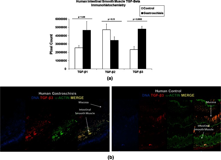
To investigate our hypothesis that intestinal smooth muscle cells were capable of expressing TGF-β3 in GRID, we stained intestinal tissue from human GS infants for all TGF-β isoforms. TGF-β3 immunostaining (pixel units: control—2,324,004 ± 382,219 and GS—4,801,475 ± 330,388*) was significantly increased compared with TGF-β1 and 2 in GS infants compared with controls. Compared with control infants, TGF-β3 and α-actin (Fig. 1b) co-localized to the intestinal smooth muscle cells in infants with GS.
Fig. 1.
Expression of TGF-β isoforms in the intestinal smooth muscle. a Calculated mean pixel units of TGF-β1, 2 and 3 from intestinal tissue sections of human infants with gastroschisis (GS) and premature infant controls, expressed as mean ± SEM. Groups were compared by a Student’s t test for each TGF-β group. b Representative photomicrographs (magnification ×20) from intestinal tissue sections of human infants with gastroschisis (GS) and premature infant controls are depicted and show the immunoreactivity of TGF-β3 (green) and α-actin (red) in the intestinal smooth muscle layers of the intestine. Co-localization of TGF-β3 and α-actin (yellow) is also seen in the intestinal smooth muscle layer of GS infant intestine (n = 7)
TGF-β3 Promotes Collagen Gel Contraction in Intestinal Smooth Muscle Cells
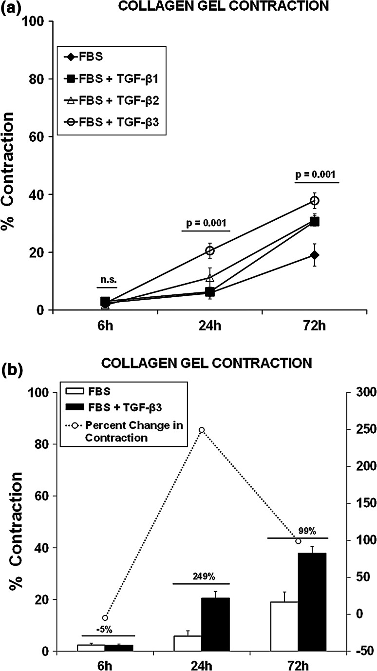
To explain the intestinal smooth muscle dysfunction seen in GRID, we hypothesized that TGF-β3 signals intestinal smooth muscle cell hyper-contractility. To determine whether TGF-β3 promotes cell contractility, we used a collagen gel matrix assay to measure intestinal smooth muscle cell contraction. As depicted in Fig. 2a, TGF-β3 contributed to more gel contraction compared with TGF-β1 or 2 (p = 0.001). At 24 h, TGF-β3 caused a 249 % (p = 0.008) increase in gel contraction compared with cells not exposed to TGF-β3 (Fig. 2b). By 72 h, TGF-β3 continued to cause intestinal smooth muscle cell contraction at a 99 % increase over cells not exposed to TGF-β3 (p = 0.005).
Fig. 2.
TGF-β3 promotes increased contraction of the collagen gel matrix. a Line diagrams show cell contraction in a collagen gel matrix (mean ± SEM) with FBS ± TGF-β1, 2 or 3 over 6, 24 and 72 h, respectively. Intestinal smooth muscle cell contraction is significantly increased in a time-dependent manner on exposure to TGF-β3. Groups were compared by ANOVA for each TGF-β group at each time point. b Line diagrams show percentage of cell contraction in a collagen gel matrix (mean ± SEM) with FBS ± TGF-β3 over 6, 24 and 72 h. Intestinal smooth muscle cells exposed to TGF-β3 became more contracted as time increased. Experimental groups were compared by a Student’s t test
TGF-β3 Promotes Intestinal Dysmotility
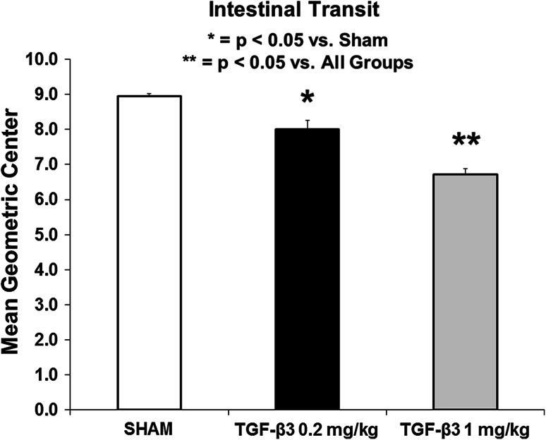
Next, we investigated the dose-dependent effects of TGF-β3 on intestinal motility in rats using FITC-Dextran intestinal transit. The data depicted in Fig. 3 demonstrate that the average mean geometric center (MGC) in high-dose TGF-β3 rats (6.7 ± 0.2**, p < 0.05) was significantly decreased compared with Sham (9.0 ± 0.1) and low-dose TGF-β3 (8.0 ± 0.3*) rats. Low-dose TGF-β3 rats had significantly decreased intestinal transit compared with Sham rats (p < 0.05).
Fig. 3.
Administration of TGF-β3 into the peritoneal cavity decreases intestinal motility. Bar diagrams show intestinal transit as the mean geometric center in the small intestine (mean ± SEM). At 12 h after i.p. injection of TGF-β3, intestinal transit is significantly impaired in a dose–response manner in the animals given i.p. TGF-β3. Experimental groups were compared by ANOVA with a Tukey–Kramer test
TGF-β3 Up-Regulates the Contractile Phenotypic Gene Expression in Intestinal Smooth Muscle Tissue and Cell Culture
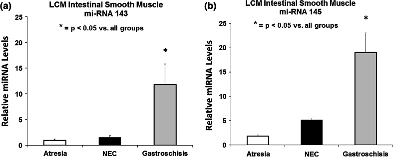
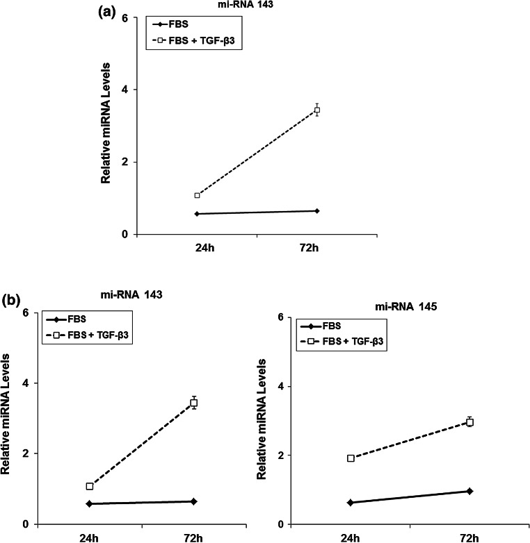
Because miRNA is known to survive the degradation process that occurs from embedding tissue in paraffin, we used LCM to isolate the intestinal smooth muscle layer from infants with and without GS. As shown in Fig. 4, smooth muscle contractile markers miRNA 143 & 145 (Relative miRNA levels l: 12 ± 4* & 19 ± 5*) are elevated in the GS infants compared to infants with Atresia (relative miRNA levels: 1 ± 0.2* & 2 ± 0.6) and NEC (relative miRNA levels: 12 ± 0.4* & 5 ± 2). When intestinal smooth muscle cells are exposed to TGF-β3 in culture, they also demonstrated a significant increase in the contractile miRNA markers 143 & 145 (Fig. 5a).
Fig. 4.
Expression of contractile micro-RNA markers in human intestinal smooth muscle. Calculated mean relative miRNA levels of miRNA-143 (a) and miRNA-145 (b) from intestinal tissue sections of human infants with gastroschisis (GS), intestinal atresia (IA), necrotizing enterocolitis (NEC) and age-matched premature infant control tissue, expressed as mean ± SEM. Groups were compared by ANOVA. Intestinal tissue from the GS patients had significantly elevated levels of miRNA 143 & 145 compared with the other groups
Fig. 5.
TGF-β3 promotes the expression of contractile micro-RNA markers in human intestinal smooth muscle. a Line diagrams show the contractile phenotype gene expression (mean ± standard error of the mean [SEM])) with FBS (a) and FBS ± TGF-β3 (b) over 6, 24 and 72 h. Contractile marker: ϒ-smooth muscle actin (ACTG2), calponin (CNN1), smooth muscle myosin heavy chain (MYH11) and smooth muscle-22α (TAGLN). Intestinal smooth muscles exposed to TGF-β3 have a significantly higher expression of contractile genes at all-time points. These data support that hISMCs exposed to TGF-β3 have a contractile phenotype
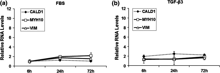
It is known that intestinal smooth muscle cells in culture over time switch from a contractile to a synthetic phenotype. To investigate whether our intestinal smooth muscle cells underwent this phenotypic gene change after TGF-β3 exposure, we evaluated known contractile and synthetic smooth muscle markers. Intestinal smooth muscle cells exposed to TGF-β3 expressed higher contractile protein markers compared with cells exposed to FBS only (Fig. 5b). As depicted in Fig. 6, there was no change in the intestinal smooth muscle cell synthetic gene expression on exposure to TGF-β3.
Fig. 6.
TGF-β3 does not exhibit a synthetic protein gene expression profile. Line diagrams show the synthetic phenotype gene expression (mean ± standard error of the mean [SEM])) with FBS (a) and FBS ± TGF-β3 (b) over 6, 24 and 72 h. Synthetic markers: L-caldesmon (CALD1), non-muscle myosin heavy chain IIB (MYH10) and vimentin (VIM). There is no significant difference in the synthetic phenotypic gene expression when hISMCs are exposed to TGF-β3
Discussion
We previously demonstrated that non-occlusive mesenteric hypertension (NMH) causes intestinal dysfunction and inflammation similar to infants with GRID. However, there is a paucity of reports on the mediators that contribute to the smooth muscle dysfunction in GRID. In this report, we show for the first time that the pro-inflammatory cytokine, TGF-β3, was significantly elevated in the intestinal smooth muscle layer in human GS compared with control subjects. Our data further demonstrated that TGF-β3 was associated with increased intestinal smooth muscle cell contractility, global intestinal dysfunction and increased smooth muscle cell markers of contractile gene expression.
The tissue-level biomechanical force from shear stress, strain or hypertension is known to signal inflammation and has recently been shown to participate in smooth muscle remodeling and phenotypic switching [14, 17]. Smooth muscle migration, hypertrophy and hyperplasia are prominent features in organ remodeling processes after injury in smooth muscle-related diseases such as asthma and atherosclerosis remodeling [18, 19]. The GS abdominal wall defect and NMH model (NMH-simulated model of the abdominal wall defect in animals) are biomechanical forces that produce mesenteric venous hypertension that results in intestinal inflammation and dysfunction [1, 8, 20]. In this report, we provide new evidence that TGF-β3 impairs intestinal transit in a dose-dependent manner similar to that seen in infants and our animal model. Our group has previously demonstrated that NMH contributed to a significant decrease in intestinal contractility prior to stimulation of the enteric nervous system with carbachol [8]. The addition of carbachol resulted in minimal improvement in NMH-induced intestinal contractile dysfunction. These findings suggested that NMH may contribute to a primary smooth muscle dysfunction that impaired transit and contractile function. Although our NMH model had decreased intestinal contractility that partially recovered, our current cell culture experiments using TGF-β3 stimulation showed increased intestinal smooth muscle cell contraction. Gawaziuk et al. [21] reported that TGF-β was responsible for super-contractile smooth muscle cell states that produced non-specific hyper-reactivity of airways in asthmatic patients. In the erectile dysfunction literature, abnormal contractile events in vascular smooth muscle cells of the penis produce end-organ dysfunction [22, 23]. Elevated intestinal smooth muscle cell contraction may explain why the NMH model exhibited decreased intestinal contractility without full recovery and decreased intestinal length.
Cellular signaling and regulation of calcium and myosin light chain kinase are important in generation and maintenance of smooth muscle cell contraction [24, 25]. Well-known signaling pathways that participate in smooth muscle contraction include RhoA/Rho-kinase and G-protein-coupled receptor proteins [23, 26]. While a paucity of data exists on TGF-β3’s role in intestinal smooth muscle contraction, several studies have demonstrated the ability of myofibroblasts and smooth muscle cells to express TGF-β3 [27–29]. Recent data support that TGF-β is capable of autocrine signaling which has been shown to produce airway smooth muscle hyperplasia [30, 31]. Our results show that TGF-β3 co-localized with smooth muscle actin, which suggests that the intestinal smooth muscle is capable of producing TGF-β3. The intestinal smooth muscle cell could serve as a source of TGF-β3 in the intestinal smooth muscle layer of GS infants acting in an autocrine/paracrine fashion to further exacerbate GRID.
In summary, we demonstrated that the TGF-β3 contributed to intestinal dysfunction at the organ level, increased contraction at the cellular level and elevated contractile gene expression at the molecular level. This hyper-contractile response may play a role in the persistent intestinal dysfunction seen in GRID.
Acknowledgments
NIH Grant T32 GM 0879201, and Robert Woods Johnson Foundation Grant 68517.
Conflict of interest
None.
References
- 1.Langer JC, Longaker MT, Crombleholme TM, Bond SJ, Finkbeiner WE, Rudolph CA, et al. Etiology of intestinal damage in gastroschisis. I: effects of amniotic fluid exposure and bowel constriction in a fetal lamb model. J Pediatr Surg. 1989;24:992–997. doi: 10.1016/S0022-3468(89)80200-3. [DOI] [PubMed] [Google Scholar]
- 2.Lao OB, Healey PJ, Perkins JD, Horslen S, Reyes JD, Goldin AB. Outcomes in children after intestinal transplant. Pediatrics. 2010;125:e550–e558. doi: 10.1542/peds.2009-1713. [DOI] [PMC free article] [PubMed] [Google Scholar]
- 3.Farmer DG, Venick RS, Colangelo J, Esmailian Y, Yersiz H, Duffy JP, et al. Pretransplant predictors of survival after intestinal transplantation: analysis of a single-center experience of more than 100 transplants. Transplantation. 2010;90:1574–1580. doi: 10.1097/TP.0b013e31820000a1. [DOI] [PubMed] [Google Scholar]
- 4.Gregersen H. A mechanical perspective on intestinal tone and gas motion. Neurogastroenterol Motility. 2006;18:873–875. doi: 10.1111/j.1365-2982.2006.00828.x. [DOI] [PubMed] [Google Scholar]
- 5.Spencer NJ, Smith CB, Smith TK. Role of muscle tone in peristalsis in guinea-pig small intestine. J Physiol. 2001;530:295–306. doi: 10.1111/j.1469-7793.2001.0295l.x. [DOI] [PMC free article] [PubMed] [Google Scholar]
- 6.Srinathan SK, Langer JC, Blennerhassett MG, Harrison MR, Pelletier GJ, Lagunoff D. Etiology of intestinal damage in gastroschisis. III: morphometric analysis of the smooth muscle and submucosa. J Pediatr Surg. 1995;30:379–383. doi: 10.1016/0022-3468(95)90036-5. [DOI] [PubMed] [Google Scholar]
- 7.Midrio P, Faussone-Pellegrini MS, Vannucchi MG, Flake AW. Gastroschisis in the rat model is associated with a delayed maturation of intestinal pacemaker cells and smooth muscle cells. J Pediatr Surg. 2004;39:1541–1547. doi: 10.1016/j.jpedsurg.2004.06.017. [DOI] [PubMed] [Google Scholar]
- 8.Shah SK, Aroom KR, Walker PA, Xue H, Jimenez F, Gill BS, et al. Effects of non-occlusive mesenteric hypertension on intestinal function: implications for gastroschisis-related intestinal dysfunction. Pediatr Res. 2012;71:668–674. doi: 10.1038/pr.2012.20. [DOI] [PMC free article] [PubMed] [Google Scholar]
- 9.Schwarz NT, Kalff JC, Turler A, Speidel N, Grandis JR, Billiar TR, et al. Selective jejunal manipulation causes postoperative pan-enteric inflammation and dysmotility. Gastroenterology. 2004;126:159–169. doi: 10.1053/j.gastro.2003.10.060. [DOI] [PubMed] [Google Scholar]
- 10.Hassoun HT, Weisbrodt NW, Mercer DW, Kozar RA, Moody FG, Moore FA. Inducible nitric oxide synthase mediates gut ischemia/reperfusion-induced ileus only after severe insults. J Surg Res. 2001;97:150–154. doi: 10.1006/jsre.2001.6140. [DOI] [PubMed] [Google Scholar]
- 11.Hassoun JT, Zou L, Moore FA, Kozar RA, Weisbrodt NW, Kone BC. α-Melanocyte stimulating hormone protects against mesenteric ischemia-reperfusion injury. Am J Physiol. 2002;282:G1059–G1068. doi: 10.1152/ajpgi.00073.2001. [DOI] [PubMed] [Google Scholar]
- 12.Attuwaybi B, Kozar RA, Gates KS, Moore-Olufemi S, Sato N, Weisbrodt NW, et al. Hypertonic saline prevents inflammation, injury, and impaired intestinal transit after gut ischemia/reperfusion by inducing heme oxygenase 1 enzyme. J Trauma. 2004;56:749–758. doi: 10.1097/01.TA.0000119686.33487.65. [DOI] [PubMed] [Google Scholar]
- 13.Beamish JA, He P, Kottke-Marchant K, Marchant RE. Molecular regulation of contractile smooth muscle cell phenotype: implications for vascular tissue engineering. Tissue Eng Part B Rev. 2010; 16: 467–491. [DOI] [PMC free article] [PubMed]
- 14.Davis-Dusenbery BN, Wu C, Hata A. Micromanaging vascular smooth muscle cell differentiation and phenotypic modulation. Arterioscler Thromb Vasc Biol. 2011;31:2370–2377. doi: 10.1161/ATVBAHA.111.226670. [DOI] [PMC free article] [PubMed] [Google Scholar]
- 15.Sperveslage J, Hoffmeister M, Henopp T. Establishment of robust controls for the normalization of miRNA expression in neuroendocrine tumors of the ileum and pancreas. Endocrine. 2014. Epub. 02/18/2014. [DOI] [PubMed]
- 16.Schaefer A, Jung M, Miller K. Suitable reference genes for relative quantification of miRNA expression in prostate cancer. Exp Mol Med. 2010;42:749–758. doi: 10.3858/emm.2010.42.11.076. [DOI] [PMC free article] [PubMed] [Google Scholar]
- 17.Shao J, Xia ZW, Li YZ, Yu SC, Deng WW. Expression of cellular phenotype switching markers-matrix protein Gla, mRNA and collagen I, III and V of human airway smooth muscle cells in vitro after TGF-beta1 stimulation. Zhonghua Er Ke Za Zhi. 2006;44:531–534. [PubMed] [Google Scholar]
- 18.Yang SP, Woolf AS, Quinn F, Winyard PJ. Deregulation of renal transforming growth factor-beta1 after experimental short-term ureteric obstruction in fetal sheep. Am J Pathol. 2001;159:109–117. doi: 10.1016/S0002-9440(10)61678-1. [DOI] [PMC free article] [PubMed] [Google Scholar]
- 19.Kurata M, Okura T, Watanabe S, Fukuoka T, Higaki J. Osteopontin and carotid atherosclerosis in patients with essential hypertension. Clin Sci (Lond). 2006;111:319–324. doi: 10.1042/CS20060074. [DOI] [PubMed] [Google Scholar]
- 20.Radhakrishnan RS, Radhakrishnan HR, Xue H, Moore-Olufemi SD, Mathur AB, Weisbrodt NW, et al. Hypertonic saline reverses stiffness in a Sprague-Dawley rat model of acute intestinal edema, leading to improved intestinal function. Crit Care Med. 2007;35:538–543. doi: 10.1097/01.CCM.0000254330.39804.9C. [DOI] [PubMed] [Google Scholar]
- 21.Gawaziuk JP, Sheikh F, Cheng ZQ, Cattini PA, Stephens NL. Transforming growth factor-beta as a differentiating factor for cultured smooth muscle cells. Eur Respir J. 2007; 4:643–652. [DOI] [PubMed]
- 22.Jin L, Linder AE, Mills TM, Webb RC. Inhibition of the tonic contraction in the treatment of erectile dysfunction. Exp Opin Therapeutic Targets. 2003;7:265–276. doi: 10.1517/14728222.7.2.265. [DOI] [PubMed] [Google Scholar]
- 23.Mills TM, Lewis RW, Wingard CJ, Chitaley K, Webb RC. Inhibition of tonic contraction—a novel way to approach erectile dysfunction. J Androl. 2002;23:S5–S9. [PubMed] [Google Scholar]
- 24.Kamm KE, Stull JT. Dedicated myosin light chain kinases with diverse cellular functions. J Biol Chem. 2001;276:4527–4530. [DOI] [PubMed]
- 25.Bivalacqua TJ, Champion HC, Usta MF, Cellek S, Chitaley K, Webb RC, et al. RhoA/Rho-kinase suppresses endothelial nitric oxide synthase in the penis: a mechanism for diabetes-associated erectile dysfunction. Proc Natl Acad Sci U S A. 2004;101:9121–9126. doi: 10.1073/pnas.0400520101. [DOI] [PMC free article] [PubMed] [Google Scholar]
- 26.Yang Z, Balenga N, Cooper PR, Damera G, Edwards R, Brightling CE, et al. Regulator of G-protein signaling-5 inhibits bronchial smooth muscle contraction in severe asthma. Am J Respir Cell Mol Biol. 2012;46:823–832. doi: 10.1165/rcmb.2011-0110OC. [DOI] [PMC free article] [PubMed] [Google Scholar]
- 27.Lee BS, Nowak RA. Human leiomyoma smooth muscle cells show increased expression of transforming growth factor-beta 3 (TGF beta 3) and altered responses to the antiproliferative effects of TGF beta. J Clin Endocrinol Metab. 2001;86:913–920. doi: 10.1210/jcem.86.2.7237. [DOI] [PubMed] [Google Scholar]
- 28.Arici A, Sozen I. Transforming growth factor-beta3 is expressed at high levels in leiomyoma where it stimulates fibronectin expression and cell proliferation. Fertil Steril. 2000;73:1006–1011. doi: 10.1016/S0015-0282(00)00418-0. [DOI] [PubMed] [Google Scholar]
- 29.McKaig BC, Hughes K, Tighe PJ, Mahida YR. Differential expression of TGF-beta isoforms by normal and inflammatory bowel disease intestinal myofibroblasts. Am J Physiol Cell Physiol. 2002;282:C172–C182. doi: 10.1152/ajpcell.00048.2001. [DOI] [PubMed] [Google Scholar]
- 30.Kajihara I, Jinnin M, Makino T, Masuguchi S, Sakai K, Fukushima S, et al. Overexpression of hepatocyte growth factor receptor in scleroderma dermal fibroblasts is caused by autocrine transforming growth factor β signaling. Biosci Trends. 2012;6:136–142. doi: 10.5582/bst.2012.v6.3.136. [DOI] [PubMed] [Google Scholar]
- 31.Lazaar AL, Panettieri RA., Jr Airway smooth muscle: a modulator of airway remodeling in asthma. J Allergy Clin Immunol. 2005;116:488–495. doi: 10.1016/j.jaci.2005.06.030. [DOI] [PubMed] [Google Scholar]


