Vol. 206 No. 1, July 7, 2014. Pages 129–143.
In the original version of Figure 7, the y axis unit labels were incorrect in panels B and C.
Figure 7.
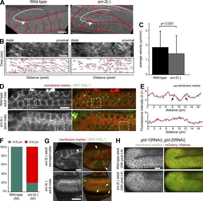
Cytoplasmic streaming in the rachis may be responsible for germline disorganization in ani-2 mutants. (A) DIC images of the germlines of wild-type (left) and ani-2(−) (right) young adult animals. Some membrane partitions are outlined in red. The white arrow depicts the direction of cytoplasmic streaming. (B) DIC images (top) and schematic representations (bottom) of kymographs of cytoplasmic streaming in the gonads of animals depicted in A. Kymographs were made along the white line shown in A. The total duration of the movie is 45 min. (C) Average velocity of cytoplasmic streaming in the rachis of wild-type (black) and ani-2(−) (gray) animals. Error bars represent SD over 9 animals analyzed for each genotype. (D) Mid-section confocal images of a wild-type (top) and an ani-2(−) (bottom) male adult germline expressing a membrane marker (red) and GFP::PGL-1 (green). (E) Measured fluorescence intensities (in arbitrary units) for the membrane marker along the lateral and apical cortices of the germ cells of each male genetic background delineated by a dashed square in D. Arrows point to peaks of membrane marker fluorescence intensity bordering a minimum. (F) Proportion of germ cells showing rachis bridges with a diameter >0.8 µm (turquoise) or <0.8 µm (red) in wild-type and ani-2(−) animals at the adult stage, as measured by membrane marker distribution. The numbers in brackets represent the total number of germ cells analyzed. (G) Mid-section confocal images of the germlines of an ani-2(−) adult hermaphrodite (top) and an ani-2(−) adult male (bottom) expressing a membrane marker (red) and GFP::PGL-1 (green). Arrows point to multinucleated germ cells, whose number is significantly reduced in ani-2(−) males. (H) Mid-section confocal images of the gonads of wild-type (top) and ani-2(−) (bottom) adult hermaphrodites expressing a membrane marker (green) and mCherry::Histone H2B (red) and depleted of GLD-1 and GLD-2 by RNAi. Bars (A, B, D, G, and H), 10 µm.
A corrected version of Figure 7 is shown below. The HTML and PDF versions of this article have been corrected. The error remains only in the print version.


