Abstract
Objective:
The aim of the present study was to determine the prevalence of supernumerary teeth with by evaluating a large group of adult patients in Turkey and to investigate the characteristics of supernumerary teeth with their complications and treatment options.
Study Design:
This descriptive and retrospective study was carried out in 7348 adult patients aged over 18 years (3212 females and 4136 males). The characteristics of the supernumerary teeth were noted and the diagnosis was made during clinical and radiographic examination with the help of panaromic, periapical, and occlusal radiography. Information on the demographic variables for each patient, including age and gender, were colleceted.
Materials and Methods:
All supernumerary teeth were classfied under several titles such as location, position, morphology, eruption, clinical complications, and treatment protocols. The data obtained were subjected to statistical analysis. Chi-squared test was used to determine differences in distribution of supernumerary teeth when stratified by gender. The statistical significance was established by confidence interval of 95% (P ≤ 0.05).
Results:
123 (2.14%) affected patients (69 females and 54 males) were observed with a female:male ratio of 1.28:1 (P < 0.05). One hundred and fifty-six supernumerary teeth were detected in all affected patients.
Conclusion:
Supernumerary teeth may be observed in adults patients with a similar frequency (2.14%) as in children and young adolescents, and clinicians should take measures and examine all patients carefully even at older ages.
Keywords: Adult population, prevalence, supernumerary teeth
INTRODUCTION
Supernumerary teeth are defined as any extra tooth formed in normal dentition and this condition is also called “hyperdontia.” The prevalence of supernumerary teeth for permanent dentition is between 0.5 and 5.3% and in primary dentition is between 0.2 and 0.8% in various populations.[1,2,3,4] In addition, the gender wise prevalence of supernumerary teeth or hyperdontia shows that these teeth are predominantly seen in males than in females.[5,6,7] Supernumerary teeth may be associated with several syndromes such as cleidocranial dysplasia, Gardner's syndrome, Ehlers–Danlos syndrome, and Fabry–Anderson syndrome.[3,7] In these cases, supernumerary teeth often appear in multiple forms. However, these teeth may occur in patients with no sydrome and may appear as single, double, or multiple and as unilateral or bilateral.[3,8,9]
Although the main etiology of supernumerary teeth is not certainly explained, several theories have been proposed and the most common view is that these teeth develop as a result of horizontal proliferation or hyperactivity of the dental lamina.[3,9,10] Supernumerary teeth may be located in any region of the dental arch; however, they are usually found on the maxillary midline between two maxillary central teeth and these teeth are called mesiodens. This is commonly followed by maxillary molar, maxillary lateral incissor, mandibular premolar, and mandibular molar.[10,11] Supernumerary teeth may be classified morphologically according to their shape as conical, tuberculate, supplemental, and odontomatous.[9]
Supernumerary teeth may erupt normally or may be impacted, and they may cause some complications such as failure of eruption, displacement, crowding, diastemas, development of odontogenic cyst, and resorption of neighboring teeth.[5,10,11] Radiological examinations have been used for locating the position of supernumerary tooth. Treatment options change according to the severity of complication. Thus, the treatment options include clinical follow-up for a particular period, surgical removal, and orthodontic intervention.[4,5,7,12,13]
The objective of the present study was to determine the prevalence of the condition by evaluating a large group of adult patients in Turkey and to investigate the characteristics of supernumerary teeth. In addition to these, it was aimed to evaluate the associated complications and treatment protocols.
MATERIALS AND METHODS
This descriptive and retrospective study was carried out in 7348 adult patients (3212 females and 4136 males), aged over 18, attending the Department of Oral and Maxillofacial Surgery (OMFS), Faculty of Dentistry, Bülent Ecevit University over a period of 5 years. One hundred and twenty-three patients (69 females and 54 males) with supernumerary teeth were included in the present study. Patients were reffered to the OMFS clinic for different reasons such as tooth extraction, undergoing major surgeries, and routine clinical examination. Patients diagnosed with any syndrome were excluded from the study. The characteristics of supernemerary teeth were noted and the diagnosis was made during clinical and radiographic examination with the help of panaromic, periapical, and occlusal radiography.
We collected information on the demographic variables for each patient, including age and gender. Following the clinical and radiographic examination, supernumerary teeth were classfied under several titles such as location (maxilla or mandible, specifying the region from anterior to posterior), position (vertical, angled, horizontal, or inverted), morphology (conical, tuberculated, supplemental, and odontoma), and eruption (unerupted or erupted). Furthermore, clinical complications and treatment protocols were analyzed. The data obtained were subjected to statistical analysis. Chi-squared test was used to determine differences in distribution of supernumerary teeth when stratified by gender. The statistical significance was established by confidence interval of 95% (P ≤ 0.05).
RESULTS
During the study, supernumerary teeth were detected in 123 patients (2.14%). Sixty-nine of these patients were females and 54 patients were males with a female: male ratio of 1.28:1 (P < 0.05) [Table 1].
Table 1.
Distribution of the supernumerary teeth according to the gender

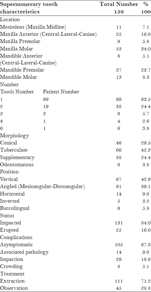
Table 2 shows the characteristics of supernumerary teeth. Totally 156 supernumerary teeth were detected in 123 patients and 80.5% (n = 99) of these patients had single supernumeraries. Double supernumeraries were observed in 19 patients (15.5%) and multiple supernumeraries in five patients. Among these five patients, one patient (0.8%) had six supernumerary teeth.
Table 2.
Characteristics of the supernumerary teeth

A total of 156 supernumerary teeth were observed, of which 62.8% (n = 98) were located in the maxillary arch while 37.2% (n = 58) were found in the mandible. Fifty-three teeth (34.0%) were found in the maxillary molar region and 37 teeth (23.7%) were found in the mandibular premolar region. These regions were followed by maxillary anterior (16.0%), mandibular molar (8.3%), maxillary premolar (5.8%), and mandibular anterior (5.1%) regions. Of the 156 supernumerary teeth examined, 42.3% (n = 66) were tuberculate, 29.5% (n = 46) were conical, 24.4% (n = 38) were supplementary, and 3.8% (n = 6) were odontomatous. When the positions were examined, 42.9% (n = 66) of the supernumerary teeth were in a vertical position. This was followed by angled position [39.1% (n = 61)]. Regarding their status within the arch, 131 supernumerary teeth (84.0%) were in impacted condition. Also, 105 supernumerary teeth (67.3%) did not cause any complications. However, 29 teeth (18.6%) caused impaction and 8 teeth (5.1%) caused displacement of the adjacent teeth. In addition to these, 14 teeth (9.0%) were associated with a pathology such as cystic lesion or pericoronitis. One hundred and eleven supernumerary teeth (28.8%) were extracted and most of them caused some complications, whereas periodical observation was chosen as a treatment option in 45 teeth (71.2%).
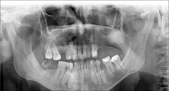
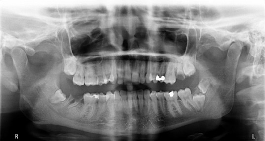
Moreover, unusual multiple bilaterally supernumerary case and one kissing molars case were diagnosed among our patients [Figures 1 and 2].
Figure 1.

Radiographic appearence of multiple bilateral supernumerary teeth
Figure 2.

Radiographic appearence of kissing molars
DISCUSSION
Supernumerary teeth are infrequent developmental alterations that may manifest in any region of the dental arches and may involve any tooth. These teeth may be associated with a syndrome or they may be found in non-sydromic patients.[5] Although various theories have been suggested, such as atavism, dichotomy of the tooth germ, and excessive growth of dental lamina, the main etiologic factor of these teeth has not been identified.[14] Supernumerary teeth have been reported in both the primary and permanent dentition; however, a higher incidence of the anomaly is noted in the permanent dentition. Thus, the prevalence in permanent dentition varies from 0.5 to 3.8% when compared to a prevalence of 0.3–0.6% in primary dentition.[7,8,9,10,11,12,13,14,15] In the present study, non-syndromic patients with permanent supernumerary teeth were evaluated. In the literature, the frequency of supernumerary teeth in the general population is reported to be between 0.1 and 3.8%.[5,12,16,17,18] In our study, it was observed that 156 patients were affected among 7348 non-syndromic patients and the frequency was 2.14%. Our findings showed that females were more significantly affected than males (P < 0.001) with a 1.28:1 ratio and this finding is not in agreement with the results reported in the literature[3,5,6,7,9,17,19] showing that supernumerary teeth appear with a higher frequency in men than in women. This situation may be explained by performing the present study on the population whose ages were over 18 because some patients, who had a surgical extraction history in young ages, might have been the reason of the difference observed.
Age of patients with supernumerary teeth ranges from 5 to 70 in the literature; however, most cases are observed to be between 7 and 10 years of age.[9,20,21,22,23] In the study of Esenlik et al.,[12] 69 patients with age ranging from 6 to 16 years were detected among 2599 subjects and the frequency was 2.7%. In a similar study by Küchler et al.,[16] the frequency of supernumerary teeth was found to be 2.3% among 1166 patients and the age of the population ranged from 6 to 12 years. Unlike these studies, in the present study, 123 affected cases were found among 7348 patients (2.14%) whose age ranged from 19 to 61 years and this result showed that supernumerary teeth may be detected in adolescents at a similar frequency as in children and young adolescents.
Supernumerary teeth can appear in any region of the jaws; however, they most commonly involve the premaxilla, which has also been identified as the predominant location by many researchers.[3,10,12,15,24] Leco et al.[6] explained that mesiodens were the most frequent supernumerary teeth found in the maxilla, occurrring at a rate of 80.0%. In the study of Ferres-Padro et al.,[3] the patient age ranged between 5 and 19 years and mesiodens were the most frequently found supernumerary teeth (53.16%). Sharma and Singh[24] performed a similar study on a population ranging in age from 4 to 14 years and found that 81.2% of the supernumerary teeth were located in the central incisor region, of which 30.0% were mesiodens. According to these results, mesiodens are the most commonly seen supernumerary teeth in children and young adolescents. In our study, the results changed in favor of the maxillary molars (34.0%), whereas the incidence of mesiodens remained at 7.1% only. This difference may be related to the range of age of the patients examined. The previous studies[3,9,10,12,24] focused on children and young adolescents ranging in age from 4 to 19 and mesiodens teeth were the most frequently seen supernumerary teeth. However, our study was performed on adults only and 34% of all supernumerary teeth were maxillary molar teeth. So, the age difference in the patients studied and the different diagnostic methods used may explain the differences observed between the results of previous and present studies.
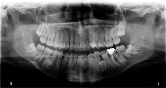
Supernumerary teeth may be observed singly or in multiples, in any region of the jaws in the same person.[9] It is well established that supernumerary teeth are more frequently single tooth, while multiple supernumerary teeth appear frequently as two teeth.[5,9,10,12,24] However, it is rare to find multiple supernumerary teeth with no associated diseases or syndromes.[25] According to our results, 63.5% (n = 99) of the supernumerary teeth were found to be single and 24.4% (n = 38) were found as two teeth [Figure 3], and our findings are in agreement with the results reported in the literature. Nevertheless, six supernumerary teeth were observed at the same time in one (0.8%) non-syndromic patient in the present study.
Figure 3.

Radiographic appearence of bilateral supernumerary premolars
In many studies,[3,7,10,12,16,24] the shape of supernumerary teeth appears conical in the permanent dentition. Nevertheless, in the present study, tuberculate form was the most (42.3%) commonly observed because of the presence of majority of supernumerary teeth in the premolar and molar zones. When assessing eruption status, we found that 84.0% (n = 131) of the supernumerary teeth were impacted generally; the presence of premolars and molars is detected coincidentally on a radiograph, given that these supernumerary teeth are usually impacted.[1,13]
In the present study, clinical complications were observed in 32.7% (n = 51) of the supernumerary teeth and impaction was the most frequently found complication, followed by associated pathology and displacement, respectively, whereas 67.3% (n = 105) of the supernumerary teeth were asymptomatic. According to Acikgöz et al.,[25] 75% of supernumerary teeth are asymptomatic and most cases are diagnosed by during routine radiographic inspection prior to the commencement of dental treatment.[1,13] In cases where supernumerary tooth does not cause any symptoms or when there is a risk of damaging the adjacent permanent tooth, it is advisable to avoid therapeutic approach and instead adhere to periodic clinical and radiological examinations.[7,26,27] In our study, 28.8% (n = 45) of the supernumerary teeth were left without any surgical intervention for periodical follow-up. Also, 71.2% (n = 111) of the supernumerary teeth were extracted surgically for avoiding the complications or eliminating the risk factors that could cause possible complications in the future.
CONCLUSION
In the literature, children and young adolescents were the most evaluated populations, and most of these patients are treated by surgical extraction before the age of 18 whereas some patients are followed up periodically until the supernumerary tooth creates a risk for any complication and this situation may occur after many years. On the other hand, some supernumerary teeth can be detected in older ages first because of some reasons such as the supernumerary tooth staying without any complications or creating a complication after many years, or due to inadequate clinical and radiographical examinations. In our research, we focused on adult patients to highlight the similar frequency of supernumerary teeth as in children and young adolescent patients and our results supported the aim of the present study. In addition, most of these supernumerary teeth were detected by the orthopantomographic radiographs taken for routine examination. Consequently, these situations should be perceived as a warning for all clinicians to take measures and examine all patients carefully even at older ages before charting out any treatment plan.
Footnotes
Source of Support: Nil
Conflict of Interest: None declared.
REFERENCES
- 1.Kaya GŞ, Yapıcı G, Ömezli MM, Dayı E. Non-syndromic supernumerary premolars. Med Oral Patol Cir Bucal. 2011;16:e522–5. [PubMed] [Google Scholar]
- 2.Sasaki H, Funao J, Morinaga H, Nakano K, Ooshima T. Multiple supernumerary teeth in the maxillary canine and mandibular premolar regions: A case in the postpermanent dentition. Int J Paediatr Dent. 2007;17:304–8. doi: 10.1111/j.1365-263X.2006.00791.x. [DOI] [PubMed] [Google Scholar]
- 3.Ferrés-Padró E, Prats-Armengol J, Ferrés-Amat E. A descriptive study of 113 unerupted supernumerary teeth in 79 pediatric patients in Barcelona. Med Oral Patol Oral Cir Bucal. 2009;14:E146–52. [PubMed] [Google Scholar]
- 4.Diaz A, Orozco J, Fonseca M. Multiple hyperodontia: Report of a case with 17 supernumerary teeth with non syndromic association. Med Oral Patol Oral Cir Bucal. 2009;14:E229–31. [PubMed] [Google Scholar]
- 5.Çelikoğlu M, Kamak H, Oktay H. Prevalence and characteristics of supernumerary teeth in a non-syndrome Turkish population: Associated pathologies and proposed treatment. Med Oral Patol Oral Cir Bucal. 2010;15:e575–8. doi: 10.4317/medoral.15.e575. [DOI] [PubMed] [Google Scholar]
- 6.Leco Berrocal M, Martin Morales JF, Martinez González JM. An observational study of the frequency of supernumerary teeth in a population of 2000 patients. Med Oral Patol Oral Cir Bucal. 2007;12:E134–8. [PubMed] [Google Scholar]
- 7.Fernández Montenegro P, Valmaseda Castellón E, Berini Aytés L, Gay Escoda C. Retrospective study of 145 supernumerary teeth. Med Oral Patol Oral Cir Bucal. 2006;11:E339–44. [PubMed] [Google Scholar]
- 8.Moore SR, Wilson DF, Kibble J. Sequential development of multiple supernumerary teeth in the mandibular premolar region-a radiographic case report. Int J Paediatr Dent. 2002;12:143–5. doi: 10.1046/j.1365-263x.2002.00336.x. [DOI] [PubMed] [Google Scholar]
- 9.Rajab LD, Hamdan MA. Supernumerary teeth: Review of the literature and a survey of 152 cases. Int J Paediatr Dent. 2002;12:244–54. doi: 10.1046/j.1365-263x.2002.00366.x. [DOI] [PubMed] [Google Scholar]
- 10.De Oliveira Gomes C, Drummond SN, Jham BC, Abdo EN, Mesquita RA. A survey of 460 supernumerary teeth in Brazilian children and adolescents. Int J Paediatr Dent. 2008;18:98–106. doi: 10.1111/j.1365-263X.2007.00862.x. [DOI] [PubMed] [Google Scholar]
- 11.Kara MI, Aktan AM, Ay S, Bereket C, Şener İ, Bülbül M, et al. Characteristics of 351 supernumerary molar teeth in Turkish population. Med Oral Patol Oral Cir Bucal. 2012;17:e395–400. doi: 10.4317/medoral.17605. [DOI] [PMC free article] [PubMed] [Google Scholar]
- 12.Esenlik E, Sayin MO, Atilla AO, Ozen T, Altun C, Başak F. Supernumerary teeth in a Turkish population. Am J Orthod Dentofacial Orthop. 2009;136:848–52. doi: 10.1016/j.ajodo.2007.10.055. [DOI] [PubMed] [Google Scholar]
- 13.Martínez-González JM, Cortés-Bretón Brinkmann J, Calvo-Guirado JL, Arias Irimia O, Barona-Dorado C. Clinical epidemiological analysis of 173 supernumerary molars. Acta Odontol Scand. 2012;70:398–404. doi: 10.3109/00016357.2011.629629. [DOI] [PubMed] [Google Scholar]
- 14.Kumar DK, Gopal KS. An epidemiological study on supernumerary teeth: A survey on 5,000 people. J Clin Diagn Res. 2013;7:1504–7. doi: 10.7860/JCDR/2013/4373.3174. [DOI] [PMC free article] [PubMed] [Google Scholar]
- 15.Mukhopadhyay S. Mesiodens: A clinical and radiographic study in children. J Indian Soc Pedod Prev Dent. 2011;29:34–8. doi: 10.4103/0970-4388.79928. [DOI] [PubMed] [Google Scholar]
- 16.Küchler EC, Costa AG, Costa Mde C, Vieira AR, Granjeiro JM. Supernumerary teeth vary depending on gender. Braz Oral Res. 2011;25:76–9. doi: 10.1590/s1806-83242011000100013. [DOI] [PubMed] [Google Scholar]
- 17.Mason C, Azam N, Holt RD, Rule DC. A retrospective study of unerupted maxillary incisors associated with supernumerary teeth. Br J Oral Maxillofac Surg. 2000;38:62–5. doi: 10.1054/bjom.1999.0210. [DOI] [PubMed] [Google Scholar]
- 18.Carvalho JC, Vinker F, Declerck D. Malocclusion, dental injures and dental anomalies in the primary dentition of Belgian children. Int J Paediatr Dent. 1998;8:137–41. doi: 10.1046/j.1365-263x.1998.00070.x. [DOI] [PubMed] [Google Scholar]
- 19.Hattab FN, Yasin OM, Rawashdeh MA. Supernumerary teeth: Report of three cases and review of the literature. ASDC J Dent Child. 1994;61:382–93. [PubMed] [Google Scholar]
- 20.Zilberman Y, Malron M, Shteyer A. Assessment of 100 children in Jerusalem with supernumerary teeth in the premaxillary region. ASDC J Dent Child. 1992;59:44–7. [PubMed] [Google Scholar]
- 21.Tay F, Pang A, Yuen S. Unerupted maxillary anterior supernumerary teeth: Report of 204 cases. ASDC J Dent Child. 1984;51:289–94. [PubMed] [Google Scholar]
- 22.Leyland L, Batra P, Wong F, Llewelyn R. A retrospective evaluation of the eruption of impacted permanent incisors after extraction of supernumerary teeth. J Clin Periatr Dent. 2006;30:225–31. doi: 10.17796/jcpd.30.3.60p6533732v56827. [DOI] [PubMed] [Google Scholar]
- 23.Koch H, Schwartz O, Klausen B. Indications for surgical removal of supernumerary teeth in the premaxilla. Int J Oral Maxillofac Surg. 1986;15:273–81. doi: 10.1016/s0300-9785(86)80085-0. [DOI] [PubMed] [Google Scholar]
- 24.Sharma A, Singh VP. Supernumerary teeth in Indian children: A survey of 300 cases. Int J Dent 2012. 2012 doi: 10.1155/2012/745265. 745265. [DOI] [PMC free article] [PubMed] [Google Scholar]
- 25.Açikgöz A, Açikgöz G, Tunga U, Otan F. Characteristics and prevalence of non-syndrome multiple supernumerary teeth: A retrospective study. Dentomaxillofac Radiol. 2006;35:185–90. doi: 10.1259/dmfr/21956432. [DOI] [PubMed] [Google Scholar]
- 26.Mason C, Rule DC, Hopper C. Multiple supernumeraries: The importance of clinical and radiographic follow-up. Dentomaxillofac Radiol. 1996;25:109–13. doi: 10.1259/dmfr.25.2.9446982. [DOI] [PubMed] [Google Scholar]
- 27.Garvey MT, Barry HJ, Blake M. Supernumerary teeth - an overview of classification, diagnosis and management. J Can Dent Assoc. 1999;65:612–6. [PubMed] [Google Scholar]


