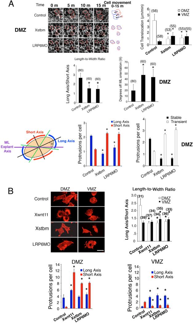
Fig. 3. Lrp6 controls the morphology and motility of mesodermal cells.
(A) Frames from time-lapse movies of DMZ and VMZ cells in shaved Keller explants from embryos injected with Lrp6MO (LRP6MO; 40 ng) and Stbm (Xstbm; 200 pg) show alterations in motility, shape, orientation, protrusion distribution along the cell periphery and protrusion stability as indicated in the bar charts. Outlines of randomly chosen cells were traced throughout each movie, and traces from the first (red) and last (blue) frame were superimposed to assess cell translocation. For measuring protrusion distributions, each cell was divided into four quadrants emanating from the center (black lines), and each quadrant was subdivided into two sectors by marking the midpoint along the cell membrane (green lines). Each protrusion was assigned to the sector to which it most closely localized (long axis in blue, short axis in red). The angle (θ) between the long axis of the cell and the mediolateral axis (purple line) of the explant was used to calculate its orientation in the explant. Protrusion stability was measured by counting the number of stable (present throughout the 15 minute movie) and transient protrusions (appearing after the first frame and/or disappearing before the last). (B) Similar cellular changes were observed in isolated DMZ cells of embryos injected with Lrp6MO. DMZ and VMZ cells from stage 10.5 embryos were dissociated, plated on fibronectin, immediately fixed and stained with Rhodamine-phalloidin. DMZ cells from wnt11 mRNA (Xwnt; 160 pg)-, stbm mRNA (Xstbm; 200 pg)-, or Lrp6MO (LRP6MO; 40 ng)-injected embryos have decreased length-to-width ratios, increased number of total cytoplasmic protrusions, and increased numbers of cytoplasmic protrusions along the short axes versus the long axes compared to GFP-injected (200 pg) control. VMZ cells from wnt11-, stbm-, or Lrp6MO-injected embryos show no obvious length-to-width ratio changes, but show a greater number of protrusions along their long axes and fewer protrusions along their short axes compared to controls. Statistical analyses in A and B were done using Student's t-test. Error bars indicate standard deviation. Asterisks mark differences that are statistically significant from control (P<0.01). Numbers of cells analyzed are indicated in parentheses. DMZ and VMZ cells used to determine length-to-width ratios were also used to determine number of protrusions per cell. Cells and explants were co-injected with myristoylated GFP (200 pg) to highlight plasma membranes and trace injected cells. Scale bars: 25 μm in A; 20 μm in B.

