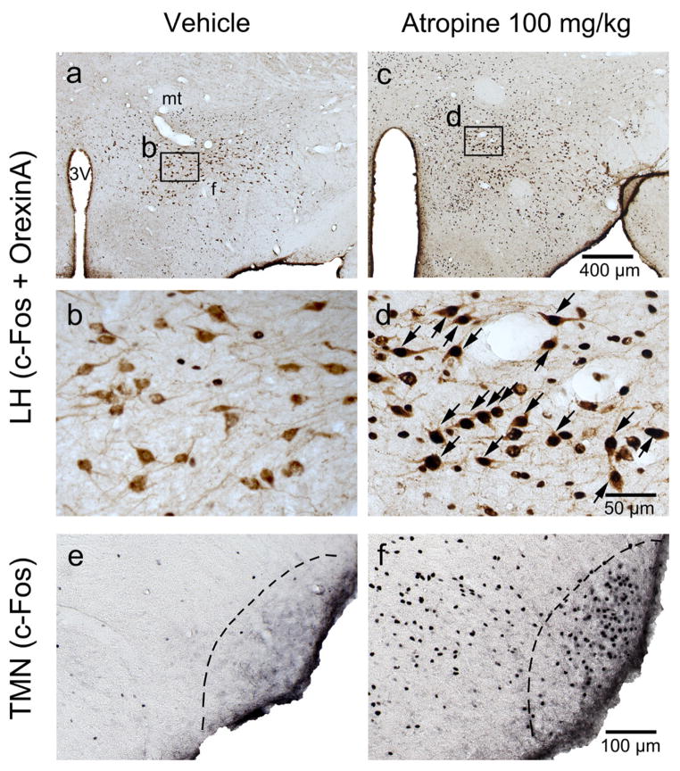
Figure 6.
Atropine induces c-Fos expression in lateral hypothalamic orexin neurons and histaminergic neurons.
a, b, c and d: representative photomicrographs of c-Fos (black) and orexin A (brown) double immunostaining in the LH of vehicle- (a and b) and 100mg/kg atropine (c and d) administered rats (b and d: high-magnification views of the rectangular areas marked in “a”, and “c” respectively). Arrows indicate the c-Fos and orexin A double-stained cells. e and f: representative photomicrographs of c-Fos (black) immunostaining in the TMN of vehicle-(e) and 100 mg/kg atropine (f) administrated rats. Scale bars: a and c, 400 μm; b and d, 50 μm. e and f, 100μm. 3V: third ventricle; f: fornix; LH: lateral hypothalamus; mt: mammillothalamic tract. TMN: tuberomammillary nucleus.

