

Sir,
A 65-year-old woman, nondiabetic and normotensive, presented with a chief complaint of cough for three months. It was associated with generalized weakness and fatigue for the last one month. She had lost over 10 kg of weight over the last one month. She had no history of dysphagia, nausea, vomiting or melena.
On physical examination, the lady was nutritionally poor with moderate pallor. No lymph nodes were palpable. Chest examination revealed bilateral diffuse crepitations. Examination of the other systems was within normal limits.
A chest X-ray, posterior-anterior (PA) view, revealed multiple pulmonary nodules, which were highly suggestive of malignancy [Figure 1]. A contrast-enhanced computed tomography (CT) scan of chest revealed ‘cannon-ball’ pulmonary metastases [Figure 2]. Ultrasonography (USG) of the abdomen revealed thickened gastric cardia and fundic areas, with multiple enlarged perigastric and pericoeliac lymph nodes. Contrast-enhanced computed tomography (CECT) of the abdomen was ordered, to look for an abdominal source of metastases. It confirmed the sonographic findings [Figure 3]. A Barium Meal X-ray revealed ‘cannon-ball’ pulmonary metastases, with free flow of dye in the stomach [Figure 4]. Upper gastrointestinal (GI) endoscopy with a guided biopsy was ordered. The histopathological examination (HPE) revealed a well-differentiated adenocarcinoma of the stomach [Figure 5]. However, the patient decided not to undergo treatment and chose hospice and palliative care at her home.
Figure 1.
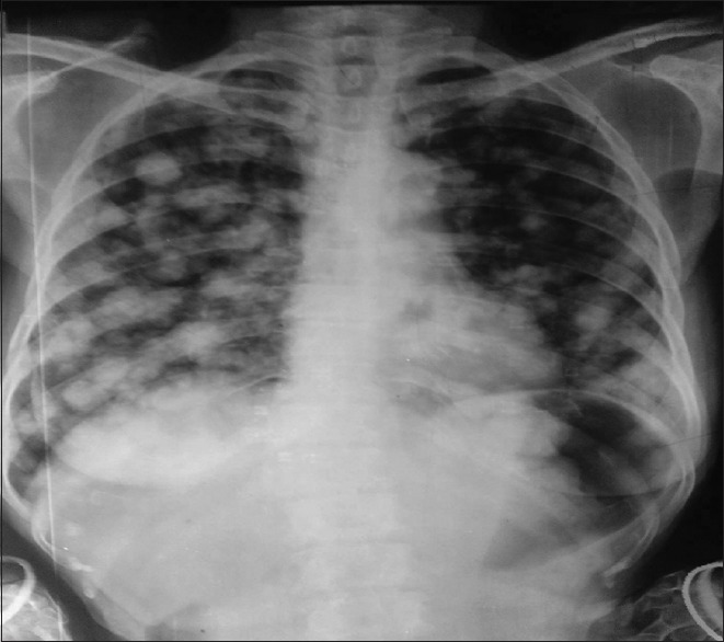
Chest X-ray showing cannon-ball pulmonary metastases
Figure 2.

CECT chest showing cannon-ball metastases
Figure 3.

CECT abdomen showing growth involving the stomach
Figure 4.

Barium Meal showing cannon-ball metastases and free flow of dye into the stomach
Figure 5.

HPE showing well-differentiated adenocarcinoma involving the stomach
Multiple pulmonary nodules on the chest X-ray have multiple causes, including, metastases (cannon-ball secondaries), various infections, immunological diseases, and arteriovenous malformations. Pulmonary metastasis is seen in 20-54% of the extrathoracic malignancies.[1] Lungs are the second most frequent site of metastases from extrathoracic malignancies. Development of pulmonary metastases implies a disseminated disease, with poor prognosis, although a few cases with favorable outcomes have been reported.[2] Breast, colorectal, lung, kidney, head and neck, and uterus cancers are the most common primary tumors with lung metastasis. However the frequency of gastric cancers metastasizing to the lungs is <1%.[3] This case highlights a rare presentation of gastric cancer, where the patient had no symptoms related to the malignancy itself, but presented with symptoms related to pulmonary involvement.
REFERENCES
- 1.Mohammed TL, Chowdhry A, Reddy GP, Amorosa JK, Brown K, Dyer DS, et al. Expert Panel on Thoracic Imaging. ACR appropriatenesscriteria® screening for pulmonary metastases. J Thorac Imaging. 2011;26:W1–3. doi: 10.1097/RTI.0b013e3182010bf9. [DOI] [PubMed] [Google Scholar]
- 2.Flavin R, Finn S, McErlean A, Smyth P, Meaney J, O’Connell F, et al. Cannonball metastases with favorable prognosis. Ir J Med Sci. 2005;174:61–4. doi: 10.1007/BF03168522. [DOI] [PubMed] [Google Scholar]
- 3.Patel T, Rajiah P. Lung Metastases Imaging. [Last accessed on 2014 Jun 15]. Available from: http://www.emedicine.medscape.com/article/358090-overview .