Abstract
Aim:
Putrefaction of the human body with its rate and stages of the various changes occurring in this entire process have been explored widely by the forensic medicine experts to estimate the time elapsed since death. However, experimental data reported in literature pertaining to rates of putrefaction of the dental pulp retrieved from jaws of the dead is scarce. This study makes an attempt to find out the series of various changes which occur during the process of putrefaction of the dental pulp in a coastal environment like that of Southern India. An attempt has also been made to estimate the time elapsed since the death by assessing the duration for which dental pulp remains microscopically intact.
Materials and Methods:
Three different study setups at different times, followed one by other were created. In each setup, 10 specimens of porcine jaws with teeth were buried in surface soil and 10 specimens in subsurface soil. Dental pulp was retrieved at an interval of every 24 h to see for the various changes. All the environmental parameters including average daily rainfall precipitation, temperature, soil humidity, soil temperature, and soil pH were recorded.
Results:
A specific series of morphological changes in terms of changes in size, color, consistency, and odor; and a sequence of histological changes were observed from both surface and subsurface samples.
Conclusion:
Dental pulp buried in a coastal environment goes through a specific series of morphological and histological changes which can be interpreted up to 144 h from burial, after which pulp ceases to exist.
Keywords: Death, dental pulp, histology, morphology, putrefaction, time elapsed
Introduction
The estimation of time elapsed since death has an important role in the investigation of cases from forensic viewpoint. But estimating postmortem time period becomes difficult when great numbers of environmental variables are involved especially in coastal regions.[1] Tooth can serve as very informative forensic evidence as it is the hardest substance in the human body which comprises of the multitude of tissues and encases often uncontaminated and well preserved pulp.[2] Dental pulp is a soft connective tissue of the tooth which undergoes putrefaction after death. This occurs in a series of morphological and histological changes of pulpal tissue. So far not much is known about employing these time-related changes of the dental pulp for estimation of time since death. Thus, this study was undertaken to identify morphological and histological time-related changes in dental pulp samples from porcine teeth buried in surface and subsurface soil and to assess its utility in the estimation of time elapsed since the death of an individual.
Materials and Methods
For this study, three similar study setups at different times, one followed by another were created. Porcine teeth were taken as the study required a large sample size. Teeth intact within the jaws were considered in order to simulate the natural conditions [Figure 1]. Porcine jaws with teeth were obtained from local meat packers in Mangalore city from freshly slaughtered <1-year old pigs (Sus scrofa). In each setup, 10 porcine jaws with teeth were buried in surface soil and 10 specimens in subsurface soil at depth of 30 cm [Figure 2].
Figure 1.
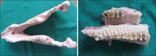
Porcine jaws with intact teeth from freshly slaughtered Sus scrofa
Figure 2.
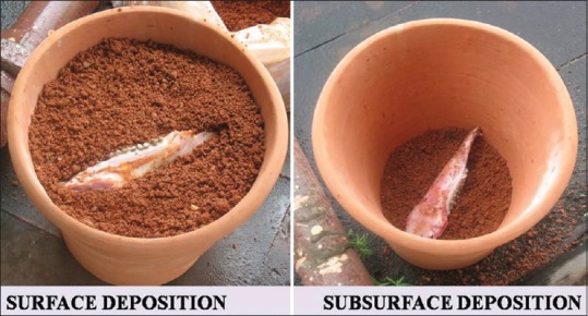
Surface and subsurface soil deposition at depth of 30 cm, of porcine jaws with teeth in mud pots
The samples were deposited in a mixture of soil and sand (1:1), kept in a custom-made mud flower pots of 40 cm depth. Five porcine jaws with teeth were directly exposed to the environment on a bed of soil and sand (surface burial) and five others were deposited 30 cm beneath the surface (subsurface burial). The study was conducted on both surface and subsurface levels, since dead bodies in a forensic situation (criminal or civil) can be left on the ground or in a hurry (as in a criminal case) are generally buried at a shallow depth in the earth. The buried specimens were kept on the roof of Manipal College of Dental Sciences, Mangalore in the month of August-September' 2007 to be exposed to natural outdoor environmental variations of South Kanara, that is, temperature and humidity.
Sufficient amount of the dental pulp was retrieved every day from both surface and subsurface samples for 7 consecutive days at an interval of every 24 h till samples showed signs of complete decomposition of pulp. Dental pulp retrieval was accomplished by placing a tooth in a polythene bag on a wooden block with shallow depressions and fracturing it by a sharp blow with a chisel and a wooden mallet. Polythene bags helped in prevention of scattering of pulp fragments. Shallow depressions on the wooden block were whittled to accommodate the tooth shape [Figure 3]. Thereafter, pulpal tissue was gently detached from the walls of the pulp chamber using a Lacron Carver and a pair of forceps. Morphological changes were recorded in terms of changes in size, color, consistency, and odor of the dental pulp. Histological changes in dental pulp were assessed using tissue processing and hematoxylin and eosin staining of retrieved dental pulp samples.
Figure 3.
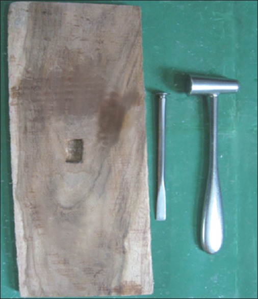
Whittled wooden block with shallow depressions to accommodate the tooth snugly
All the environmental parameters including average daily rainfall precipitation, temperature, soil humidity, soil temperature, and soil pH were recorded. Average daily rainfall and temperature were recorded from the daily newspapers in the month of September and October. The soil humidity was measured weekly as the amount of water absorbed by 1 g of soil. Soil temperature was recorded with atmospheric thermometer at the surface and at depth of 30 cm. The soil pH was recorded at the beginning of the experimental setup as well as at the end of the experiment from the soil samples submitted to Regional Agricultural Department, Mangalore.
Results
The maximum time period for which the pulp tissue remained was 144 h. From the 2nd day of the experiment, small white to brown maggots were seen which eventually increased in size with the progression of experiment [Figure 4].
Figure 4.
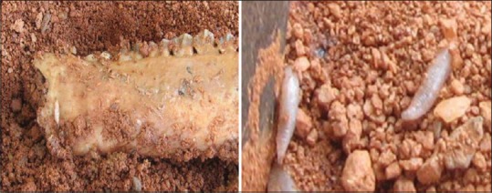
Appearance of small white to brown maggots in the soil
Morphologically, the pulp tissue gradually changed from pink, soft, vascular tissue to dirty pink, friable and collapsed tissue. Lately, it turned into jelly-like consistency followed by fluid consistency along with a fetid putrefying odor. At the end of experiment, only a semi-viscid fluid was left in the pulp chamber as remaining pulp tissue [Figure 5].
Figure 5.
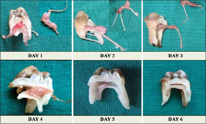
Sequence of morphological changes in pulp tissue at 24-72h pink, soft, vascular tissue to dirty pink, friable and collapsed tissue, at 96-120h jelly-like consistency followed by fluid consistency, at 144 h semi-viscid fluid as remaining pulp tissue
Histologically, fresh pulp retrieved showed intact, plump fibroblastic nuclei in a primitive ectomesenchyme like stroma with delicate fibers. Few scattered white blood cells (WBCs) with deeply basophilic nuclei and normal blood vessels with intact endothelial cells were observed [Figure 6]. Changes in the odontoblasts and their nuclei could not be assessed as the periphery of the pulp tissue could not be preserved.
Figure 6.
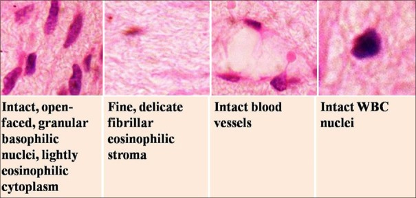
Photomicrograph showing histological appearance of freshly removed dental pulp (H and E, ×40)
At the end of 24 h, the pulp retrieved from the surface samples showed fibroblast with intact nuclei except a few cells showing chromatin margination and nuclear hyperchromatism. The stroma was homogenously fibrous with condensed appearing eosinophilic areas. The pulp retrieved from subsurface samples revealed predominantly intact, normal appearing plump fibroblastic nuclei. Both surface and subsurface pulp samples revealed intact WBCs and blood vessels [Figure 7]. After 48 h, pulp showed dramatic nuclear degenerative changes which varied from an undamaged or intact chromatin arrangement to a mosaic form with nuclear hyperchromasia, pyknosis, karryorhexis, and chromatin margination in surface pulp. The subsurface pulp showed relatively lesser number of fibroblasts and less evident changes. Both surface and subsurface pulp revealed intact WBCs and blood vessels in a characteristically vacuolated intercellular matrix [Figure 8]. After 72 h, surface and subsurface pulp exhibited the continued spectrum of nuclear degenerative changes along with the evidence of faint nuclear outlines and nuclear remnants depicting the process of karyolysis. However, the stroma varied from condensed eosinophilic, fibrillar to vacuolated type with intact WBCs and blood vessels [Figure 9]. After 96 h, both surface and subsurface pulp showed predominantly faint, ghost outlines of fibroblasts with nuclear debris in few areas. Few bizarre nuclei, which appeared to be WBC's, were still evident in a highly vacuolated stroma. However, surface pulp revealed faint outlines of blood vessels, but without endothelial cells whereas in subsurface samples, few endothelial cells remained [Figure 10]. After 120 days, both surface and subsurface pulp showed predominantly vacuolated, slightly basophilic, loose matrix with lesser number of cell ghosts and no evidence of any nuclear debris of endothelial cells. Some of the degenerating WBCs were evident [Figure 11]. After 144 h, surface pulp completely putrefied to semi-viscid material, which did not reveal any significant change. The subsurface pulp exhibited slightly basophilic vacuolated, fragmented to fibrous stroma with fragmented nuclear debris [Figure 12].
Figure 7.
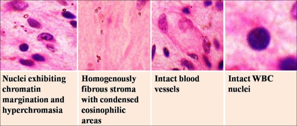
Photomicrograph showing histological changes in dental pulp at the end of 24 h (H and E, ×40)
Figure 8.
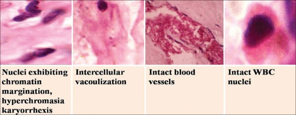
Photomicrograph showing histological changes in dental pulp at the end of 48 h (H and E, ×40)
Figure 9.
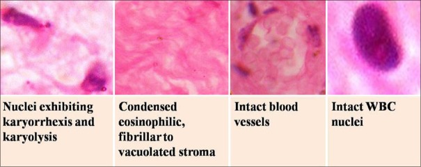
Photomicrograph showing histological changes in dental pulp at the end of 72 h (H and E, ×40)
Figure 10.
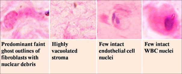
Photomicrograph showing histological changes in dental pulp at the end of 96 h (H and E, ×40)
Figure 11.
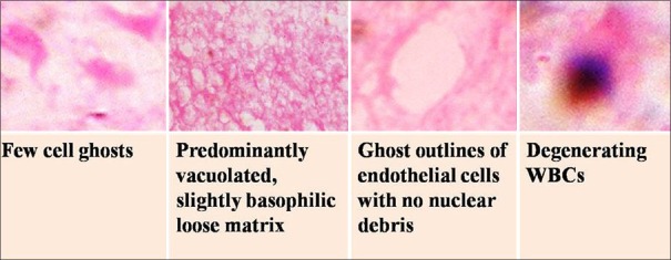
Photomicrograph showing histological changes in dental pulp at the end of 120 h (H and E, ×40)
Figure 12.
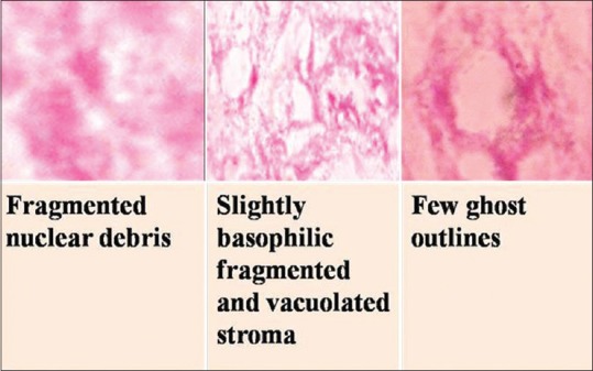
Photomicrograph showing histological changes in dental pulp at the end of 144 h (H and E, ×40)
Average daily temperature recorded in the months of August and September varied between 27°C and 29°C. Average daily rainfall recorded for 2 months varied between 13 mm and 13.2 mm. Soil temperature for these months varied from 25°C to 32°C. Average soil humidity was found to be 3.5 ml/g, whereas soil pH varied from 6.7 to 6.8 during these months.
Discussion
Shortly after the death of an individual, decomposition of tissue starts leading to a series of changes which can be observed morphologically and histologically. These changes occurring in a specific sequence can also be observed in dental pulp tissue retrieved from the jaws of buried dead bodies.[1] There are a number of factors which have their interplay in this complete process of decomposition of tissue. Release of intracellular hydrolytic enzymes initiates the process of cellular disintegration.[3] The rate of decomposition increases as the microorganisms invade these tissues. Soon after, multiplication of these microorganisms especially, anaerobic bacteria occur and various gases are released such as hydrogen sulfide, methane, cadaverine, and putrescine.[3,4] The byproducts of their growth lead to production of a putrid odor from the dead tissue. This entire process is called as putrefaction. Pressure built up inside tissues by these gases cause movement of fluids out of the cells and blood vessels. Foul smelling gases further attract a variety of insects. In the absence of normal body defenses, blowflies and house flies take an opportunity to invade the putrefying tissues and lay eggs. Young maggots from the eggs secrete digestive enzymes, aid in the spread of microorganisms and causes tearing of tissues with their mouth hooks. Hence, intracellular enzymes, insect activity along with various environmental parameters such as temperature, rainfall, and depth at which the dead body is buried, soil humidity, soil temperature and soil pH of that particular area are some of the important factors which have their effect on the rate of putrefaction.
Various studies have been done to determine the effect of these factors on the rate of putrefaction of dental pulp[5,6,7,8] in different experimental setups. The present study is a unique study done in the coastal area of southern India, South Kanara in which all these factors have been taken into account to observe their effect on the rates of putrefaction of the dental pulp and also to determine the series of changes in the pulp morphologically and histologically. The effect of decomposition of tissues through external factors causes a series of morphological changes. The sequence thus comprises of fresh unaffected light pink and soft pulp tissue changing to disintegrating dark colored and slimy pulp tissue. Finally, it becomes extremely friable with jelly-like consistency and a putrefying odor toward the end of decomposition.
In this study, histological changes in dental pulp started after 48 h to almost complete putrefaction by the end of 144 h. Only faint outlines of the cells were evident at the end of the experiment. In a study by Duffy et al. 1991, stability of pulp nuclei was found to be ranging from 4 days to 2 weeks in the pulp tissue retrieved from coastal environment. The lesser time elapsed since the death in the present study could be due to the difference in the environmental parameters and thus their effects on the rate of putrefaction of pulp. In the present study, daily temperature was either 28°C or 29°C as compared to 15°C observed by Duffy in his study. Higher temperature and moist conditions are conducive to necrotic autolysis by endogenous hydrolytic enzymes and to putrefaction, in that they favor bacterial and fungal proliferation. On the contrary, cold temperatures inhibit microbial activity as does desiccation of tissue.[9] Damp environment and acidic soil also attribute to rapid decomposition rate as is seen near coastal places.[1]
The putrefaction revealed varied changes in nuclei of cells of pulp. The initial changes varied from normal nuclei to pyknotic nuclei with nuclear debris and karyorrhexis, while the late changes included karyolysis and finally leading to complete putrefaction of the dental pulp. The matrix in earlier stages showed increased eosinophilia and condensation in which progressive putrefaction created lacunae/vacuolation. These changes were similar to those observed in another study on dental pulp in the coastal environment.[1]
The subsurface environment appeared to delay the rate of putrefaction as pulp tissue from subsurface samples showed comparatively slower rate of histological changes as compared to those in surface pulp samples. This could be because of the lower temperature and the inaccessibility of flesh eating insects at subsurface level when compared to surface. Early disintegration of fibroblastic nuclei was observed as compared to nuclei of WBCs and endothelial cells in both surface and subsurface samples.
Conclusion
Putrefaction is a continual process that can take from weeks to years, depending on the surrounding environment. The pivotal role is played by the deposition environment, and the climatic conditions, like temperature and moisture, as well as accessibility to insects. Thus, the present study concludes that a buried pulp in the above described coastal environment goes through a specific series of morphological and histological changes, which can be interpreted up to 144 h from burial, after which dental pulp ceases to exist.
Footnotes
Source of Support: Nil
Conflict of Interest: None declared
References
- 1.Duffy JB, Skinner MF, Waterfield JD. Rates of putrefaction of dental pulp in the Northwest coast environment. J Forensic Sci. 1991;36:1492–502. [PubMed] [Google Scholar]
- 2.Gawande M, Chaudhary M, Das A. Estimation of the time of death of an individual by evaluating histological changes in the pulp. Indian J Forensic Med Toxicol. 2012;6:80–2. [Google Scholar]
- 3.Paczkowski S, Schütz S. Post-mortem volatiles of vertebrate tissue. Appl Microbiol Biotechnol. 2011;91:917–35. doi: 10.1007/s00253-011-3417-x. [DOI] [PMC free article] [PubMed] [Google Scholar]
- 4.Saraswat PK, Nirwan PS, Saraswat S, Mathur P. Biodegradation of dead bodies including human cadavers and their safe disposal with reference to mortuary practice. J Indian Acad Forensic Med. 2008;30:273–80. [Google Scholar]
- 5.Yamamoto K. Experimental study on changes in extracted human teeth in relation to time elapsed. Shikwa Gakuho. 1959;58:1–21. [Google Scholar]
- 6.Rodriguez WC, Bass WM. Insect activity and its relationship to decay rates of human cadevers in East Tennessee. J Forensic Sci. 1983;28:423–32. [Google Scholar]
- 7.Galloway A, Birkby WH, Jones AM, Henry TE, Parks BO. Decay rates of human remains in an arid environment. J Forensic Sci. 1989;34:607–16. [PubMed] [Google Scholar]
- 8.Vavpotic M, Turk T, Martincic DS, Balazic J. Characteristics of the number of odontoblasts in human dental pulp post-mortem. Forensic Sci Int. 2009;193:122–6. doi: 10.1016/j.forsciint.2009.09.023. [DOI] [PubMed] [Google Scholar]
- 9.Gordon I, Shapiro HA, Berson SD, editors. Diagnosis and Early Signs of Death. New York: Churchill Livingstone; 1988. [Google Scholar]


