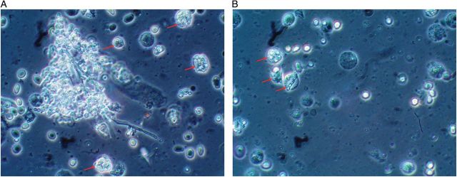
A 56-year-old woman who underwent a lung transplant followed 2 years later by a deceased donor kidney transplant presented with pyuria. Enterococcus bacteremia was previously treated with amoxicillin/sulbactam for 14 days. Subsequent to this therapy, urinalysis was carried out, which revealed the following: specific gravity 1.010; pH 7.5; nitrite negative; albumin 1+; hemoglobin 3+. Urine microscopy revealed one to two squamous epithelial cells/high power field (HPF); ∼20 white blood cells/HPF; >50 red blood cells/HPF; 0–2 hyaline casts/low power field; and blastoconidia and pseudomycelia (Candida sp.). Interestingly, numerous macrophages phagocytosing blastoconidia were noted throughout the specimen (Figure 1).
Fig. 1.

(A) Candida blastoconidia and pseudomycelia are seen on urine microscopy. Several macrophages have phagocytosed blastoconidia (arrows). (B) Several macrophages containing blastoconidia are noted (arrows).
Candida sp. is a human fungal pathogen. The major host defense against systemic Candida infection is ingestion of fungi by cells of the immune system, in particular macrophages and neutrophils. The fungal cell membrane is the initial contact with the immune system and plays an important role in host immune cell recognition and phagocytosis [1]. It is a dynamic organelle that determines both the shape of the fungus and its viability. Depending on the environment, these organisms are able to undergo reversible morphological changes moving between yeast, pseudohyphal and hyphal forms [2]. This plasticity allows fungi to successfully infect different anatomical sites. Hyphae have invasive properties that allow tissue penetration and escape from immune cells [3], whereas yeast forms are suited to disseminate via the bloodstream [4]. Phagocytic clearance of fungal pathogens consists of four stages: (i) accumulation of phagocytes where fungi reside; (ii) recognition of fungal molecular patterns; (iii) ingestion of fungi attached to phagocyte cell membranes and (iv) phagocyte processing of engulfed fungi by fusion [5]. The urinary findings of macrophages ingesting fungi in our patient support the importance of this host defense against such invading microorganisms.
Conflict of interest statement
None declared.
References
- 1.Netea MG, Brown GD, Kullberg BJ, et al. An integrated model of the recognition of Candida albicans by the innate immune system. Nat Rev Microbiol. 2008;6:67–78. doi: 10.1038/nrmicro1815. doi:10.1038/nrmicro1815. [DOI] [PubMed] [Google Scholar]
- 2.Moyes DL, Runglall M, Murciano C, et al. A biphasic innate immune MAPK response discriminates between the yeast and hyphal forms of Candida albicans in epithelial cells. Cell Host Microbe. 2010;8:225–235. doi: 10.1016/j.chom.2010.08.002. doi:10.1016/j.chom.2010.08.002. [DOI] [PMC free article] [PubMed] [Google Scholar]
- 3.McKenzie CG, Koser U, Lewis LE, et al. Contribution of Candida albicans cell wall components to recognition by and escape from murine macrophages. Infect Immun. 2010;78:1650–1658. doi: 10.1128/IAI.00001-10. doi:10.1128/IAI.00001-10. [DOI] [PMC free article] [PubMed] [Google Scholar]
- 4.Kumamoto CA, Vinces MD. Contributions of hyphae and hypha-coregulated genes to Candida albicans virulence. Cell Microbiol. 2005;7:1546–1554. doi: 10.1111/j.1462-5822.2005.00616.x. doi:10.1111/j.1462-5822.2005.00616.x. [DOI] [PubMed] [Google Scholar]
- 5.Lewis LE, Bain JM, Lowes C, et al. Stage specific assessment of Candida albicans phagocytosis by macrophages identifies cell wall composition and morphogenesis as key determinants. PLoS Pathogens. 2012;8:1–15. doi: 10.1371/journal.ppat.1002578. [DOI] [PMC free article] [PubMed] [Google Scholar]


