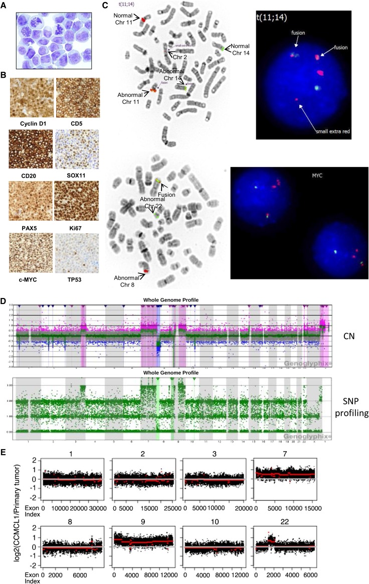
Figure 1.
Characterization of CCMCL1 cells. (A) Wright staining (original magnification ×1000) of CCMCL1 cells. (B) An NSG mouse was injected with 10 million first passage CCMCL1 cells via tail vein and sacrificed 5 weeks later. The spleen was collected and fixed in 10% formalin overnight, and a paraffin block was prepared. Immunostaining was performed using an automated stainer. (C) FISH of CCND1 (upper panels) and MYC (lower panels). FISH assay was performed on metaphases with an IGH/CCND1 dual-color, dual-fusion probe set (Abbott Molecular, Des Plaines, IL). Rearrangement of the MYC locus (8q24) was tested with the Break Apart probe from Abbott Molecular. FISH data were analyzed with ASI software. (D) Comparative genomic hybridization (CGH) + single nucleotide polymorphism (SNP) array (detailed information available in supplemental Methods, found on the Blood Web site). Upper panel: chromosome copy number (CN); lower panel: SNP profiling. The log ratio and allele difference plots are shown on the y-axis. (E) Whole-exome sequencing and CNV analysis were performed as described elsewhere.2 The duplication or amplification of specific chromosome regions in CCMCL1 cells (red line) compared with primary tumor cells (white line) was shown. The complete CNV analysis is shown in supplemental Figure 3. Chr, chromosome.

