Abstract
Purpose
The early onset of menarche is related to the adulthood risk of cardiovascular (CV) disease. This study examines the relation of early onset of menarche to carotid artery intima-media thickness (IMT), which is a surrogate marker of cardiovascular (CV) disease, among asymptomatic young adult women in a black-white community.
Methods
A cohort of 461 women (31% black, 69 % white) aged 24-43 years (mean of 35.6 years) were participants in the Bogalusa Heart Study. The age at menarche was retrospectively collected. In addition to cardiovascular risk factor variable measurements B-mode ultrasound images of the far walls of carotid artery segments were obtained. The multivariate linear regression model along with mediating effect by Sobel test was applied to analyze menarcheal age effect on carotid artery IMT, adjusting for covariates.
Results
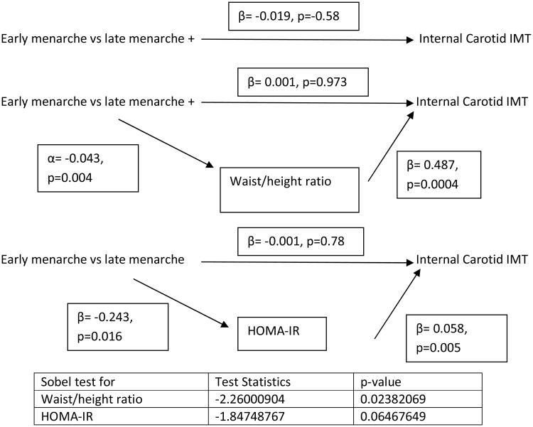
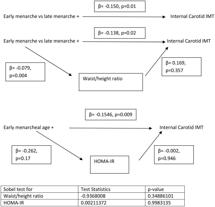
Waist/height ratio was significantly greater (p-value=0.01) in early menarcheal age (< 11 yrs) vs menarcheal age (≥11yrs) in both black and white women. Homeostasis model assessment of insulin resistance (HOMA-IR) was significantly greater (p-value=0.01) in early menarcheal age (< 11 yrs) vs menarcheal age (≥11yrs) in white women and also similar direction in black women. Internal carotid artery IMT was same in early menarcheal age (< 11 yrs) vs menarcheal age (≥11yrs) in white women but higher (p-value=0.02) in black women. Given above these different associations, the mediation analysis by race was performed. The effect of early menarcheal age (< 11 yrs) vs menarcheal age (≥11yrs) was mediated by waist/height ratio and HOMA-IR in white women after adjusting for parental education and age. The mediating effect of waist/height ratio (Sobel test-2.26 and p-value 0.02) and HOMA-IR (Sobel test -1.85 and p-valie 0.06) on internal carotid artery IMT was noted in white women. The direct effect of early menarcheal age (< 11 yrs) vs menarcheal age (≥11yrs) on internal carotid artery IMT (β= -0.150, p=0.01) was observed in black women.
Conclusions
The observed deleterious effect of early onset of menarche on carotid artery IMT in asymptomatic black and white younger adult women has biological, social and public health implications.
Keywords: Early onset of menarche, Internal carotid artery intima-media thickness, young adult women
Background
The menarcheal age is used to assess sexual maturation among females. There is no standard definition of early menarcheal age and various definitions exist within the literature (1-3). According to American Academy of Pediatrics, menarcheal age range is between 9-16 years of age. Most studies define menarcheal age at or below 12 years while others consider race specific 25th percentile of menarcheal age as early menarche. Early menarche is a widely used indicator of sexual maturation, more prevalent among US black girls than white girls (1-6). For example, in the Bogalusa Heart Study, the mean menarcheal ages of whites and blacks were similar (12.8 vs 12.9) but blacks were more likely (30% vs 25%), p= 0.10) to undergo a relatively early menarche below 12 years (7). Observation from a cross- sectional study in the Bogalusa Heart Study shows that early menarche is inversely associated with increased obesity in adulthood (8). Further, a longitudinal study within the Bogalusa Heart Study shows that early menarche is associated with metabolic syndrome in adulthood (8). Although it is known that early menarche is associated with metabolic risk factors for cardiovascular disease (CV) disease (7-9), there is a paucity of information in relation to early menarche and carotid artery intima media thickness (IMT) as a precursor of CV disease after consideration of the mediation effects of metabolic risk factors for CV disease in black-white young women (10-12). Racial composition of black-white reflects social constructs and proxy indicators of socioeconomic status (SES) (Ref 13-15). Even though race is social construct it has a large impact on biological mechanisms and health status (16, 17). Carotid artery IMT is a surrogate marker of CV disease morbidity and mortality (18, 19). B-mode ultrasonography is used to measure non-invasively the IMT of different segments of carotid artery. As part of Bogalusa Heart Study, a community-based study of the early natural history of CV disease (20), the present non-concurrent cohort study examines the direct and indirect effect of early menarche on carotid artery IMT in black-white women after controlling for parental education and age.
Materials and Methods
Study Population
During the 2001-2002 survey of black and white young adults, aged 18 to 45 years (n=1203; 70% white), ultrasonography of carotid artery IMT along with measurements of CV risk factor variables were performed as part of a longitudinal cohort study. Of these, 513 female participants (69% white, 31% black; mean age 35.6 years) who had data on menarcheal age were initially included in this study. The Tulane University Medical Center Institutional Review Board approved the study. Informed consent was obtained from all participants.
Menstrual History
As previously described (4), information on menarcheal age was obtained through interviews conducted by a nurse. Women were asked to identify the month and year when of first period occurred, with the help of questions such as, “Do you remember what grade you were in when you started having periods?” Participants who did not provide the month of menarche were considered menarche experienced at the midpoint of that specified year. Self-reported age of menarche was then calculated by subtracting month and year of birth from month and year of menstrual period. The reliability of self-reported age of menarche was reported in earlier studies (1, 7). Six participants were excluded from the analysis as their menstrual year was missing (n=3) or below 6 years (n=2) or above 20 yrs (n=1). They had similar distribution with the participants in terms of age distribution, education and parental education. The participants were also excluded if they were diabetes (n=11) or/both on antihypertensive (n=29) or/both cholesterol medications (n=11). Of 513 participants, 461 participants were included in the present study.
Participants were divided into two groups based on their menarcheal age: < 11 and ≥11 years as early maturers are found to be shorter but heavier in adulthood (1).
Carotid Artery Intima-Media Thickness
As previously described (21), carotid ultrasound measurements were recorded bilaterally using a Toshiba ultrasound instrument (Power Vision Toshiba SSH-380 ultrasound system, Toshiba America Medical System, Carrollton, TX) and a 7.5 MHz linear array transducer according to the protocol developed for the Atherosclerosis Risk in Communities Study (22). Images recorded on S-VHS tapes were read by certified readers from the Division of Vascular Ultrasound Research at Wake Forest University School of Medicine using a semi-automatic ultrasound image processing program developed by the California Institute of Technology and Jet Propulsion Laboratory (22, 23). The mean of the maximum carotid artery IMT readings of 3 right and 3 left far walls of common, bulb, and internal segments was used. If bilateral images could not be obtained, one side was used in the calculation.
General Examination
Standardized techniques and protocols were used by trained field observers (24). Replicate measurements of waist and height were made twice and the mean values were used to calculate waist/height ratio as a measure of visceral adiposity. Systolic and diastolic blood pressures were measured three times using a mercury sphygmomanometer by each of two randomly assigned observers. Mean arterial pressure was calculated as diastolic blood pressure plus one third pulse pressure. Information on smoking status was obtained using health habit questionnaires. The SES was assessed using both participants' and parental education. Those who smoked at least one cigarette per week during the past one year were identified as current smokers; those who smoked before past one year considered as ex-smoker and those who never smoked considered as non-smokers.
Laboratory Measurements
Serum total cholesterol and triglycerides were assayed on fasting samples using an enzymatic procedure on the Hitachi 902 Automatic Analyzer (Roche Diagnostics, Indianapolis, IN). Serum lipoprotein cholesterol levels were analyzed using a combination of heparin-calcium precipitation and agar-agarose gel electrophoresis procedures (25). The laboratory is being monitored for precision and accuracy of lipid measurements by the surveillance program of the Center for the Disease Control and Prevention (Atlanta, GA). A commercial radioimmunoassay kit was used for measuring plasma immunoreactive insulin levels (Phadebas, Pharmacia Diagnostics, Piscataway, NJ). Plasma glucose levels were measured by a glucose oxidase method as part of multiple chemistry profile (SMA20, Laboratory Corporation of America, Burlington, NC). Homeostasis model assessment of insulin resistance (HOMA-IR) was calculated based on the following formula: HOMA-IR=fasting insulin (μU/mL) × fasting glucose (mmol/L)/22.5. This model is considered useful to assess insulin resistance in epidemiologic studies (26).
Statistical Analyses
All analyses were conducted using SAS software, version 9.1 (SAS institute, Cary, NC). For all comparisons, participants were divided into two groups based on their menarcheal age: < 11 and ≥11 years. A race specific t-test was used to determine differences in mean values of risk factor variables and carotid artery IMT by age of menarche: <11vs ≥11 years. The chi-square test and Fisher's exact were applied as needed for categorical variables. Log transformation was employed if continuous variables were not normally distributed. Normal distribution was examined using the Shapiro-Wilk normality test statistic. The test of effect modification by race was assessed between the relationship of menarcheal age and carotid artery IMT. To evaluate the mediator effect of wasit/height ratio or HOMA-IR in the causal pathway, race specific several simple and multiple regression analysis were performed (27). In model 1, dependent variable was carotid artery IMT and independent variables was early menarcheal age (< 11 yrs) vs menarcheal age (≥11yrs) adjusted for parental education and age. In model 2, dependent variable was waist/height ratio or HOMA-IR and independent variable was early menarcheal age (< 11 yrs) vs menarcheal age (≥11yrs) adjusted for parental education and age. In model 3, dependent variable was carotid artery IMT and independent variables were early menarcheal age (< 11 yrs) vs menarcheal age (≥11yrs), waist/height ratio or HOMA-IR adjusted for parental education and age. The Sobel test (28, 29) was conducted to test whether waist/height ratio or HOMA-IR carried the influence of early menarche on carotid artery IMT.
Results
The effect modification of race was significant (p-value=0.05). Therefore analysis was done separately by race. Table 1 shows race-specific risk factor variables and internal carotid artery IMT by menarcheal age. Early menarcheal age (< 11 yrs) vs menarcheal age (≥11yrs) related significantly to higher waist/height ratio in both white and black women. Furthermore, early menarcheal age (< 11 yrs) vs menarcheal age (≥11yrs) related significantly to higher HOMA-IR in white only. The early menarcheal age was significantly related to only higher internal carotid artery IMT in black women (p=0.02).
Table 1. Levels of risk factor variables by status of menarcheal age in whites vs blacks.
| Menarcheal age < 11 yrs | Menarcheal age ≥11 yrs | p-value | |
|---|---|---|---|
|
| |||
| White | |||
| Age (yrs) | 34.9±5.1 | 36.0±4.2 | 0.11 |
| Participants' education (College or post graduate) | 28 (15%) | 157 (85%) | 0.58 |
| Parental education (College or post graduate) | 16 (15%) | 88 (85%) | 0.87 |
| Waist/ height ratio | 0.56±0.1 | 0.52±0.1 | 0.01 |
| Mean blood pressure (mmHg) | 87.0±7.8 | 85.8±8.5 | 0.37 |
| Triglycerides/HDL cholesterol ratio | 2.7±1.4 | 2.6±2.2 | 0.18 |
| HOMA-IR | 2.9±2.3 | 2.2±1.6 | 0.01 |
| Current smoker | 9 (15.6%) | 84 (12.5%) | .32 |
| Ex smoker | 7 (20%) | 34 (31%) | |
| Internal carotid artery IMT (mm) | 0.67±0.15 | 0.66±0.16 | 0.74 |
| Carotid bulb IMT (mm) | 0.89±0.10 | 0.90±0.20 | 0.96 |
| Common carotid artery IMT (mm) | 0.70±0.10 | 0.70±0.10 | 0.70 |
| Black | |||
| Age (yrs) | 35.1±4.9 | 34.8±4.7 | 0.81 |
| Participants' education (College or post graduate) | 6 (15%) | 46 (85%) | 0.88 |
| Parental education (College or post graduate) | 2 (12%) | 15 (88%) | 0.72 |
| Waist/ height ratio | 0.63±0.13 | 0.56±0.10 | 0.006 |
| Mean blood pressure (mmHg) | 94.1±9.8 | 90.8±11.3 | 0.24 |
| Triglycerides/HDL cholesterol ratio | 2.0±0.7 | 1.8±1.0 | 0.18 |
| HOMA-IR | 3.9±3.3 | 3.4±5.6 | 0.19 |
| Current smoker | 9 (7.1%) | 38 (30.1) | 0.44 |
| Ex smoker | 1 (5.6) | 3 (16.7%) | |
| Internal carotid artery IMT (mm) | 0.79±0.20 | 0.67±0.14 | 0.02 |
| Carotid bulb IMT (mm) | 0.86±0.17 | 0.89±0.17 | 0.54 |
| Common carotid artery IMT (mm) | 0.72±0.11 | 0.74±0.10 | 0.46 |
Mean±SD for continuous variables
HOMA-IR=homeostasis model assessment of insulin resistance; ns=not significant; IMT=Intima media thickness
Figure 2 displays the mediating effect of waist/height ratio and HOMA-IR adjusted for age and parental education in white women. The early menarcheal age (< 11 yrs) vs menarcheal age (≥11yrs) influenced the effect on internal carotid artery IMT through the causal pathway of waist/height ratio (p-value for sobel test 0.02) and HOMA-IR ((p-value for sobel test 0.06).
Figure 2. Testing Mediating Effect on Internal Carotid Artery IMT for White Women.
In figure 3 displays no mediating effect of waist/height ratio ((p-value for sobel test 0.35) and HOMA-IR ((p-value for sobel test 0.99) for the effect of menarcheal age on internal carotid artery IMT in black women. The early menarcheal age (< 11 yrs) vs menarcheal age (≥11yrs) direcly influenced the internal carotid artery IMT (p-value-0.01) after controlling for age and parental education
Figure 3. Testing Mediating Effect on Internal Carotid artery IMT for Black Women.
Discussion
The present community-based study demonstrates an inverse and independent association between early menarcheal age and internal carotid artery IMT in black women and indirect effect in white women. On average, internal carotid artery IMT decreased by 0.15 mm, (p-value 0.01) early menarcheal age (< 11 yrs) vs menarcheal age (≥11yrs) in black women after controlling for parental education and age. In white women, the early vs late menarcheal age has no direct effect (β= -0.019, p=-0.58), mediating through waist/height ratio (p-value= 0.02) and HOMA-IR (p-value= 0.06). This finding is consistent with recently-published longitudinal study with larger sample size measuring CV disease and mortality after 10.6-12.0 years follow-up (12). However, that study did not show the mediating effect of obesity and insulin resistance in relation to early menarchal age and CV disease. The mediating effect of obesity and insulin resistance was also observed in the literature (8, 10, 11). These studies explained the effect of obesity and insulin resistance in relation to early menarche with CV risk factors and carotid atherosclerosis.
The present study shows the relationship of menarcheal age with internal carotid artery IMT although menarcheal age was not related to other segments of carotid artery IMT. The atherosclerotic changes initiated at the internal carotid and bulb segments are more reflective of atherosclerosis (30). Therefore, including these segments are more sensitive and useful for early changes (21).
This observed deleterious association of early menarche with internal carotid artery IMT in black women was noted from a sub-sample of community-based cohort study. Early menarcheal age is associated with early life circumstances, namely birthweight, childhood obesity, childhood SES and psychological factors (31,32). On average, black girls experience increased levels of early life conditions when compared to white girls. The SES is inversely associated with menarcheal age in both black and white girls in the United States (33) although we did not find both participant's and parental education effect on menarcheal age. This finding is an agreement with previous study in terms of parental education (34). Race specific effect of carotid artery IMT by early menarcheal age may be also explained by genetic influence.
The major strength of this study includes being community-based study of young adults of racial difference (white-.black women). However, our study has some limitations. For instance, this is a reconstructed non-concurrent longitudinal study. Concurrent cohort studies are needed to determine the effect of early menarche on carotid artery IMT. Moreover, the age at which menarche was self reported was recalled via memory by those who are 18-43 years of age which leaves the results subject to bias. However, self-reported menarcheal age is the most commonly used method in epidemiologic studies. Black-white differences in education may subject to differential misclassification bias that underestimates the prevalence of early menarcheal age in black women. Furthermore, we could not assess parental SES by income status which is inversely related to obesity but positively related to menarcheal age in both white and black girls in the United States (33-36). Lastly, the measurement of carotid artery IMT was assessed only once in each subject in their current individual states of adulthood.
In conclusion, the observed deleterious effect of early onset of menarche on carotid artery IMT in asymptomatic black young adult women has biological, socioeconomic and public health implications as black girls experience early menarche, which is related to obesity, CV disease morbidity and mortality (6,37,38). A recent study shows that the black/white difference in early life conditions such as childhood BMI explained about 18% of the overall difference in age of menarche while birth weight differences accounted for another 11%. (15) Further, longitudinal studies are needed to draw conclusions regarding this effect of early onset of menarche on carotid artery IMT after carefully considering early life conditions. The current observations show that timing of menarche can relate to early atherosclerosis directly to black young women and indirectly to white young women
Figure 1. Diagram of reconstructed non-concurrent cohort study.
Acknowledgments
The Bogalusa Heart Study is a joint effort of many individuals whose cooperation is gratefully acknowledged. We are especially grateful to the study participants.
Funding source: Supported by grants 546145G1 from Tulane University, 0855082E from the American Heart Association, AG-16592 from the National Institute on Aging, HL-38844 from the National Heart, Lung, and Blood Institute, R01-AG-16592-12 from National Institute on Aging and 1R01ES021724-01from National Institute of Environmental Health Sciences.
List of abbreviations
- SES
Socioeconomic Status
- CV
Cardiovascular
- IMT
Carotid artery intima-media thickness
Footnotes
Financial Disclosures: Authors have no financial relationships relevant to this article to disclose
Conflict of Interest: Authors have no conflict of interest to disclose
Publisher's Disclaimer: This is a PDF file of an unedited manuscript that has been accepted for publication. As a service to our customers we are providing this early version of the manuscript. The manuscript will undergo copyediting, typesetting, and review of the resulting proof before it is published in its final citable form. Please note that during the production process errors may be discovered which could affect the content, and all legal disclaimers that apply to the journal pertain.
References
- 1.Freedman DS, Khan LK, Serdula MK, Dietz WH, Srinivasan SR, Berenson GS. Relation of age at menarche to race, time period, and anthropometric dimensions: the Bogalusa Heart Study. Pediatrics. 2002;110(4):e43. doi: 10.1542/peds.110.4.e43. [DOI] [PubMed] [Google Scholar]
- 2.Adair LS, Gordon-Larsen P. Maturational timing and overweight prevalence in US adolescent girls. Am J Public Health. 2001;91(4):642–644. doi: 10.2105/ajph.91.4.642. [DOI] [PMC free article] [PubMed] [Google Scholar]
- 3.Biro FM, McMahon RP, Striegel-Moore R, Crawford PB, Obarzanek E, Morrison JA, Barton BA, Falkner F. Impact of timing of pubertal maturation on growth in black and white female adolescents: The National Heart, Lung, and Blood Institute Growth and Health Study. J Pediatr. 2001;138:636–643. doi: 10.1067/mpd.2001.114476. [DOI] [PubMed] [Google Scholar]
- 4.Wattigney WA, Srinivasan SR, Chen W, Greenlund KJ, Berenson GS. Secular trend of earlier onset of menarche with increasing obesity in black and white girls: the Bogalusa Heart Study. Ethn Dis. 1999;9(2):181–9. [PubMed] [Google Scholar]
- 5.Salsberry PJ, Reagan PB, Pajer K. Growth differences by age of menarche in African American and White girls. Nurs Res. 2009;58:382–390. doi: 10.1097/NNR.0b013e3181b4b921. [DOI] [PMC free article] [PubMed] [Google Scholar]
- 6.Euling SY, Herman-Giddens ME, Lee PA, Selevan SG, Juul A, Sørensen TI, Dunkel L, Himes JH, Teilmann G, Swan SH. Examination of US puberty-timing data from 1940 to 1994 for secular trends: panel findings. Pediatrics. 2008;121(Suppl 3):S172–191. doi: 10.1542/peds.2007-1813D. [DOI] [PubMed] [Google Scholar]
- 7.Freedman DS, Khan LK, Serdula MK, Dietz WH, Srinivasan SR, Berenson GS. The relation of menarcheal age to obesity in childhood and adulthood: the Bogalusa heart study. BMC Pediatr. 2003;3:3. doi: 10.1186/1471-2431-3-3. [DOI] [PMC free article] [PubMed] [Google Scholar]
- 8.Frontini MG, Srinivasan SR, Berenson GS. Longitudinal changes in risk variables underlying metabolic Syndrome X from childhood to young adulthood in female subjects with a history of early menarche: the Bogalusa Heart Study. Int J Obes Relat Metab Disord. 2003;27:1398–404. doi: 10.1038/sj.ijo.0802422. [DOI] [PubMed] [Google Scholar]
- 9.Feng Y, Hong X, Wilker E, Li Z, Zhang W, Jin D, Liu X, Zang T, Xu X, Xu X. Effects of age at menarche, reproductive years, and menopause on metabolic risk factors for cardiovascular diseases. Atherosclerosis. 2008;196:590–7. doi: 10.1016/j.atherosclerosis.2007.06.016. [DOI] [PMC free article] [PubMed] [Google Scholar]
- 10.Stöckl D, Peters A, Thorand B, Heier M, Koenig W, Seissler J, Thiery J, Rathmann W, Meisinger C. Reproductive factors, intima media thickness and carotid plaques in a cross-sectional study of postmenopausal women enrolled in the population-based KORA F4 study. BMC Womens Health. 2014;14:17. doi: 10.1186/1472-6874-14-17. [DOI] [PMC free article] [PubMed] [Google Scholar]
- 11.Kivimäki M, Lawlor DA, Smith GD, Elovainio M, Jokela M, Keltikangas-Järvinen L, Vahtera J, Taittonen L, Juonala M, Viikari JS, Raitakari OT. Association of age at menarche with cardiovascular risk factors, vascular structure, and function in adulthood: the Cardiovascular Risk in Young Finns study. Am J Clin Nutr. 2008;87:1876–1882. doi: 10.1093/ajcn/87.6.1876. [DOI] [PubMed] [Google Scholar]
- 12.Lakshman R, Forouhi NG, Sharp SJ, Luben R, Bingham SA, Khaw KT, Wareham NJ, Ong KK. Early age at menarche associated with cardiovascular disease and mortality. J Clin Endocrinol Metab. 2009;94:4953–60. doi: 10.1210/jc.2009-1789. Epub 2009 Oct 30. [DOI] [PubMed] [Google Scholar]
- 13.LaVeist TA, Isaac LA. Editors Race, ethnicity and Health. San Francisco, CA: Jossey-Bass; 2013. [Google Scholar]
- 14.Braithwaite D, Moore DH, Lustig RH, et al. Socioeconomic status in relation to early menarche among black and white girls. Cancer Causes Control. 2009;20:713–20. doi: 10.1007/s10552-008-9284-9. [DOI] [PubMed] [Google Scholar]
- 15.Reagan PB, Salsberry PJ, Fang MZ, Gardner WP, Pajer K. African-American/white differences in the age of menarche: accounting for the difference. Soc Sci Med. 2012;75:1263–70. doi: 10.1016/j.socscimed.2012.05.018. Epub 2012 Jun 8. [DOI] [PMC free article] [PubMed] [Google Scholar]
- 16.Goodman AH. Why genes don't count (for racial differences in health) Am J Public Health. 2000;90(11):1699–702. doi: 10.2105/ajph.90.11.1699. [DOI] [PMC free article] [PubMed] [Google Scholar]
- 17.Jones CP, Truman BI, Elam-Evans LD, Jones CA, Jones CY, Jiles R, Rumisha SF, Perry GS. Using “socially assigned race” to probe white advantages in health status. Ethn Dis. 2008;18:496–504. [PubMed] [Google Scholar]
- 18.Grobbee DE, Bots ML. Carotid artery intima-media thickness as an indicator of generalized atherosclerosis. J Intern Med. 1994;236:567–573. doi: 10.1111/j.1365-2796.1994.tb00847.x. [DOI] [PubMed] [Google Scholar]
- 19.Sarzynska-Dlugosz I, Nowaczenko M. Common carotid artery intima-media thickness: the role in evaluation of atherosclerosis progression. Neurol Neurochir Pol. 2001;35:1093–1102. [PubMed] [Google Scholar]
- 20.The Bogalusa Heart Study 20th anniversary symposium. Am J Med Sci. 1995;310(suppl 1):S1–S138. doi: 10.1097/00000441-199512000-00001. [DOI] [PubMed] [Google Scholar]
- 21.Urbina EM, Srinivasan SR, Tang R, Bond MG, Kieltyka L, Berenson GS Bogalusa Heart Study. Impact of multiple coronary risk factors on the intima-media thickness of different segments of carotid artery in healthy young adults (The Bogalusa Heart Study) Am J Cardiol. 2002;90:953–958. doi: 10.1016/s0002-9149(02)02660-7. [DOI] [PubMed] [Google Scholar]
- 22.Bond MG, Barnes RW, Riley WA, et al. ARIC Study Group High resolution B- mode ultrasound scanning methods in the Atherosclerosis Risk in Communities Study (ARIC) J Neuroimaging. 1991;1:68–73. [PubMed] [Google Scholar]
- 23.Tang R, Hennig M, Thomasson B, et al. Baseline reproducibility of B-mode ultrasonic measurement of carotid artery intima-media thickness: the European Lacidipine Study on Atherosclerosis (ELSA) J Hypertens. 2000;18:197–201. doi: 10.1097/00004872-200018020-00010. [DOI] [PubMed] [Google Scholar]
- 24.Berenson GS. Editor Cardiovascular risk factors in children. New York: Oxford University Press; 1980. [Google Scholar]
- 25.Srinivasan SR, Berenson GS. Serum lipoproteins in children and methods for study. In: Lewis LA, editor. CRC Handbook of Electrophoresis v III Lipoprotein Methodology and Human Studies. Boca Raton, FL: CRC Press; 1983. pp. 185–204. [Google Scholar]
- 26.Matthews DR, Hosker JP, Rudenski AS, Naylor BA, Treacher DF, Turner RC. Homeostasis model assessment: insulin resistance and beta-cell function from Fasting plasma glucose and insulin concentrations in man. Diabetologia. 1985;28:412–419. doi: 10.1007/BF00280883. [DOI] [PubMed] [Google Scholar]
- 27.Neter J, Wasserman W, Kutner MH. Applied Linear Statistical Models: Regression, Analysis of Variance and Experimental Designs. Third. IRWIN; Homewood, IL and Boston, MA: 1990. pp. 225–236.pp. 730–750. [Google Scholar]
- 28.Baron RM, Kenny DA. The moderator-mediator variable distinction in social psychological research: conceptual, strategic, and statistical considerations. J Pers Soc Psychol. 1986;51:1173–82. doi: 10.1037//0022-3514.51.6.1173. [DOI] [PubMed] [Google Scholar]
- 29.Sobel test calculator. [accessed 01.05.14]; http://www.quantpsy.org/sobel/sobel.htm.
- 30.Stensland-Bugge E, Bonaa KH, Joakimsen O, Njolstad I. Sex differences in the relationship of risk factors to subclinical carotid atherosclerosis measured 15 years later: the Tromso study. Stroke. 2000;31:574–581. doi: 10.1161/01.str.31.3.574. [DOI] [PubMed] [Google Scholar]
- 31.Mishra GD, Cooper R, Tom SE, Kuh D. Early life circumstances and their impact on menarche and menopause. Womens Health (Lond Engl) 2009;5(2):175–90. doi: 10.2217/17455057.5.2.175. Review. [DOI] [PMC free article] [PubMed] [Google Scholar]
- 32.Mishra GD, Cooper R, Kuh D. A life course approach to reproductive health: theory and methods. Maturitas. 2010;65(2):92–7. doi: 10.1016/j.maturitas.2009.12.009. Epub 2010 Jan 15. Review. [DOI] [PMC free article] [PubMed] [Google Scholar]
- 33.Braithwaite D, Moore DH, Lustig RH, Epel ES, Ong KK, Rehkopf DH, Wang MC, Miller SM, Hiatt RA. Socioeconomic status in relation to early menarche among black and white girls. Cancer Causes Control. 2009;20:713–720. doi: 10.1007/s10552-008-9284-9. [DOI] [PubMed] [Google Scholar]
- 34.James-Todd T, Tehranifar P, Rich-Edwards J, Titievsky L, Terry MB. The impact of socioeconomic status across early life on age at menarche among a racially diverse population of girls. Ann Epidemiol. 2010;20(11):836–42. doi: 10.1016/j.annepidem.2010.08.006. [DOI] [PMC free article] [PubMed] [Google Scholar]
- 35.Braveman PA, Cubbin C, Egerter S, Williams DR, Pamuk E. Socioeconomic disparities in health in the United States: what the patterns tell us. Am J Public Health. 2010;100(Suppl 1):S186–96. doi: 10.2105/AJPH.2009.166082. [DOI] [PMC free article] [PubMed] [Google Scholar]
- 36.Baum CL, Ruhm CJ. Age, socioeconomic status and obesity growth. J Health Econ. 2009 May;28(3):635–48. doi: 10.1016/j.jhealeco.2009.01.004. [DOI] [PubMed] [Google Scholar]
- 37.Karapanou O, Papadimitriou A. Determinants of menarche. Reprod Biol Endocrinol. 2010;8:115. doi: 10.1186/1477-7827-8-115. [DOI] [PMC free article] [PubMed] [Google Scholar]
- 38.Prentice P, Viner RM. Pubertal timing and adult obesity and cardiometabolic risk in women and men: a systematic review and meta-analysis. Int J Obes (Lond) 2013;37:1036–1043. doi: 10.1038/ijo.2012.177. [DOI] [PubMed] [Google Scholar]


