Abstract
Multiple sclerosis (MS) is an autoimmune disease of the central nervous system (CNS). Studies in animal models of MS have shown that granulocyte-macrophage colony-stimulating factor (GM-CSF) produced by T cells is necessary for development of autoimmune CNS inflammation. This suggests that GM-CSF may have a pathogenic role in MS as well, and a clinical trial testing its blockade is ongoing. However, there have been few reports on GM-CSF production by T cells in MS. The objective of this study was to characterize GM-CSF production by T cells of MS patients, and to determine the effect of interferon-beta (IFN-β) therapy on its production. GM-CSF production by peripheral blood (PB) T cells and the effects of IFN-β were characterized in samples of untreated and IFN-β-treated MS patients vs. healthy subjects. GM-CSF production by T cells in MS brain lesions was analyzed by immunofluorescence. Untreated MS patients had significantly greater numbers of GM-CSF+ CD4+ and CD8+ T cells in PB compared to healthy controls and IFN-β-treated MS patients. IFN-β significantly suppressed GM-CSF production by T cells in vitro. A number of CD4+ and CD8+ T cells in MS brain lesions expressed GM-CSF. Elevated GM-CSF production by PB T cells in MS is indicative of aberrant hyperactivation of the immune system. Given its essential role in animal models, abundant GM-CSF production at the sites of CNS inflammation suggests that GM-CSF contributes to MS pathogenesis. Our findings also reveal a potential mechanism of IFN-β therapy, namely suppression of GM-CSF production.
INTRODUCTION
MS is an autoimmune disease characterized by the accumulation of immune cells in the CNS, leading to focal inflammation, demyelination, axonal/neuronal loss and disability (1). In a widely held view, T cells specific for CNS antigens play a pivotal role in MS pathogenesis. During the disease course in both MS and its animal model, experimental autoimmune encephalomyelitis (EAE), autoreactive T cells facilitate CNS inflammation by secreting a variety of proinflammatory cytokines, such as GM-CSF (2–5). GM-CSF is essential for the development and progression of EAE. Mice deficient in GM-CSF are resistant to EAE induction (6), and blockade of GM-CSF in wild-type (WT) mice suppresses ongoing disease (5). Although few studies have been devoted to GM-CSF in MS, convincing findings from EAE established GM-CSF as a promising therapeutic target in MS and led to a clinical trial testing its blockade (NCT01517282, ClinicalTrials.gov).
GM-CSF can be produced by either bone-marrow-derived cells, such as T cells (4, 5) and monocytes/macrophages (1), or by resident tissue cells (7–10). While various cell types produce it, GM-CSF from myelin-specific CD4+ T cells is essential to EAE development (2, 4, 5). These insights were obtained from CD4+ T cell-driven EAE models, and the role of GM-CSF in encephalitogenicity of CD8+ T cells is not known. CD8+ T cells can produce large quantities of GM-CSF (11), suggesting that it may contribute to their pathogenicity in EAE and MS.
IFN-β is a widely used treatment for MS, but its mechanisms of disease modulation are not well understood (12). It is known, however, that IFN-β upregulates HLA Class I expression and downregulates expression of HLA Class II, thereby inhibiting Th cell activation. IFN-β also modulates expression of proinflammatory cytokines and of molecules that facilitate extravasation, such as matrix metalloproteases and VLA4 (13). Interestingly, IFN-β was effective in ameliorating EAE driven by Th1 cells, but exacerbated disease induced by Th17 cells (14). Recently, a novel mechanism of IFN-β therapy in MS was proposed, whereby IFN-β promotes development of FoxA1+ regulatory T cells (15). Nevertheless, there is a lack of data on the effects of IFN-β on GM-CSF production by T cells.
In this study, we found significantly greater numbers of GM-CSF-producing CD4+ and CD8+ T cells in peripheral blood (PB) of untreated MS patients compared with patients under IFN-β therapy and healthy controls. In agreement with this ex vivo analysis, IFN-β suppressed GM-CSF production by T cells in vitro. A number of T cells in MS lesions expressed GM-CSF, strengthening the possibility that GM-CSF contributes to disease pathogenesis in the CNS. These findings suggest involvement of GM-CSF in MS, supporting the view that targeting it may be therapeutic. In addition, we also identify a potential mechanism by which IFN-β reduces relapse rate in MS.
MATERIALS AND METHODS
Blood samples and cell culture
All subjects provided informed consent prior their participation in current study. All human studies were approved by Institutional Review Board (IRB) of Thomas Jefferson University. Blood was obtained from ten IFN-β-treated MS patients, fifteen untreated MS patients, and fifteen healthy donors at the Department of Neurology, Thomas Jefferson University. Untreated MS patients did not receive any immunosuppressive or immunomodulatory treatment before venipuncture. Information on healthy donors and MS patients is given in Table I. PBMC were collected by Ficoll-Paque Plus density gradient centrifugation. PBMCs were washed and cultured at a density of 2×10⁶ cells/ml in X-VIVO 15 serum-free medium. Cells were stimulated with 1 μg/ml of anti-CD3 (HIT3a, BD Biosciences) and 1 μg/ml anti-CD28 (CD28.2, BD Biosciences) for five days in the presence of 1000 IU/ml of human recombinant IFN-β (R&D Systems). Concentration of 1000 IU/ml of IFN-β was chosen based on our serial dilution curve (Supplemental Fig. 1) and the study by Ramgolam et al. (16).
Table I.
Clinical characteristics of MS patients.
| Healthy donors | Untreated MS | IFN-β-treated MS | |
|---|---|---|---|
| Number | 15 | 15 | 10 |
| Female/Male | 9/6 | 11/4 | 9/1 |
| Age | 43.0 ± 3.5 | 49.5 ± 3.3 | 51.7 ± 3.9 |
| Rebif/Avonex | - | - | 4/6 |
| MS Type | - | RRMS | RRMS |
| Years Since Diagnosis | - | 10.6 ± 2.5 | 12.7 ± 2.5 |
MS = multiple sclerosis, IFN-β = interferon beta, RRMS = relapse- remitting MS
CD45RA−CD4+ T cells were purified from PBMCs by negative selection with CD4 Microbeads kit followed by negative selection with CD45RA Microbeads (Miltenyi Biotec) according to the manufacturer’s instructions. Purity of CD45RA−CD4+ T cells was checked by flow cytometry and samples that were ≥ 95% pure were used for analysis. CD45RA−CD4+ T cells were cultured in X-VIVO 15 serum-free medium for five days at a density of 1×10⁶ cells/ml. Cells were stimulated with 1 μg/ml of anti-CD3 (HIT3a, BD Biosciences) and 1 μg/ml anti-CD28 (CD28.2, BD Biosciences) with or without addition of IFN-β.
Flow cytometry
For cytokine detection, PBMCs and CD45RA−CD4+ T cells were activated with 50 ng/ml Phorbol 12-myristate 13-acetate (PMA) (Sigma), 500 ng/ml ionomycin (Sigma), and GolgiPlug (1 μg per 1×10⁶ cells, BD Biosciences) for five hours. Cells were washed in staining buffer (1% FBS and 0.1% sodium azide in PBS) and stained with following antibodies: anti-human CD4 PacificBlue (RPA-T4, BD Biosciences), CD4 PerCP-Cy5.5 (OKT-4, eBioscience), CD8 PacificBlue (RPA-T8, BD Biosciences), GM-CSF PE (BVD2-21C11, BD Biosciences), IL-17A Alexa Fluor 488 (eBio64DEC17, eBioscience), IFN-γ APC (4S.B3, eBioscience), T-bet PerCP-Cy5.5 (eBio4B10, eBioscience), RORγt PE (B2D, eBioscience), IL-4 FITC (8D4-8, eBioscience), GATA3 PE (TWAJ, eBioscience), Foxp3 FITC (206D, Biolegend), IL-22 APC (IL22JOP, eBioscience), CCR10 PE (1B5, BD Biosciences), CD45RA PE (HI100, BD Biosciences), and CD45RO PE-Cy7 (UCHL1, BD Biosciences). Cells were fixed and permeabilized with Caltag Fix/Perm reagents (Invitrogen) following the manufacturer’s instructions. Data were acquired on a FACSAria (BD Biosciences) and analyzed using FlowJo software (TreeStar).
Cytokine quantification
PBMCs and purified CD4+ T cells were cultured for five days and supernatants were collected and stored at −20°C until the day of analysis. GM-CSF, IL-10, IL-17A and IFN-γ concentrations were quantified by ELISA (R&D Systems) according to the manufacturer’s instructions.
Immunohistochemistry
Multiple sclerosis brain tissues were obtained from the Rocky Mountain Multiple Sclerosis Center Tissue Bank (Aurora, CO). Tissues were cryosectioned (10 μm thick) for immunohistochemistry at −25°C (Cryotome SME, Thermo Fisher Scientific, Pittsburgh, PA), and stored at −80°C.
Brain sections were fixed in acetone, washed in PBS and blocked with 8% horse serum, 3% BSA in PBS. Subsequently, sections were incubated with primary antibodies (5 μg/ml in 8% horse serum, 3% BSA, and 0.03% triton X-100 in PBS) overnight at 4°C. Mouse monoclonal anti-human CD4 (BC/1F6), rat monoclonal anti-human CD4 (YNB46.1.8), mouse monoclonal anti-human CD8 (BL-Ts 8/2), rat monoclonal anti-human CD8 (YTC182.20), rat monoclonal anti-human GM-CSF (BVD2-21C11), rabbit polyclonal anti human IL-17A, and rabbit polyclonal anti-human IFN-γ antibodies were purchased from Abcam, Inc. (Cambridge, MA). Sections were then washed in PBS and incubated with secondary antibodies at room temperature for one hour. Alexa Fluor 488 donkey anti-mouse IgG, Alexa Fluor 594 donkey anti-mouse IgG, Alexa Fluor 594 donkey anti-rat IgG, and Alexa Fluor 647 donkey anti-rabbit IgG were purchased from Life Technologies Corporation (Philadelphia, PA). To quench autofluorescence, sections were washed in PBS, rinsed in distilled water and incubated for 10 minutes in 10 mM CuSO4 in 50 mM ammonium acetate pH 5.0. Sections were then rinsed in distilled water and mounted with Prolong Diamond Anitifade Mountant with DAPI (Life Technologies Corp., Philadelphia, PA). Sections serving as controls for specificity of staining (negative controls) were generated from serial sections of each sample by staining with secondary antibodies only. Immunofluorescent images were captured using a Zeiss LSM 510 UV META confocal microscope with a 20X Plan-Apochromat 20x/0.75 NA objective, or a Plan-Neo 40x/1.3 NA oil objective. Z-stacks were acquired at a thickness of 1 μm and images were analyzed using the ImageJ software (NIH). Representative confocal images were generated by maximum or average projection through the Z axis of the image stack. Resulting projections of each channel were then overlaid in Photoshop CS6 software (Adobe System). For quantification, lesion tissues from three MS brains were examined. Nine sections (three per lesion) were randomly selected and quantitated. The number of stained cells per section was counted under 20x magnification. Data are expressed as mean ± SEM.
Statistics
Statistical analysis was performed by GraphPad Prism 6 software. One-way ANOVA was used to determine significance between multiple groups. The paired, two-tailed student t-test was used to analyze datasets before and after in vitro treatment with IFN-β. Parametric data were analyzed using an unpaired, two-tailed student t-test. The Bonferroni correction was applied for adjustment of the significance values for multiple comparisons; adjusted p ≤ 0.05 was considered significant. Data represent mean ± SEM.
RESULTS
IFN-γ+ and IFN-γ− CD4+ and CD8+ T cells in PB are major producers of GM-CSF
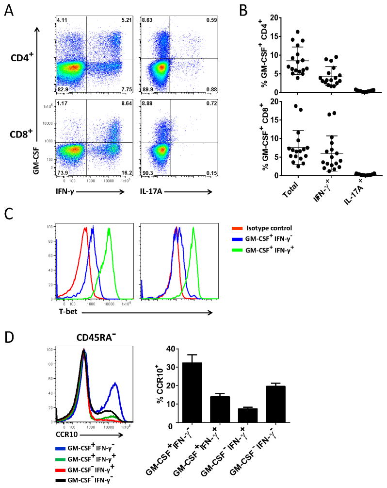
We first characterized the phenotype of GM-CSF-producing T cells in PB of healthy individuals. Subsets of both CD4+ and CD8+ T cells produced GM-CSF upon stimulation with PMA and ionomycin (Fig. 1A). GM-CSF was produced by both IFN-γ+ and IFN-γ− T cells, while the percentages of IL-17A+ and IL-22+ T cells were low (Fig. 1B and Supplemental Table I). A negligible number of CD4+ and CD8+ GM-CSF+ T cells expressed RORγt, IL-4, GATA3 and Foxp3 (data not shown). GM-CSF+IFN-γ− T cells did not express lineage-specific cytokines or transcription factors (Supplemental Table I) and we designated them “GM-CSF-only” T cells. These GM-CSF-only producing T cells either did not express T-bet, or expressed it at a low level, whereas the majority of GM-CSF+IFN-γ+ T cells were clearly T-bet+ (Fig. 1C and Supplemental Table I), a phenotype consistent with Th1/Tc1 lineage. Hence, GM-CSF+IFN-γ+ and GM-CSF-only producing T cells are the main GM-CSF-producing T cell subpopulations in PB of healthy individuals.
Figure 1. Human PB T cells express GM-CSF.
PBMCs from healthy donors (n=15) were activated with PMA + ionomycin + Golgiplug (PMA/iono/GP), stained and analyzed by flow cytometry. A) Representative flow cytometry dot-plots for gated CD4+ and CD8+ T cells from one donor. B) Summary (%) for GM-CSF+ CD4+ and CD8+ T cells among total CD4+ and CD8+ T cells. C) Representative histogram for T-bet expression in GM-CSF+IFN-γ+ and GM-CSF+IFN-γ− CD4+ and CD8+ T cells from one donor (n=5). D) Representative histogram for CCR10 expression in GM-CSF+IFN-γ−, GM-CSF+IFN-γ+, GM-CSF−IFN-γ+, and GM-CSF−IFN-γ− CD45RA−CD4+ cells. Graph on the right shows the percentage of CCR10+ cells in CD45RA−CD4+ T cells (mean ± SEM, n=6).
We then characterized GM-CSF-producing T cells in more detail. Staining for CD45RA and CD45RO stratified CD4+ T cells in naïve CD45RA+CD45RO− and effector memory CD45RA−CD45RO+ subpopulations, which were similar in size (45–50% each) (Supplemental Fig. 2A and C). The majority (> 75%) of CD8+ T cells were CD45RA+CD45RO− (Supplemental Fig. 2B and D). Among both CD4+ and CD8+ T cells, CD45RA−CD45RO+ subpopulations were predominant producers of GM-CSF and IFN-γ, while a small number of CD45RA+CD45RO− produced these cytokines (Supplemental Fig. 2). In a recent publication, Noster et al. sorted CD45RA−CD4+ T cells based on surface expression of chemokine receptors and analyzed their cytokine production (17). CXCR3−CCR4+CCR6−CCR10+ cells produced GM-CSF but not IFN-γ, IL-17A or IL-22. The authors concluded that the above combination of chemokine receptors is characteristic of GM-CSF-only producing cells. We attempted to determine expression of the chemokine receptors on all GM-CSF-only CD4+ T cells. CCR6 was expressed on a small number of cells, and we discontinued staining for this marker, deeming it uninformative in this context. Both CXCR3 and CCR4 were present on substantial portions of CD4+ T cells before they had been exposed to PMA and ionomycin, but after exposure the percentage of CXCR3+ and CCR4+ cells dropped several fold, precluding reliable correlation between cytokine and the chemokine receptors expression. However, numbers of CCR10+CD4+ T cells were not altered by PMA and ionomycin, and we continued analyses using this marker. Only a third of CD45RA−GM-CSF+IFN-γ−CD4+ T cells stained for CCR10 (Fig. 1D). Hence, CXCR3−CCR4+CCR6−CCR10+ phenotype identifies a minority of GM-CSF-only CD4+ T cells.
Untreated MS patients have increased numbers of GM-CSF+ T cells in PB, while patients undergoing IFN-β therapy have normal numbers
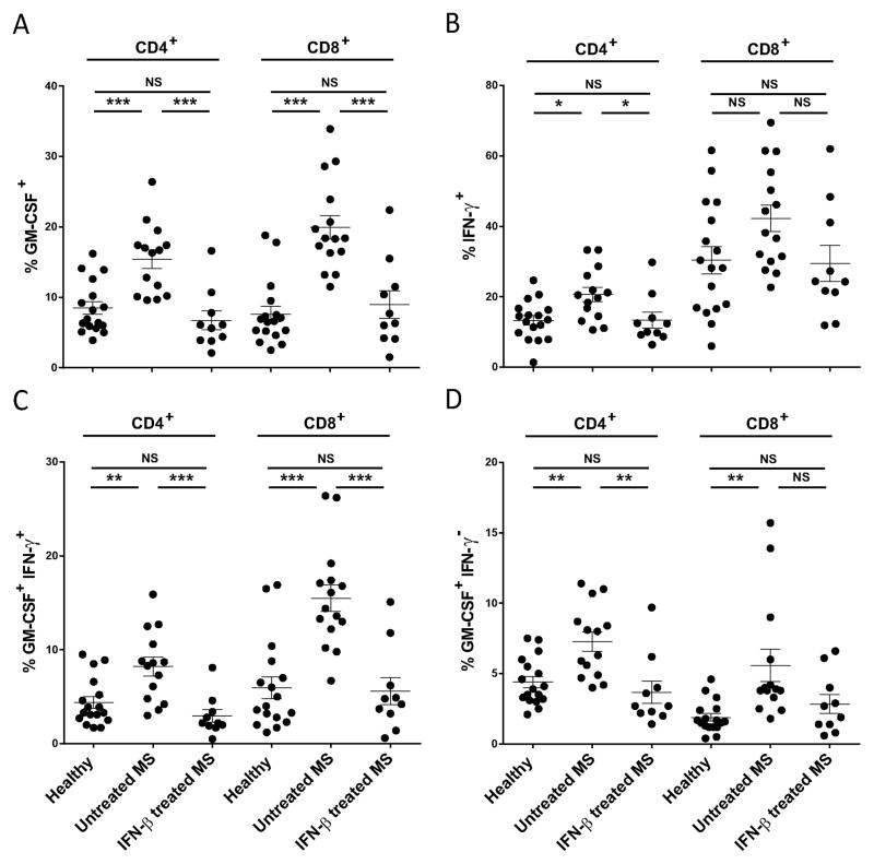
We then compared GM-CSF production by T cells of untreated and IFN-β-treated MS patients, and of healthy donors. Untreated MS patients had on average twice as many GM-CSF+CD4+ and three times more GM-CSF+CD8+ T cells compared to both healthy donors and IFN-β-treated MS patients, while the latter two did not differ (Fig. 2A). Untreated MS patients also had significantly increased numbers of IFN-γ+CD4+ T cells compared to healthy donors and IFN-βtreated patients, but the increase was less pronounced than in the case of GM-CSF+ cells (Fig. 2B). There was a trend toward a higher percentage of IFN-γ+CD8+ T cells in samples of untreated MS-patients, but the difference did not reach statistical significance (Fig. 2B). IL-17A+ and IL-22+ T cells were present in low numbers in all three tested groups and did not contribute substantially to the pool of GM-CSF+ T cells (data not shown). Frequencies of both GM-CSF+IFN-γ+ and GM-CSF-only CD4+ and CD8+ T cells were significantly greater in the case of untreated MS patients when compared with healthy donors and IFN-β-treated patients (Figs. 2C, D). In addition, numbers of GM-CSF+IFN-γ+ and GM-CSF-only CD4+ T cells were similar, while among CD8+ T cells GM-CSF+IFN-γ+ cells were approximately twice as abundant as GM-CSF-only cells. These data demonstrate that untreated MS patients have increased numbers of GM-CSF-producing T cells in their PB, and that IFN-β therapy normalized their numbers.
Figure 2. Untreated MS patients have more GM-CSF-producing T cells than healthy individuals and IFN-β-treated MS patients.
PBMCs from healthy donors (n=15), untreated MS patients (n= 15), and IFN-β-treated MS patients (n=10) were activated with PMA/iono/GP, stained and analyzed by flow cytometry. A) Collective data (%) of GM-CSF+ CD4+ and CD8+ T cells among total CD4+ and CD8+ T cells. B) IFN-γ expression by CD4+ and CD8+ T cells. C) and D) flow cytometry analysis of GM-CSF expression in IFN-γ+ and IFN-γ− T cells. * p < 0.05, ** p < 0.01, *** p < 0.001, NS: not significant, (One-way ANOVA).
IFN-β suppresses GM-CSF production by T cells of healthy individuals in vitro
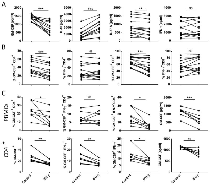
IFN-β-treated MS patients had fewer GM-CSF+ T cells compared to untreated patients, which prompted us to examine the effect of IFN-β on GM-CSF production by T cells. Peripheral blood mononuclear cells (PBMCs) from healthy donors were stimulated with anti-CD3 and anti-CD28 Abs in the presence of IFN-β and their cytokine production was analyzed after five days. IFN-β significantly reduced GM-CSF and increased IL-10 concentrations in cell culture supernatants, while it caused no change in concentrations of IFN-γ (Fig. 3A). Concentrations of IL-17A were also significantly reduced, but only to a small extent. IFN-β reduced frequency of GM-CSF+ CD4+ and CD8+ T cells, while numbers of IFN-γ+ T cells remained unchanged (Fig. 3B). These data show that IFN-β inhibits GM-CSF production by T cells in vitro, while IFN-γ production remains unaffected.
Figure 3. IFN-β suppresses GM-CSF production by T cells from healthy donors.
PBMCs and purified CD4+CD45RA− T cells from healthy donors were stimulated with anti-CD3 and anti-CD28 Abs for five days, with or without addition of IFN-β. A) Concentrations of GM-CSF, IL-10, IL-17A and IFN-γ in cell culture supernatants from PBMCs (n=15) assayed by ELISA. B) Percentages of GM-CSF+ and IFN-γ+ CD4+ and CD8+ cells in total CD4+ and CD8+ T cells from above PBMC cultures (n=15). C) To evaluate direct effect of IFN-β on CD4+ T cells, purified CD45RA−CD4+ T cells and PBMCs from the same donors (n=8) were cultured in the presence of IFN-β. Percentages of GM-CSF+, GM-CSF+IFN-γ+ and GM-CSF+IFN-γ− CD4+ cells in total CD4+ cells. GM-CSF concentrations measured by ELISA in cell culture supernatants. * p<0.05, ** p<0.01, *** p<0.001, NS: not significant (Paired, two-tailed Student t-test).
To test whether IFN-β suppresses GM-CSF production by directly acting on T cells, we purified CD45RA−CD4+ T cells and activated them with anti-CD3 and anti-CD28 Abs in the presence of IFN-β. IFN-β significantly decreased the percentage of GM-CSF+ cells in cultures of CD45RA− CD4+ T cells, similar to PBMCs (Fig. 3C). IFN-β also reduced the percentage of both GM-CSF-only and GM-CSF+IFN-γ+ cells in purified CD45RA−CD4+ T cells, while it had no significant effect on GM-CSF+IFN-γ+CD4+ T cells in PBMC cultures. Furthermore, IFN-β reduced GM-CSF concentrations in culture supernatants of CD45RA−CD4+ T cells and PBMCs (Fig. 3C). These data show that IFN-β suppresses GM-CSF production by CD4+ T cells by acting directly on them.
IFN-β reduces GM-CSF production but not numbers of GM-CSF+ T cells of MS patients in vitro
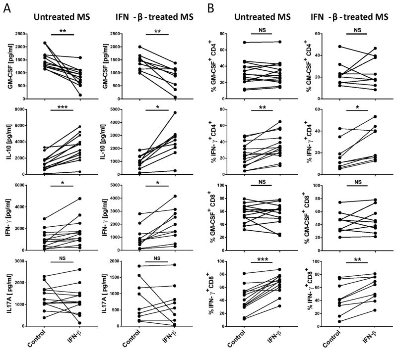
We then investigated the effect of IFN-β on GM-CSF production by PBMCs of MS patients. IFN-β significantly reduced GM-CSF concentrations in samples of both untreated and IFN-β-treated MS patients, while increasing IL-10 and IFN-γ concentrations (Fig. 4A). IL-17A production remained unchanged. In contrast to reduced GM-CSF concentrations in culture supernatants, IFN-β did not reduce numbers of GM-CSF+ CD4+ and CD8+ T cells (Fig. 4B). This was also in contrast to its suppressive effect on frequency of GM-CSF+ T cells from healthy donors. IFN-β increased the percentages of IFN-γ+ CD4+ and CD8+ T cells from MS patients in vitro (Fig. 4B) and the vast majority of these IFN-γ+ T cells simultaneously expressed GM-CSF. These data demonstrate that IFN-β has different effects on T cells of healthy individuals and MS patients, irrespective of whether they come from IFN-β-treated or untreated patients. Most notably, in contrast to healthy individuals, IFN-β did not reduce numbers of GM-CSF+ T cells in the samples from MS patients, but it increased numbers of IFN-γ+ T cells. However, even though IFN-β did not reduce numbers of T cells having the capacity to produce GM-CSF, it reduced GM-CSF concentrations in cell culture supernatants.
Figure 4. IFN-β reduces GM-CSF production by T cells from MS patients.
PBMCs of IFN-β-treated and untreated MS patients were cultured with anti-CD3 and anti-CD28 Abs for five days with or without addition of IFN-β. A) GM-CSF, IL-10, IL-17A, and IFN-γ concentrations in above cell culture supernatants. B) Percentages of GM-CSF+ and IFN-γ+ CD4+ and CD8+ cells in total CD4+ and CD8+ T cells from above PBMC cultures. * p<0.05, ** p<0.01, *** p<0.001, NS: not significant (Paired, two-tailed Student t-test).
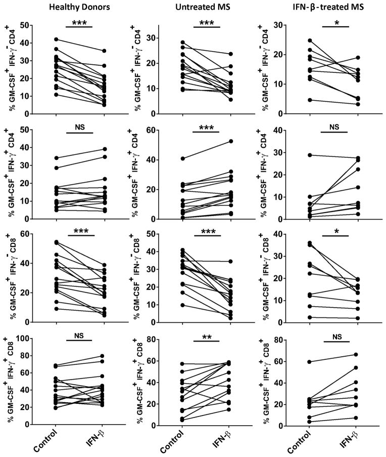
To identify subpopulations of GM-CSF+ T cells affected by IFN-β treatment in vitro, PBMCs were cultured in the presence of IFN-β for five days and analyzed by flow cytometry. IFN-β significantly decreased the frequency of GM-CSF-only CD4+ and CD8+ T cells from healthy donors and from untreated and IFN-β-treated MS patients (Fig. 5). However, IFN-β either had no effect on, or it increased numbers of GM-CSF+IFN-γ+ CD4+ and CD8+ T cells in all three groups tested. These data suggest that only IFN-γ− T cells are susceptible to suppression by IFN-β, and that reduction in total numbers of GM-CSF+ T cells in samples from healthy donors (Fig. 3) is due to reduction in numbers of GM-CSF-only T cells. However, in samples from MS patients a similar reduction in GM-CSF-only T cells was compensated for by a pronounced increase in GM-CSF+IFN-γ+ T cells, resulting in a lack of effect of IFN-β on total numbers of GM-CSF+ T cells (Fig. 4B). These data also suggest that reduced concentrations of GM-CSF in cell culture supernatants of IFN-β-treated samples are mostly due to inhibition of GM-CSF only T cells.
Figure 5. IFN-β reduces numbers of GM-CSF only CD4+ and CD8+ T cells.
PBMCs from healthy donors (n=15) and MS patients (untreated MS n=13; IFN-β treated n=9) were stimulated with anti-CD3/anti-CD28 Abs in the presence or absence of IFN-β. Percentages of GM-CSF+IFN-γ+ and GM-CSF only CD4+ and CD8+ T cells in total CD4+ and CD8+ cells are shown. * p<0.05, ** p<0.01, *** p<0.001, NS: not significant (Paired, two-tailed Student t-test).
CD4+ and CD8+ T cells express GM-CSF in MS lesions
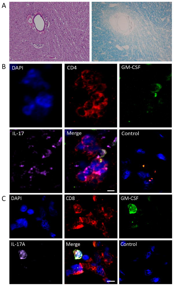
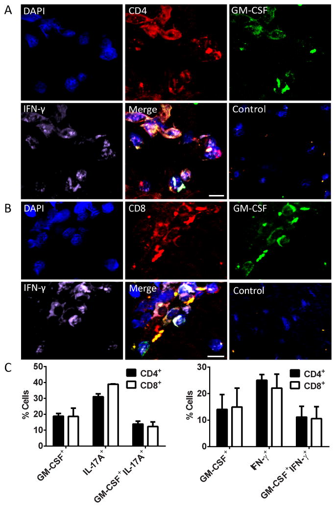
In order to expand the relevance of our findings obtained with PB T cells, we investigated GM-CSF expression by T cells in active MS lesions. Approximately 15–20% of both CD4+ and CD8+ T cells expressed GM-CSF, and the majority co-expressed IL-17A or INF-γ (Figs. 6 and 7). Although the frequencies of GM-CSF+ T cells in MS lesions appeared to be similar to those in PB, it is likely that a substantially higher proportion of T cells in MS lesions had the capacity to express GM-CSF given that they had not been stimulated with PMA and ionomycin prior to tissue staining, as was the case with PBMCs. Hence, due to important methodological differences in analyses a direct comparison of frequencies of cytokine-producing T cells in PB and MS lesions would be false. Nonetheless, it is obvious that higher proportions of T cells in MS lesions express, or have the capacity to express cytokines compared with T cells in PB. This is especially notable in the case of IL-17A. Typically less than 1% of PB T cells expressed IL-17A, while in MS lesions over 30% of T cells expressed this cytokine even without prior stimulation with PMA and ionomycin. Taken together, these data show expression of GM-CSF by T cells at CNS inflammation sites, supporting the hypothesis that GM-CSF contributes to MS pathogenesis.
Figure 6. CD4+ and CD8+ T cells co-express GM-CSF and IL-17A in MS brain lesions.
Serial sections from MS brain were stained with A) H&E and LFB to demonstrate the infiltration of T cells and demyelination in the MS lesion. B) CD4+ and C) CD8+ T cells were stained for GM-CSF and IL-17A and analyzed by confocal microscopy. Merged images show co-localization of GM-CSF and IL-17A in CD4+ and CD8+ T cells. Negative controls are virtually devoid of staining. Scale bars= 10μm.
Figure 7. CD4+ and CD8+ T cells co-express GM-CSF and INF-γ in MS brain lesions.
Stained sections from three MS patients were analyzed by confocal microscopy. A) CD4+ and B) CD8+ T cells in brain lesions were stained for GM-CSF and INF-γ. Merged images show co-localization of GM-CSF and INF-γ in CD4+ and CD8+ T cells. Negative controls are virtually devoid of staining. C) Quantification of CD4+ and CD8+ T cells performed by counting average of 200 cells per group from three MS brains at 20X magnification. Scale bars = 10 μm.
DISSCUSION
Although GM-CSF was discovered over forty years ago (18), and its essential role in EAE was demonstrated fourteen years ago (6), this cytokine has been little studied in MS. It is likely that in MS, as in EAE, myelin-specific T cells are the principal source of pathogenic GM-CSF. We found that CD4+ and CD8+ T cells in MS lesions produce GM-CSF, which likely facilitates disease pathogenesis. Treatment-naïve MS patients had greater numbers of GM-CSF+ T cells in PB compared to healthy individuals and patients undergoing IFN-β therapy. Notably, IFN-β therapy reduced numbers of GM-CSF+ T cells, an effect that is consistent with our in vitro data. Possibly, IFN-β also suppresses GM-CSF production by other cell types, which may add to its therapeutic efficacy in MS.
Our data show that a number of CD4+ and CD8+ T cells in MS brain lesions express GM-CSF, while the frequency of GM-CSF+ T cells among PBMCs was lower. In addition, a large proportion of GM-CSF+ T cells in MS lesions also expressed IFN-γ and IL-17A, while their frequency at the periphery was smaller, especially in the case of IL-17A+ cells. The enrichment in cytokine-producing T cells in MS lesions has been described previously, as the majority (~80%) of T cells, both CD4+ and CD8+, in MS lesions expressed IL-17A, while in the periphery only a fraction of T cells expressed this cytokine (19). Given that a variety of cell types, both resident CNS cells (20–23) and peripheral myeloid cells that infiltrate the CNS, express GM-CSF receptor, the abundant GM-CSF production in MS lesions suggests its contribution to pathogenesis.
Although healthy individuals and MS patients have similar overall frequency of myelin-specific T cells in PB (1, 24–27) more T cells of MS patients have activated effector/memory phenotype (28–30). Our ex vivo analysis showed that IFN-γ+ CD4+ and CD8+ T cells represent a majority of GM-CSF-producing cells, but a substantial proportion of IFN-γ− T cells also expresses GM-CSF. Both IL-22+ and IL-17A+ T cells were present in much smaller numbers than IFN-γ+ and GM-CSF+ cells. This was true for healthy controls, and treated and untreated MS patients. Consistent with previous findings that T-bet is not required for GM-CSF expression (4), both T-bet+ and T-bet− T cells expressed GM-CSF. GM-CSF-only T cells either did not express T-bet or its levels were low, while among GM-CSF+IFN-γ+ T cells most cells were T-bet+, which is typical Th1/Tc1 phenotype. We could not classify GM-CSF-only T cells into any conventional lineage of effector T cells, because they did not express hallmark cytokines or transcription factors of those lineages.
Noster et al. have recently described a CD4+ T cell subpopulation from PB that produces GM-CSF, but does not express any of the Th lineage-specific cytokines or transcription factors (17). These GM-CSF-only expressing CD4+ T cells had CD45RA−CCR7− effector memory phenotype and the characteristic combination of chemokine receptors expressed on their surface (CXCR3−CCR4+CCR6−CCR10+). However, our analyses, based on staining for CCR10, showed that only a portion of GM-CSF-only cells have the above combination of surface markers. Our data showing that this surface phenotype is not characteristic of GM-CSF-only CD4+ T cells is consistent with those of Noster et al. showing that the majority of sorted CXCR3−CCR4+CCR6−CCR10+CD4+ T cells did not produce GM-CSF upon activation. The same authors also described that MS patients had normal numbers of GM-CSF+CD4+ T cells in PB. Information on whether the patients were treated with IFN-β has not been provided, which precludes determining whether this result is in agreement with our findings that IFN-β-treated MS patients have normal numbers of GM-CSF+CD4+ T cells in their PB (17). Consistent with our data, a recent publication describes that MS patients have significantly greater frequencies of GM-CSF+ and IFN-γ+ CD4+ T cell in PB compared with patients with other neurologic diseases (31). Furthermore, MS patients treated with one of the immunomodulatory therapies (IFN-β, methylprednisolone, Natalizumab, Glatiramer acetate) had a significantly lower percentage of GM-CSF+CD4+ T cells compared with untreated patients. The authors compared data from treated MS patients, irrespective of therapy, as a single group, with untreated MS patients, which precludes insight into whether IFN-β-treated patients had reduced frequencies of GM-CSF+CD4+ T cells. In contrast to our data, they did not find an increased frequency of GM-CSF+CD8+ T cells in PB of MS patients (31). The reason for this discrepancy between our data is unclear.
Even though samples of untreated MS patients had 2–3 times more GM-CSF+ T cells than those from healthy donors, activated PBMCs of all three test groups produced similar quantities of GM-CSF after five days of culture, and IFN-β reduced concentrations of GM-CSF in culture supernatants to a similar extent. However, IFN-β reduced the percentage of GM-CSF+ CD4+ and CD8+ T cells only in the case of healthy individuals, while this percentage remained unchanged in the case of MS patients, both untreated and undergoing IFN-β therapy. It appears that the main reason for this differential effect is an increase in GM-CSF+IFN-γ+ T cells in samples from MS patients treated with IFN-β, while in samples from healthy donors there was no such increase. This increase in MS patient samples canceled out the decrease in percentage of GM-CSF-only T cells, so the net effect was no change in overall number of GM-CSF+ T cells. Concentrations of IFN-γ in cell culture supernatants were in agreement with these observations, as they remained unchanged in the case of healthy donors, while in MS patient samples they were increased. It should be noted that the samples from healthy donors also showed a trend toward an increase in numbers of GM-CSF+IFN-γ+ T cells, but the increase was smaller than in the case of MS samples and did not reach statistical difference. Hence, it appears that the difference between healthy individuals and MS patients in this regard is not substantial.
Others have found that IFN-β has no effect on IFN-γ production by naïve human CD4+ T cells cultured in nonpolarizing conditions (14). In agreement with this, we found that IFN-β has no effect on IFN-γ production by T cells/PBMCs of healthy donors; however, in the case of MS patients, both treatment-naïve and under IFN-β therapy, there was a significant increase in percentage of IFN-γ+ CD4+ and CD8+ T cells, as well as an increase in IFN-γ concentrations in cell culture supernatants. Moreover, the same authors showed that IFN-β suppresses Th1 cell-mediated EAE, whereas it has the opposite effect on Th17 cell-mediated disease. Our data showing that IFN-β therapy reduced numbers of IFN-γ+CD4+ T cells in PB of MS patients support the hypothesis that in MS, similar to EAE, IFN-β has beneficial effects by acting on Th1 responses.
MS patients under IFN-β therapy have normal levels of IFN-γ+GM-CSF+ and GM-CSF-only T cells in PB, while they are both increased in untreated MS patients, demonstrating that the therapy reduces numbers of both IFN-γ+GM-CSF+ and GM-CSF-only T cells. This is inconsistent with our in vitro data, where IFN-β reduced only numbers of GM-CSF-only T cells, indicating that only this T cell subpopulation is susceptible to IFN-β suppression of GM-CSF production. However, it is also possible that in vitro a portion of GM-CSF only T cells did not stop GM-CSF production, but rather IFN-β induced IFN-γ expression in them, resulting in smaller GM-CSF-only and greater GM-CSF+IFN-γ+ cell numbers, while the total number of GM-CSF+ cells remained unchanged. The possibility that GM-CSF-only T cells upregulated IFN-γ production is strengthened by findings of Noster et al. that these cells start expressing IFN-γ upon exposure to IL-12 (17).
In summary, our findings demonstrate that untreated MS patients have significantly increased numbers of GM-CSF-producing CD4+ and CD8+ T cells. Whether myelin-specific T cells of MS patients produce GM-CSF, and to which extent they contribute to the overall number of GM-CSF+ T cells remains to be addressed in future studies. Likewise, whether a greater frequency of GM-CSF-producing T cells contributes to MS immunopathogenesis, or whether this is only an epiphenomenon of an abnormally activated immune system requires further studying.
The suppression of GM-CSF by IFN-β might be one of the mechanisms underlying its beneficial effects. We did not study if MS patients that are not responsive to IFN-β therapy maintain elevated numbers of GM-CSF-producing T cells despite IFN-β treatment, or if their PBMC GM-CSF production in vitro remains unaffected by IFN-β. If shown to be the case, analysis of the effects of IFN-β on GM-CSF production by T cells can potentially be predictive of a patient’s likelihood to respond to IFN-β therapy.
Supplementary Material
Acknowledgments
We thank K. Regan for editorial assistance. We especially thank Dr. Mark Curtis for his help with histopathology of the MS brain tissue, Dr. Benjamin Leiby for input on statistical analyses, and Rocky Mountain Multiple Sclerosis Center Tissue Bank for providing MS brain tissue.
Footnotes
This study was funded by a grant from the National Institutes of Health (5U19AI082726 and R01-NS048435).
POTENTIAL CONFLICT OF INTEREST
The authors have declared that no conflict of interest exists.
References
- 1.Frohman EM, Racke MK, Raine CS. Multiple sclerosis--the plaque and its pathogenesis. The New England journal of medicine. 2006;354:942–955. doi: 10.1056/NEJMra052130. [DOI] [PubMed] [Google Scholar]
- 2.Ponomarev ED, Shriver LP, Maresz K, Pedras-Vasconcelos J, Verthelyi D, Dittel BN. GM-CSF production by autoreactive T cells is required for the activation of microglial cells and the onset of experimental autoimmune encephalomyelitis. Journal of immunology. 2007;178:39–48. doi: 10.4049/jimmunol.178.1.39. [DOI] [PubMed] [Google Scholar]
- 3.El-behi M, Rostami A, Ciric B. Current views on the roles of Th1 and Th17 cells in experimental autoimmune encephalomyelitis. Journal of neuroimmune pharmacology : the official journal of the Society on NeuroImmune Pharmacology. 2010;5:189–197. doi: 10.1007/s11481-009-9188-9. [DOI] [PMC free article] [PubMed] [Google Scholar]
- 4.El-Behi M, Ciric B, Dai H, Yan Y, Cullimore M, Safavi F, Zhang GX, Dittel BN, Rostami A. The encephalitogenicity of T(H)17 cells is dependent on IL-1- and IL-23-induced production of the cytokine GM-CSF. Nature immunology. 2011;12:568–575. doi: 10.1038/ni.2031. [DOI] [PMC free article] [PubMed] [Google Scholar]
- 5.Codarri L, Gyulveszi G, Tosevski V, Hesske L, Fontana A, Magnenat L, Suter T, Becher B. RORgammat drives production of the cytokine GM-CSF in helper T cells, which is essential for the effector phase of autoimmune neuroinflammation. Nature immunology. 2011;12:560–567. doi: 10.1038/ni.2027. [DOI] [PubMed] [Google Scholar]
- 6.McQualter JL, Darwiche R, Ewing C, Onuki M, Kay TW, Hamilton JA, Reid HH, Bernard CC. Granulocyte macrophage colony-stimulating factor: a new putative therapeutic target in multiple sclerosis. The Journal of experimental medicine. 2001;194:873–882. doi: 10.1084/jem.194.7.873. [DOI] [PMC free article] [PubMed] [Google Scholar]
- 7.Leizer T, Cebon J, Layton JE, Hamilton JA. Cytokine regulation of colony-stimulating factor production in cultured human synovial fibroblasts: I. Induction of GM-CSF and G-CSF production by interleukin-1 and tumor necrosis factor. Blood. 1990;76:1989–1996. [PubMed] [Google Scholar]
- 8.Timoshanko JR, Kitching AR, Semple TJ, Holdsworth SR, Tipping PG. Granulocyte macrophage colony-stimulating factor expression by both renal parenchymal and immune cells mediates murine crescentic glomerulonephritis. Journal of the American Society of Nephrology : JASN. 2005;16:2646–2656. doi: 10.1681/ASN.2004121107. [DOI] [PubMed] [Google Scholar]
- 9.Campbell IK, Novak U, Cebon J, Layton JE, Hamilton JA. Human articular cartilage and chondrocytes produce hemopoietic colony-stimulating factors in culture in response to IL-1. Journal of immunology. 1991;147:1238–1246. [PubMed] [Google Scholar]
- 10.Zucali JR, Dinarello CA, Oblon DJ, Gross MA, Anderson L, Weiner RS. Interleukin 1 stimulates fibroblasts to produce granulocyte-macrophage colony-stimulating activity and prostaglandin E2. The Journal of clinical investigation. 1986;77:1857–1863. doi: 10.1172/JCI112512. [DOI] [PMC free article] [PubMed] [Google Scholar]
- 11.Min L, Mohammad Isa SA, Shuai W, Piang CB, Nih FW, Kotaka M, Ruedl C. Cutting edge: granulocyte-macrophage colony-stimulating factor is the major CD8+ T cell-derived licensing factor for dendritic cell activation. Journal of immunology. 2010;184:4625–4629. doi: 10.4049/jimmunol.0903873. [DOI] [PubMed] [Google Scholar]
- 12.Rudick RA, Goelz SE. Beta-interferon for multiple sclerosis. Experimental cell research. 2011;317:1301–1311. doi: 10.1016/j.yexcr.2011.03.002. [DOI] [PubMed] [Google Scholar]
- 13.Markowitz CE. Interferon-beta: mechanism of action and dosing issues. Neurology. 2007;68:S8–11. doi: 10.1212/01.wnl.0000277703.74115.d2. [DOI] [PubMed] [Google Scholar]
- 14.Axtell RC, de Jong BA, Boniface K, van der Voort LF, Bhat R, De Sarno P, Naves R, Han M, Zhong F, Castellanos JG, et al. T helper type 1 and 17 cells determine efficacy of interferon-beta in multiple sclerosis and experimental encephalomyelitis. Nature medicine. 2010;16:406–412. doi: 10.1038/nm.2110. [DOI] [PMC free article] [PubMed] [Google Scholar]
- 15.Liu Y, Carlsson R, Comabella M, Wang J, Kosicki M, Carrion B, Hasan M, Wu X, Montalban X, Dziegiel MH, et al. FoxA1 directs the lineage and immunosuppressive properties of a novel regulatory T cell population in EAE and MS. Nature medicine. 2014;20:272–282. doi: 10.1038/nm.3485. [DOI] [PubMed] [Google Scholar]
- 16.Ramgolam VS, Sha Y, Jin J, Zhang X, Markovic-Plese S. IFN-beta inhibits human Th17 cell differentiation. Journal of immunology. 2009;183:5418–5427. doi: 10.4049/jimmunol.0803227. [DOI] [PubMed] [Google Scholar]
- 17.Noster R, Riedel R, Mashreghi MF, Radbruch H, Harms L, Haftmann C, Chang HD, Radbruch A, Zielinski CE. IL-17 and GM-CSF expression are antagonistically regulated by human T helper cells. Science translational medicine. 2014;6:241ra280. doi: 10.1126/scitranslmed.3008706. [DOI] [PubMed] [Google Scholar]
- 18.Burgess AW, Metcalf D. The nature and action of granulocyte-macrophage colony stimulating factors. Blood. 1980;56:947–958. [PubMed] [Google Scholar]
- 19.Tzartos JS, Friese MA, Craner MJ, Palace J, Newcombe J, Esiri MM, Fugger L. Interleukin-17 production in central nervous system-infiltrating T cells and glial cells is associated with active disease in multiple sclerosis. The American journal of pathology. 2008;172:146–155. doi: 10.2353/ajpath.2008.070690. [DOI] [PMC free article] [PubMed] [Google Scholar]
- 20.Hamilton JA. Colony-stimulating factors in inflammation and autoimmunity. Nature reviews. Immunology. 2008;8:533–544. doi: 10.1038/nri2356. [DOI] [PubMed] [Google Scholar]
- 21.Parajuli B, Sonobe Y, Kawanokuchi J, Doi Y, Noda M, Takeuchi H, Mizuno T, Suzumura A. GM-CSF increases LPS-induced production of proinflammatory mediators via upregulation of TLR4 and CD14 in murine microglia. Journal of neuroinflammation. 2012;9:268. doi: 10.1186/1742-2094-9-268. [DOI] [PMC free article] [PubMed] [Google Scholar]
- 22.Ridwan S, Bauer H, Frauenknecht K, von Pein H, Sommer CJ. Distribution of granulocyte-monocyte colony-stimulating factor and its receptor alpha-subunit in the adult human brain with specific reference to Alzheimer’s disease. Journal of neural transmission (Vienna, Austria : 1996) 2012;119:1389–1406. doi: 10.1007/s00702-012-0794-y. [DOI] [PubMed] [Google Scholar]
- 23.Schabitz WR, Kruger C, Pitzer C, Weber D, Laage R, Gassler N, Aronowski J, Mier W, Kirsch F, Dittgen T, et al. A neuroprotective function for the hematopoietic protein granulocyte-macrophage colony stimulating factor (GM-CSF) Journal of cerebral blood flow and metabolism : official journal of the International Society of Cerebral Blood Flow and Metabolism. 2008;28:29–43. doi: 10.1038/sj.jcbfm.9600496. [DOI] [PubMed] [Google Scholar]
- 24.Crawford MP, Yan SX, Ortega SB, Mehta RS, Hewitt RE, Price DA, Stastny P, Douek DC, Koup RA, Racke MK, et al. High prevalence of autoreactive, neuroantigen-specific CD8+ T cells in multiple sclerosis revealed by novel flow cytometric assay. Blood. 2004;103:4222–4231. doi: 10.1182/blood-2003-11-4025. [DOI] [PubMed] [Google Scholar]
- 25.Jingwu Z, Medaer R, Hashim GA, Chin Y, van den Berg-Loonen E, Raus JC. Myelin basic protein-specific T lymphocytes in multiple sclerosis and controls: precursor frequency, fine specificity, and cytotoxicity. Annals of neurology. 1992;32:330–338. doi: 10.1002/ana.410320305. [DOI] [PubMed] [Google Scholar]
- 26.Joshi N, Usuku K, Hauser SL. The T-cell response to myelin basic protein in familial multiple sclerosis: diversity of fine specificity, restricting elements, and T-cell receptor usage. Annals of neurology. 1993;34:385–393. doi: 10.1002/ana.410340313. [DOI] [PubMed] [Google Scholar]
- 27.Martin R, Jaraquemada D, Flerlage M, Richert J, Whitaker J, Long EO, McFarlin DE, McFarland HF. Fine specificity and HLA restriction of myelin basic protein-specific cytotoxic T cell lines from multiple sclerosis patients and healthy individuals. Journal of immunology. 1990;145:540–548. [PubMed] [Google Scholar]
- 28.Lovett-Racke AE, Trotter JL, Lauber J, Perrin PJ, June CH, Racke MK. Decreased dependence of myelin basic protein-reactive T cells on CD28-mediated costimulation in multiple sclerosis patients. A marker of activated/memory T cells. The Journal of clinical investigation. 1998;101:725–730. doi: 10.1172/JCI1528. [DOI] [PMC free article] [PubMed] [Google Scholar]
- 29.Markovic-Plese S, Cortese I, Wandinger KP, McFarland HF, Martin R. CD4+CD28- costimulation-independent T cells in multiple sclerosis. The Journal of clinical investigation. 2001;108:1185–1194. doi: 10.1172/JCI12516. [DOI] [PMC free article] [PubMed] [Google Scholar]
- 30.Scholz C, Patton KT, Anderson DE, Freeman GJ, Hafler DA. Expansion of autoreactive T cells in multiple sclerosis is independent of exogenous B7 costimulation. Journal of immunology. 1998;160:1532–1538. [PubMed] [Google Scholar]
- 31.Hartmann FJ, Khademi M, Aram J, Ammann S, Kockum I, Constantinescu C, Gran B, Piehl F, Olsson T, Codarri L, et al. Multiple sclerosis-associated IL2RA polymorphism controls GM-CSF production in human TH cells. Nature communications. 2014;5:5056. doi: 10.1038/ncomms6056. [DOI] [PubMed] [Google Scholar]
Associated Data
This section collects any data citations, data availability statements, or supplementary materials included in this article.


