

Abstract
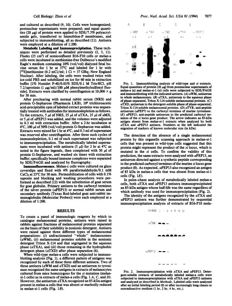
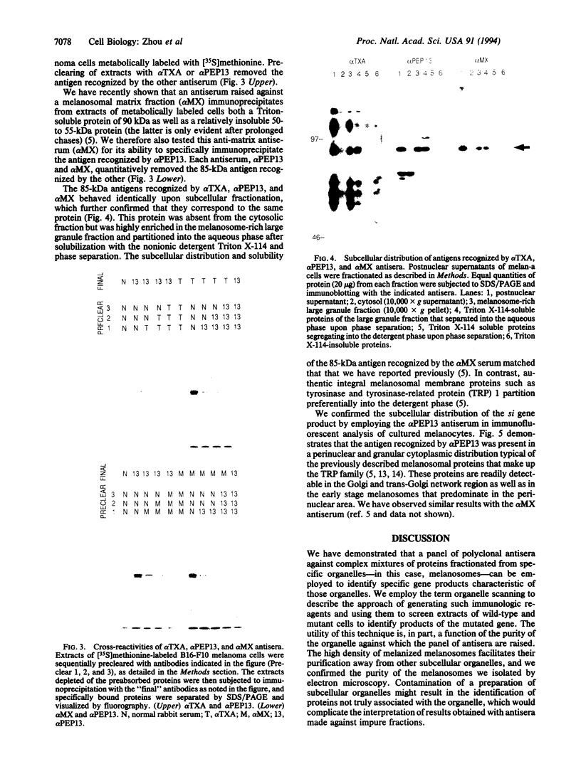
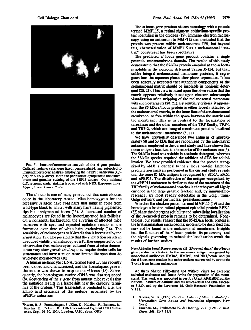
To identify a broad spectrum of melanosomal proteins, antisera were raised in rabbits against melanosomal protein fractions separated on the basis of their solubility in the nonionic detergent Triton X-114. Antisera against the different fractions each recognized a distinct set of bands when used for immunoblotting analysis with extracts of melanocytes cultured from wild-type black mice. Immunoblotting with antisera to whole melanosomes or to Triton X-114-soluble melanosomal proteins that segregated with the detergent phase gave identical patterns with protein extracts from melanocytes from wild-type mice and from mice homozygous for the si (silver) coat color mutation. By contrast, an antiserum against Triton X-114 soluble melanosomal proteins that segregated in the aqueous phase recognized an 85-kDa protein that was present in extracts from wild-type melanocytes but was absent from si melanocytes. This suggested that the protein was encoded at the si (silver) locus. This was confirmed by employing an antiserum directed against the carboxyl terminus of the predicted murine silver protein sequence. The detergent solubility, biochemical characteristics, and immunologic properties of the 85-kDa protein and of the authentic si gene product were identical. Further analysis demonstrated that this protein corresponds to a melanosomal matrix glycoprotein that we recently described. Our results suggest that employing polyclonal antisera to fractionated organelles such as melanosomes, to screen tissues from mutant mice, a technique that we call "organelle scanning", can serve as a powerful means of identifying new organellar proteins and their respective genes.
Full text
PDF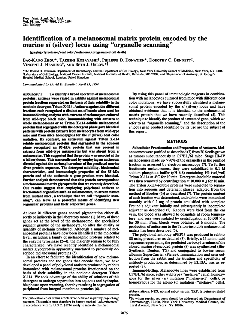
Images in this article
Selected References
These references are in PubMed. This may not be the complete list of references from this article.
- Adema G. J., de Boer A. J., van 't Hullenaar R., Denijn M., Ruiter D. J., Vogel A. M., Figdor C. G. Melanocyte lineage-specific antigens recognized by monoclonal antibodies NKI-beteb, HMB-50, and HMB-45 are encoded by a single cDNA. Am J Pathol. 1993 Dec;143(6):1579–1585. [PMC free article] [PubMed] [Google Scholar]
- Bennett D. C., Cooper P. J., Hart I. R. A line of non-tumorigenic mouse melanocytes, syngeneic with the B16 melanoma and requiring a tumour promoter for growth. Int J Cancer. 1987 Mar 15;39(3):414–418. doi: 10.1002/ijc.2910390324. [DOI] [PubMed] [Google Scholar]
- Bordier C. Phase separation of integral membrane proteins in Triton X-114 solution. J Biol Chem. 1981 Feb 25;256(4):1604–1607. [PubMed] [Google Scholar]
- Cox A. L., Skipper J., Chen Y., Henderson R. A., Darrow T. L., Shabanowitz J., Engelhard V. H., Hunt D. F., Slingluff C. L., Jr Identification of a peptide recognized by five melanoma-specific human cytotoxic T cell lines. Science. 1994 Apr 29;264(5159):716–719. doi: 10.1126/science.7513441. [DOI] [PubMed] [Google Scholar]
- Goldenthal K. L., Hedman K., Chen J. W., August J. T., Willingham M. C. Postfixation detergent treatment for immunofluorescence suppresses localization of some integral membrane proteins. J Histochem Cytochem. 1985 Aug;33(8):813–820. doi: 10.1177/33.8.3894499. [DOI] [PubMed] [Google Scholar]
- Jimbow K., Jimbow M., Chiba M. Characterization of structural properties for morphological differentiation of melanosomes: II. Electron microscopic and SDS-PAGE comparison of melanosomal matrix proteins in B16 and Harding Passey melanomas. J Invest Dermatol. 1982 Jan;78(1):76–81. doi: 10.1111/1523-1747.ep12497959. [DOI] [PubMed] [Google Scholar]
- Jiménez M., Tsukamoto K., Hearing V. J. Tyrosinases from two different loci are expressed by normal and by transformed melanocytes. J Biol Chem. 1991 Jan 15;266(2):1147–1156. [PubMed] [Google Scholar]
- Kawakami Y., Eliyahu S., Delgado C. H., Robbins P. F., Sakaguchi K., Appella E., Yannelli J. R., Adema G. J., Miki T., Rosenberg S. A. Identification of a human melanoma antigen recognized by tumor-infiltrating lymphocytes associated with in vivo tumor rejection. Proc Natl Acad Sci U S A. 1994 Jul 5;91(14):6458–6462. doi: 10.1073/pnas.91.14.6458. [DOI] [PMC free article] [PubMed] [Google Scholar]
- Kim R. Y., Wistow G. J. The cDNA RPE1 and monoclonal antibody HMB-50 define gene products preferentially expressed in retinal pigment epithelium. Exp Eye Res. 1992 Nov;55(5):657–662. doi: 10.1016/0014-4835(92)90170-w. [DOI] [PubMed] [Google Scholar]
- Kwon B. S., Chintamaneni C., Kozak C. A., Copeland N. G., Gilbert D. J., Jenkins N., Barton D., Francke U., Kobayashi Y., Kim K. K. A melanocyte-specific gene, Pmel 17, maps near the silver coat color locus on mouse chromosome 10 and is in a syntenic region on human chromosome 12. Proc Natl Acad Sci U S A. 1991 Oct 15;88(20):9228–9232. doi: 10.1073/pnas.88.20.9228. [DOI] [PMC free article] [PubMed] [Google Scholar]
- Kwon B. S. Pigmentation genes: the tyrosinase gene family and the pmel 17 gene family. J Invest Dermatol. 1993 Feb;100(2 Suppl):134S–140S. doi: 10.1111/1523-1747.ep12465022. [DOI] [PubMed] [Google Scholar]
- Mochii M., Agata K., Eguchi G. Complete sequence and expression of a cDNA encoding a chicken 115-kDa melanosomal matrix protein. Pigment Cell Res. 1991 Feb;4(1):41–47. doi: 10.1111/j.1600-0749.1991.tb00312.x. [DOI] [PubMed] [Google Scholar]
- Orlow S. J., Boissy R. E., Moran D. J., Pifko-Hirst S. Subcellular distribution of tyrosinase and tyrosinase-related protein-1: implications for melanosomal biogenesis. J Invest Dermatol. 1993 Jan;100(1):55–64. doi: 10.1111/1523-1747.ep12354138. [DOI] [PubMed] [Google Scholar]
- Orlow S. J., Chakraborty A. K., Pawelek J. M. Membrane glycoproteins common to vesicles and melanosomes in mouse melanoma cells. Pigment Cell Res. 1992;Suppl 2:162–170. doi: 10.1111/j.1600-0749.1990.tb00368.x. [DOI] [PubMed] [Google Scholar]
- Orlow S. J., Zhou B. K., Boissy R. E., Pifko-Hirst S. Identification of a mammalian melanosomal matrix glycoprotein. J Invest Dermatol. 1993 Aug;101(2):141–144. doi: 10.1111/1523-1747.ep12363626. [DOI] [PubMed] [Google Scholar]
- Spanakis E., Lamina P., Bennett D. C. Effects of the developmental colour mutations silver and recessive spotting on proliferation of diploid and immortal mouse melanocytes in culture. Development. 1992 Mar;114(3):675–680. doi: 10.1242/dev.114.3.675. [DOI] [PubMed] [Google Scholar]
- Tsukamoto K., Jackson I. J., Urabe K., Montague P. M., Hearing V. J. A second tyrosinase-related protein, TRP-2, is a melanogenic enzyme termed DOPAchrome tautomerase. EMBO J. 1992 Feb;11(2):519–526. doi: 10.1002/j.1460-2075.1992.tb05082.x. [DOI] [PMC free article] [PubMed] [Google Scholar]
- Vijayasaradhi S., Doskoch P. M., Houghton A. N. Biosynthesis and intracellular movement of the melanosomal membrane glycoprotein gp75, the human b (brown) locus product. Exp Cell Res. 1991 Oct;196(2):233–240. doi: 10.1016/0014-4827(91)90256-t. [DOI] [PubMed] [Google Scholar]
- Winder A. J., Wittbjer A., Rosengren E., Rorsman H. The mouse brown (b) locus protein has dopachrome tautomerase activity and is located in lysosomes in transfected fibroblasts. J Cell Sci. 1993 Sep;106(Pt 1):153–166. doi: 10.1242/jcs.106.1.153. [DOI] [PubMed] [Google Scholar]