

Abstract
Those born with sirenomelia, a rare congenital anomaly, have features resembling a mermaid. Characteristics of sirenomelia are a single lower limb, sacral and pelvic bone defects, and anorectal and urogenital malformations. There is an increased incidence of sirenomelia in males and twins. This case was a preterm male, dizygotic twin and product of in vitro fertilisation. The baby was born by caesarean section due to breech presentation. He was found to have a fused lower extremity and absent external genitalia and anus. The baby passed away shortly after birth due to severe respiratory failure. Radiographic findings showed small lung volume and pneumothoraces. There were multiple segmental fusions of the vertebrae. Single femur and single tibia were presented in a fused lower limb. Autopsy demonstrated large intestinal atresia, intra-abdominal testes, absence of kidney, ureter and bladder, single umbilical artery, agenesis of blood vessels at lower extremity and agenesis of sacrum and coccyx.
Background
Sirenomelia, or mermaid syndrome, was described in 1542 by Rocheus and in 1953 by Palfyn. In 1961, Duhamal defined sirenomelia as the most severe form of caudal regression syndrome (CRS).1 Sirenomelia is a very rare congenital malformation with an incidence of 0.98–4.2 per 100 000 live births.2–4 Most cases are premature births with a birth weight of less than 2500 g.2 Male-to-female ratio is approximately 3:13 4 and 8–15% of cases are in twins.2 5 The characteristics of this condition are a single midline lower limb, sacral and pelvic bone anomalies, absent external genitalia, imperforated anus and renal dysgenesis or agenesis. According to the classification from Stocker and Heifetz,6 there are seven types of sirenomelia. Each type depends on the number of femur, tibia and metatarsal bones.
The aetiology of sirenomelia remains unclear but two main hypotheses are postulated. The vascular steal hypothesis is based on an aberrant abnormal umbilical artery that comes directly from a high abdominal aorta. Blood stolen from the lower part of the body results in hypoperfusion and an underdevelopment of multiple organs below. The second hypothesis is defective blastogenesis, which is based on a deficiency of caudal mesoderm, impairing the development of the caudal embryonic body in early gestation.2 7 8
Sirenomelia has been considered to be the most severe variety of CRS. However, there are some differences between the two disorders. CRS is strongly associated with maternal diabetes mellitus. Most patients had polyhydramnios, normal umbilical vessels, non-lethal renal anomalies and hypoplastic lower limbs.9 However, sirenomelia, while associated with CRS, has distinct causality and presentation with oligohydramnios, a single umbilical artery, renal agenesis or dysgenesis and one or two fused lower limbs. In summary, sirenomelia has more severe presentation and a higher risk of death. Sirenomelia also has some shared features with VACTREL association including vertebral anomaly, anorectal malformation, cardiac defect, tracheo-oesophageal fistula, renal and limb anomaly. Both diseases are of mesodermal defect origin; however, the upper part of the body is primarily involved in VACTREL association.
Case presentation
A 27-week-gestation, first twin baby was born by caesarean section due to breech presentation. The mother was 20 years old. She had a history of two spontaneous abortions prior to this pregnancy, but was otherwise healthy. This was an in vitro fertilisation pregnancy. Her serology was negative for HIV, VDRL and HBsAg. She had no reported history of medication or substance abuse. The parents were not consanguineous. Prenatal ultrasonography identified a dichorion-diamnion twin pregnancy with one fetus displaying disrupted lower limb development. During the pregnancy, the mother had multiple spotted-vaginal bleeding. In this episode, maternal presentation included vaginal bleeding and premature contractions. The first twin was delivered following an emergency caesarean section with a birth weight of 716.5 g. The baby was found to have a short-fused lower limb, and an absence of external genitalia and anus (figure 1). Dysmorphic facial features including a sloped forehead, low set ears, abnormal ear lobulation, flattened nose, receded chin and bifid right thumb (figure 2) were also observed.
Figure 1.

Morphological appearance of the patient showed a dysmorphic face, a single fused lower limb and absence of external genitalia and anus.
Figure 2.

Bifid right thumb.
Investigations
Radiographic findings after death showed bilateral pneumothoraces, abnormal segmental fusion of the T12-L1, L2-3 and L4-5 vertebrae, and sacral dysplasia with a contracted lesser pelvis. A single lower extremity with a reduced tibia, and absence of fibula and foot was seen. Normal appearance of the visualised skull, bilateral clavicles, bilateral scapulars and bilateral upper extremities were reported (figure 3).
Figure 3.
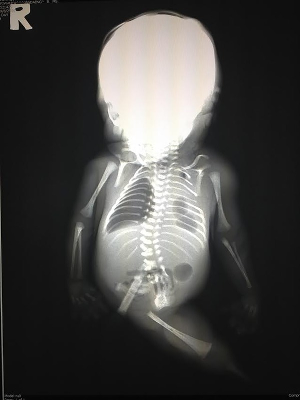
Radiograph of the body demonstrating bilateral pneumothoraces, fusion of the T12-L1, L2-3 and L4-5 vertebrae, absent left hip, rudimentary right ileum and single right lower limb with hypoplastic femur and tibia.
The autopsy demonstrated the following multiple organ abnormalities:
Skeletal abnormalities: hypoplasia of lower extremity (single lower limb), agenesis of sacrum and coccyx and bifid right thumb.
Gastrointestinal abnormalities: imperforated anus and agenesis of large intestine.
Genitourinary system abnormalities: absence of external genitalia; intra-abdominal testes were present; bilateral renal agenesis, including both ureters and urinary bladder.
Respiratory system abnormalities: bilateral pulmonary hypoplasia (combined lung weight=8 g, lung to body weight ratio=0.011, normal ratio>0.015) and hyaline membrane disease.
A single umbilical artery, agenesis of blood vessels at lower limbs.
Treatment
Intubation, positive pressure ventilation, chest compressions and administration of epinephrine were performed.
Outcome and follow-up
The baby displayed continued cyanosis and bradycardia. Apgar scores were 1 and 2 at 1 and 5 min, respectively. He passed away 30 min after birth due to an inability to ventilate. The second twin was born alive, but developed respiratory distress from surfactant deficiency. No dysmorphic features were observed.
Discussion
Sirenomelia is a very rare congenital malformation and lethal congenital anomaly. According to the physical, radiographic and autopsy results, our case diagnosis was sirenomelia. The baby was classified as sirenomelia type VI or simpus apus because single hypoplastic femur and single tibia were found in a fused limb. In addition, this baby had characteristics of Potter's sequence, which include dysmorphic facial features, pulmonary hypoplasia and renal agenesis. Potter's sequence has been reported in 12.4% of sirenomelia cases and this condition results in a poor prognosis.2 4 10 Moreover, we found a bifid thumb, which is a related upper limb defect. Radial aplasia/hypoplasia, radial club hands, polydactyly, thumb agenesis, upper limb reduction, lobster claw hands, webbed limbs and joint contracture have been reported with sirenomelia, and these upper limb defects can be found in approximately 30% of patients.2 4 11 This case was of note, as it was of an in vitro fertilisation, dizygotic twin pregnancy with sirenomelia, of which limited studies have been reported.12–15
Learning points.
Sirenomelia is a rare and fatal congenital anomaly; incidence is increased in male preterm twins.
The characteristics of this condition are single midline lower limb, sacral and pelvic bone defect, absence of external genitalia and anus, and renal dysgenesis or agenesis.
It has been considered to be the most severe variety of caudal regression syndrome. However, sirenomelia has distinct causality and presentation with oligohydramnios, a single umbilical artery, renal agenesis or dysgenesis and one or two fused lower limbs.
The aetiology is unclear. Abnormal origination of umbilical artery vessel and defective blastogenesis are two main hypotheses.
A clear understanding of the natural history and clinical symptoms will help physicians in early recognition of the disease and prediction of the clinical prognoses of such conditions.
Footnotes
Contributors: KN wrote the proposal, collected the data, wrote the manuscript, read the final draft of the manuscript and revised the manuscript. PV wrote the radiology report. SK wrote the pathology report. VK got informed consent, wrote and edited the manuscript, read the final draft of, revised and submitted the manuscript, and overall supervised the work.
Competing interests: None declared.
Patient consent: Obtained.
Provenance and peer review: Not commissioned; externally peer reviewed.
References
- 1.Kshirsagar V, Ahmed M, Colaco S. Sirenomelia apus: a rare deformity. J Clin Neonatol 2012;1:146–8. 10.4103/2249-4847.101699 [DOI] [PMC free article] [PubMed] [Google Scholar]
- 2.Orioli IM, Amar E, Arteaga-Vazquez J et al. Sirenomelia: an epidemiologic study in a large dataset from the international clearinghouse of birth defects surveillance and research, and literature review. Am J Med Genet C Semin Med Genet 2011;157:358–73. 10.1002/ajmg.c.30324 [DOI] [PMC free article] [PubMed] [Google Scholar]
- 3.Nisenblat V, Leibovitz Z, Paz B et al. Dizygotic twin pregnancy discordant for sirenomelia. J Ultrasound Med 2007;26:97–103. [DOI] [PubMed] [Google Scholar]
- 4.Tonni G, Grisolia G. Sirenomelia: a review on embryogenic environmental theories, novel three-dimentional ultrasound imaging and first trimester diagnosis in a case of mosaic 69,XXX/46,XX fetus. Arch Gynecol Obstet 2013;288:3–11. 10.1007/s00404-013-2847-3 [DOI] [PubMed] [Google Scholar]
- 5.Di Lorenzo M, Brandt ML, Veilleux A. Sirenomelia in an identical twin: a case report. J Pediatr Surg 1991;26:1334–6. 10.1016/0022-3468(91)90614-Y [DOI] [PubMed] [Google Scholar]
- 6.Stocker JT, Heifetz SA. Sirenomelia. A morphological study of 33 cases and review of the literature. Perspect Pediatr Pathol 1987;10:7–50. [PubMed] [Google Scholar]
- 7.Monteagudo A, Mayberry P, Rebarber A et al. Sirenomelia sequence: first-trimester diagnosis with both two- and three-dimensional sonography. J Ultrasound Med 2002;21:915–20. [DOI] [PubMed] [Google Scholar]
- 8.Stevenson RE, Jones KL, Phelan MC et al. Vascular steal: the pathogenetic mechanism producing sirenomelia and associated defects of the viscera and soft tissues. Pediatrics 1986;78:451–7. [PubMed] [Google Scholar]
- 9.Bruce JH, Romaguera RL, Rodriguez MM et al. Caudal dysplasia syndrome and sirenomelia: are they part of a spectrum? Fetal Pediatr Pathol 2009;28:109–31. 10.1080/15513810902772383 [DOI] [PubMed] [Google Scholar]
- 10.Al-Haggar M, Yahia S, Abdel-Hadi D et al. Sirenomelia (symelia apus) with Potter's syndrome in connection with gestational diabetes mellitus: a case report and literature review. Afr Health Sci 2010;10:395–9. [PMC free article] [PubMed] [Google Scholar]
- 11.Lhuaire M, Jestin A, Boulangnon C et al. Sirenomelia: a new type, showing VACTERL association with Thomas syndrome and a review of literature. Birth Defects Res A Clin Mol Teratol 2013;97:123–32. 10.1002/bdra.23125 [DOI] [PubMed] [Google Scholar]
- 12.Drossou-Agakidou V, Xatzisevastou-Loukidou C, Soubasi V et al. Rare manifestations of sirenomelia syndrome: a report of five cases. Am J Perinatol 2004;21:395–401. 10.1055/s-2004-835314 [DOI] [PubMed] [Google Scholar]
- 13.Horikoshi T, Kikichi A, Tatematsu M et al. Two cases of a fetus with sirenomelia sequence. Congenit Anom (Kyoto) 2005;45:93–5. 10.1111/j.1741-4520.2005.00074.x [DOI] [PubMed] [Google Scholar]
- 14.Bakhtar O, Benirschke K, Masliah E. Sirenomelia of an intracytoplasmic sperm injection conceptus: a case report and review of mechanism. Pediatr Dev Pathol 2006;9:245–53. 10.2350/08-05-0092.1 [DOI] [PubMed] [Google Scholar]
- 15.Chen CP, Hsu CY, Lee MS et al. Magnetic resonance imaging demonstration of sirenomelia in one fetus of a dizygotic twin pregnancy conceived by intracytoplasmic sperm injection, in vitro fertilization and embryo transfer. Taiwan J Obstet Gynecol 2011;50:561–3. 10.1016/j.tjog.2011.10.034 [DOI] [PubMed] [Google Scholar]