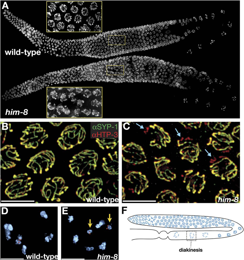
Figure 1. him-8 Mutants Display X Chromosome-Specific Defects in Synapsis and Chiasma Formation.
(A) Whole gonads from wild-type and him-8 hermaphrodites stained with DAPI. Insets magnify the pachytene region to show that him-8 nuclei retain a polarized appearance that is usually restricted to transition-zone nuclei.
(B and C) Pachytene nuclei stained with antibodies to SYP-1 (green) and HTP-3 (red).
(B) In wild-type hermaphrodites, these antibodies show very similar localization patterns at pachytene along the entire lengths of all six pairs of chromosomes.
(C) In him-8 mutant hermaphrodites we observe one pair of unsynapsed chromosomes in each nucleus. Here these are revealed as chromosomes that stain with HTP-3 (red) but not SYP-1 (green). Examples are indicated with blue arrows.
(D and E) Oocyte nuclei at diakinesis, shortly prior to the meiotic nuclear divisions.
(D) Wild-type nuclei have six DAPI staining bodies, indicating the formation of chiasmata between all six pairs of homologous chromosomes. One of these bivalents is marked by a FISH probe specific for the X chromosomes.
(E) him-8 nuclei usually reveal seven DAPI staining bodies at diakinesis. Achiasmate, or univalent, X chromosomes marked by a FISH probe are indicated by yellow arrows.
(F) Diagram showing the location of diakinesis within the worm gonad. At this stage, the SC has largely broken down and homologs are held together by chiasmata.
All images are projections of 3D images following deconvolution. Scale bars represent 5 µm.

