

Sir,
We read with great interest the case report by Morey et al. “Intraarticular osteochondroma of the knee,”1 where the authors reported that there are only three other similar cases in the literature. This is indeed a rare presentation, and I must congratulate the authors for a nice presentation of this “teaching case.”
However, we would like to point out that “intraarticular osteochondroma” is a well known entity and has been described in the literature as Trevor's disease (tarso-epiphysial aclasis).2 First described in 1926 by Mouchet and Belot (tarsomegalie),3 this condition is also known as Fairbank's disease or dysplasia epiphysealis hemimelica.4 Trevor's diseases is considered a skeletal developmental disorder characterized by asymmetric overgrowth of cartilage in the epiphyses, analogous to an “osteochondroma” of the epiphysis. The prevalence has been reported as 1 in 1 million and the etiology is unknown.5 Moreover, it needs to be differentiated from another similar entity called extraskeletal paraarticular osteochondromas, which is an osteocartilaginous lesion arising in the soft tissues, adjacent to the joint but without any bone continuity.6
Thus, an intrarticular osteochondroma is not as rare as reported by the authors, possibly because of several nomenclatures and ambiguity in literature search. In fact, apart from just the three other cases of the knee noted by the authors,7,8,9 literature review actually shows several other similar intraarticular osteochondromas (excluding extraskeletal paraarticular osteochondromas) arising in the knee joint7,8,9,10,11,12,13,14 and several of these had been removed arthroscopically in contrast to the authors’ conclusion.7,8,10,11 We have also personally resected a similar lesion in a 5-year-old male who had a full range of motion without any growth disturbance at 7 years followup [Figures 1–5]. A review of literature in 2007 revealed 57 patients with Trevor's disease with 44 of the lesions in the knee (29 of them in the distal femur and 15 in the proximal tibia).12 It has also been classified as localized, classical and generalized form.13 Multiple epiphyses may be involved in upto two-thirds of the cases, and thus a skeletal survey is recommended once the initial diagnosis is made. Most cases in the literature were treated by surgical removal of the mass and correction of any joint deformity. Recurrence is unlikely but has been reported.14 However, we do agree with the authors that Trevor's disease of the knee should be considered in the differential of an intrarticular knee mass in young patients.
Figure 1.
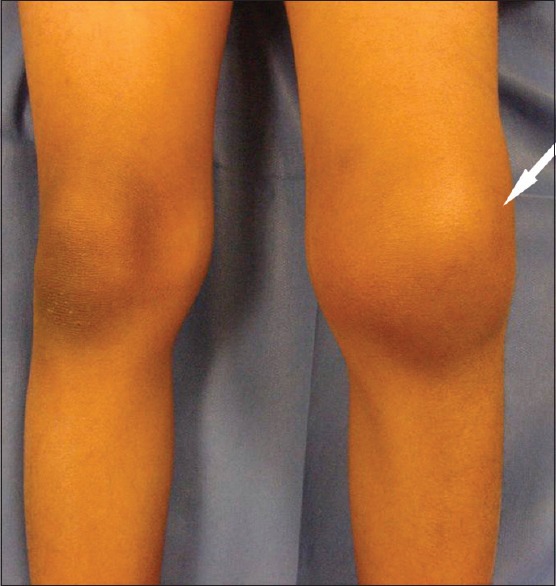
Clinical photograph of a 5-year-old boy showing diffuse swelling in the left knee (white arrow). The patient presented with increasing swelling, locking, stiffness (range of motion 30°–100°) and intermittent pain in the left knee for the past 6 months
Figure 5.

(a) Antero-posterior and (b) lateral radiographs of the same knee after 7 years followup showing no residual mass, deformity or premature closure of growth plates. The patient was totally asymptomatic with full painless range of motion
Figure 2.

(a) Antero posterior and (b) lateral radiographs of the knee showing an intraarticular mineralized mass. The rest of the bone seemed uninvolved (white arrow)
Figure 3.

A sagittal magnetic resonance imaging with proton density sequence (a) TR 3000, TE 13 showing an intraarticular mass (slight hyperintense to the muscle) in the intercondylar notch with a large effusion (white arrow). A coronal short tau inversion recovery sequence (b) TR 5680, TE 51 showing lobulation and heterogeneity in the mass
Figure 4.

Because of the symptoms, imaging studies and size of the mass, the patient was indicated for open excisional biopsy (a) Intraoperative photograph showing a large osteochondromatous mass in the intercondylar notch with bony continuity with the rest of the femur (black arrow). (b) The mass measured about 3 × 3 × 2 cm and was reported as an osteochondroma on histology, confirming the diagnosis of Trevor's disease
REFERENCES
- 1.Morey VM, Jalan D, Mittal R, Gamangatti S. Intraarticular osteochondroma of the knee. Indian J Orthop. 2014;48:332–4. doi: 10.4103/0019-5413.132532. [DOI] [PMC free article] [PubMed] [Google Scholar]
- 2.Trevor D. Tarso-epiphysial aclasis; A congenital error of epiphysial development. J Bone Joint Surg Br. 1950;32-B:204–13. doi: 10.1302/0301-620X.32B2.204. [DOI] [PubMed] [Google Scholar]
- 3.Mouchet A, Belot J. La tarsomegalie. J Radiol Electrol. 1926;10:289–93. [Google Scholar]
- 4.Fairbank TJ. Dysplasia epiphysialis hemimelica (tarso-ephiphysial aclasis) J Bone Joint Surg Br. 1956;38-B:237–57. doi: 10.1302/0301-620X.38B1.237. [DOI] [PubMed] [Google Scholar]
- 5.Araujo CR, Jr, Montandon S, Montandon C, Teixeira KI, Moraes FB, Moreira MA. Best cases from the AFIP: Dysplasia epiphysealis hemimelica of the patella. Radiographics. 2006;26:581–6. doi: 10.1148/rg.262055126. [DOI] [PubMed] [Google Scholar]
- 6.Maheshwari AV, Jain AK, Dhammi IK. Extraskeletal paraarticular osteochondroma of the knee – A case report and tumor overview. Knee. 2006;13:411–4. doi: 10.1016/j.knee.2006.05.008. [DOI] [PubMed] [Google Scholar]
- 7.Takahashi M, Nishihara A, Ohishi T, Shiga K, Yamamoto K, Nagano A. Arthroscopic resection of an intraarticular osteochondroma of the knee in the patient with multiple osteochondromatosis. Arthroscopy. 2004;20(Suppl 2):28–31. doi: 10.1016/j.arthro.2004.04.012. [DOI] [PubMed] [Google Scholar]
- 8.Schmoyer S, Ciullo JV. Arthroscopic resection of an osteochondroma of the knee. Arthroscopy. 2001;17:765–7. doi: 10.1053/jars.2001.22401. [DOI] [PubMed] [Google Scholar]
- 9.Dienst M, Schneider G, Pahl S, Ensslin S, Kohn D. Intraarticular osteochondroma of the posterior cavity of the knee. Arch Orthop Trauma Surg. 2002;122:462–5. doi: 10.1007/s004020100311. [DOI] [PubMed] [Google Scholar]
- 10.Kesgin E, Çelik C, Karaoglu S. Arthroscopic resection of osteochondroma of the knee: Two case reports. Eklem Hastalik Cerrahisi. 2013;24:112–6. doi: 10.5606/ehc.2013.25. [DOI] [PubMed] [Google Scholar]
- 11.Kim JI, Kwon JH, Park YJ, D’Almeida VR, Soni SM, Nha KW. Arthroscopic excision of solitary intraarticular osteochondroma of the Knee. Knee Surg Relat Res. 2013;25:36–9. doi: 10.5792/ksrr.2013.25.1.36. [DOI] [PMC free article] [PubMed] [Google Scholar]
- 12.Rosero VM, Kiss S, Terebessy T, Köllö K, Szöke G. Dysplasia epiphysealis hemimelica (Trevor's disease): 7 of our own cases and a review of the literature. Acta Orthop. 2007;78:856–61. doi: 10.1080/17453670710014662. [DOI] [PubMed] [Google Scholar]
- 13.Azouz EM, Slomic AM, Marton D, Rigault P, Finidori G. The variable manifestations of dysplasia epiphysealis hemimelica. Pediatr Radiol. 1985;15:44–9. doi: 10.1007/BF02387852. [DOI] [PubMed] [Google Scholar]
- 14. [Last accessed on 2014 May 18]. Available from: http://www.rarediseases.org/rare-diseaseinformation/rare-diseases/byID/572/viewFullReport .