Abstract
The central nervous system (CNS) controls food intake and energy expenditure via tight coordinations between multiple neuronal populations. Specifically, two distinct neuronal populations exist in the arcuate nucleus of hypothalamus (ARH): the anorexigenic (appetite-suppressing) pro-opiomelanocortin (POMC) neurons and the orexigenic (appetite-increasing) neuropeptide Y (NPY)/agouti-related peptide (AgRP) neurons. The coordinated regulation of neuronal circuit involving these neurons is essential in properly maintaining energy balance, and any disturbance therein may result in hyperphagia/obesity or hypophagia/starvation. Thus, adequate knowledge of the POMC and NPY/AgRP neuron physiology is mandatory to understand the pathophysiology of obesity and related metabolic diseases. This review will discuss the history and recent updates on the POMC and NPY/AgRP neuronal circuits, as well as the general anorexigenic and orexigenic circuits in the CNS. [BMB Reports 2015; 48(4): 229-233]
Keywords: GABA, Neuronal circuit, NPY/AgRP neuron, POMC neuron, α-MSH
INTRODUCTION
Hypothalamus is a key brain area that regulates homeostasis. In particular, specific areas of hypothalamus are believed to control feeding behavior. The classic experiments by Hetherington and Ranson established the ventromedial hypothalamus as the appetite-suppressing (or anorexigenic) center (1), and later experiments demonstrated that lateral hypothalamic area (LHA) is the appetite-increasing (or orexigenic) center (2). These results suggested that a specific hypothalamic area may regulate feeding, and subsequent studies attempted to revise this concept by using more refined neuroanatomical methods. In recent years, the development of mouse genetics and other techniques such as electrophysiology, optogenetics, and chemogenetics has led us to gain more detailed information on the identity of specific neurons that affect feeding behavior. Specifically, a large body of information is currently available on the appetite-regulating role of two distinct neuronal populations within the ventral medial part of hypothalamus.
THE ARCUATE NUCLEUS OF HYPOTHALAMUS
The arcuate nucleus of hypothalamus (ARH) is undoubtedly one of the best-characterized brain regions as it is related to the control of feeding behavior. This is in large part due to the presence of two distinct neuronal populations, which have opposite effects on the feeding behavior: the anorexigenic pro-opiomelanocortin (POMC) neurons and the orexigenic neuropeptide Y (NPY)/agouti-related peptide (AgRP) neurons. These neurons are well-positioned to receive information from peripheral organs; the ARH resides in the ventral medial part of hypothalamus, which receives rich blood supply due to its proximity to the median eminence. Thus, information from peripheral organs may easily access the POMC and NPY/AgRP neurons. In addition, they receive intensive input from multiple parts of the central nervous system (CNS). Therefore, POMC and NPY/AgRP neurons are in a good position to integrate peripheral and central inputs to produce a central command for feeding behavior. Indeed, the activity of POMC and NPY/AgRP neurons is modulated by multiple neurotransmitters and/or hormones. For instance, the anorexigenic effects of serotonin and the adipocyte-derived hormone leptin are considered to be at least in part mediated by excitation of POMC neurons and suppression of NPY/AgRP neurons (3-6). Ghrelin, an orexigenic hormone released from gastric mucosa, was shown to suppress POMC neurons and excite NPY/AgRP neurons by indirect mechanisms (7-9). The effects of insulin on POMC neurons and NPY/AgRP neurons need to be clarified, as the experimental results are not consistent between studies from independent groups (6, 10, 11).
THE POMC NEURONS AND THE CENTRAL MELANOCORTIN PATHWAY
The anorexigenic effect of POMC neuron was evidenced by the hyperphagia and obesity observed in the POMC knockout mice (12). Recent studies employed optogenetic and chemogenetic stimulation methods to activate specific neuronal population, and demonstrated that direct activation of POMC neurons lead to suppression of food intake (13, 14). It is believed that POMC neurons suppress appetite by releasing α-melanocyte stimulating hormone (α-MSH) which is an agonist at the anorectic melanocortin-4 receptors (MC4Rs). Consistent with this idea, MC4R deficiency results in hyperphagia and obesity in mice (15-17). Importantly, human patients with mutations in Mc4r genes are also hyperphagic and obese (18, 19). Thus, the central melanocortin pathway that involves POMC neurons and the MC4R-expressing neurons represent a key anorexigenic circuit in the CNS.
One of the key anorexigenic signals that activate the central melanocortin pathway is serotonin (5-HT). Fenfluramine (d-Fen), which increases the availability of serotonin in brain, was an effective and widely-used prescription diet pill until it was withdrawn from the market due to serious cardiovascular side effects (20). The anorexigenic effect of serotonin is largely mediated by the serotonin 2C receptors (5-HT2CRs) expressed by the POMC neurons (21-23). Stimulation of 5-HT2CRs increases the activity of POMC neurons (21, 24, 25), which presumably increases α-MSH release. Consistently, the anorexigenic effects of d-Fen were demonstrated to be mediated by the MC4Rs expressed by the single-minded 1 (Sim1) neurons within the paraventricular nucleus of hypothalamus (PVH), as well as the 5-HT2CRs expressed by POMC neurons (26). Currently, d-Fen has been replaced by lorcaserin, a novel prescription diet pill and a specific agonist at 5-HT2CRs (27), which is expected to activate the central melanocortin pathway.
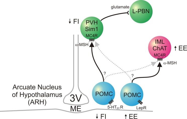
Unlike 5-HT2CRs, however, the deletion of leptin receptors (LepRs) specifically in POMC neurons does not increase food intake (28). In addition, the reactivation of LepRs specifically in POMC neurons of LepR-null mice does not rescue hyperphagia (29). Thus, while leptin activates POMC neurons, the anorexigenic effects of leptin are likely to be mediated by LepRs expressed in other parts of the brain, independent of the melanocortin system (30). In fact, leptin-induced activation of POMC neurons does not seem to activate central melanocortin pathways that suppress food intake. Instead, it seems that leptin excitation of POMC neurons activates central melanocortin pathways that increase energy expenditure (28, 29). Considering that energy expenditure is not affected by deletion or reactivation of 5-HT2CRs specifically in POMC neurons (22, 23), it is suspected that POMC neurons are heterogeneous in their response to leptin and serotonin. Consistent with this idea, the acute response of POMC neurons to leptin and mCPP (a 5-HT2CR agonist) is segregated to distinct subpopulations of POMC neurons (24). These results are intriguing since the metabolic effects of MC4Rs are mediated by different brain nuclei; MC4Rs expressed by the PVH decreases food intake, while MC4Rs expressed by the sympathetic neurons within the intermediolateral column (IML) of spinal cord increases energy expenditure (16, 31). Taken together, the “appetite-suppressing” POMC neurons may project to PVH, while the “energy-consuming” POMC neurons may project to the IML (Fig. 1).
Fig. 1. POMC neurons and the central melanocortin pathway. Two distinct populations of POMC neurons are present within the ARH; 5-HT2CR-expressing POMC neurons suppress food intake, and LepR-expressing POMC neurons increase energy expenditure. The effects of MC4Rs on energy balance are localized to different central nuclei; MC4Rs expressed by the PVH Sim1 neurons suppress food intake, and MC4Rs expressed by the IML sympathetic neurons increase energy expenditure. The solid black lines show possible connections between the specific POMC neurons and specific brain nuclei; the connections represented by the broken gray lines have not been ruled out. MC4R-expressing PVH glutamatergic neurons send axons to the neurons of the L-PBN. 3V: third ventricle, 5-HT2CR: serotonin 2C receptor, α-MSH: α-melanocyte stimulating hormone, ChAT: choline acetyltransferase, EE: energy expenditure, FI: food intake, IML: intermediolateral column of spinal cord, L-PBN: lateral parabrachial nucleus, LepR: leptin receotor, MC4R: melanocortin-4 receptor, ME: median eminence, POMC: pro-opiomelanocortin, PVH: paraventricular nucleus of hypothalamus, Sim1: single-minded 1.
THE PARAVENTRICULAR NUCLEUS OF HYPOTHALAMUS AND THE PARABRACHIAL NUCLEUS: ANOREXIGENIC CENTERS
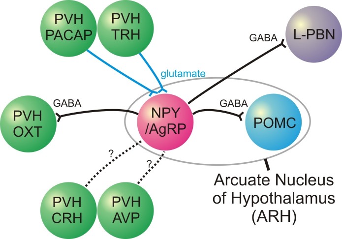
The PVH and the parabrachial nucleus (PBN) of brainstem are representative anorexigenic centers in addition to POMC neurons and the central melanocortin pathways. Sim1 is a transcription factor that is critical for PVH development, and Sim 1 haploinsufficiency is associated with hyperphagia and obesity (32). Thus, a significant population of PVH neurons is considered to suppress food intake. Specifically, the oxytocin (OXT) neurons within the PVH are known to have loss-of-function mutations in Prader-Willi syndrome (33) and as a consequence of Sim1 deficiency (34, 35), both of which are characterized by hyperphagia. The MC4R-expressing glutamatergic neurons within the PVH, which may be considered as a part of the central melanocortin system, reduce food intake (36). Intriguingly, the MC4R-expressing PVH neurons were found to be distinct subpopulation from the OXT, corticotropin-releasing hormone (CRH), arginine vasopressin (AVP), and prodynorphin (Pdyn) neurons within the PVH (36). In addition, Sim1 neurons that express either thyrotropin-releasing hormone (TRH) or pituitary adenylate cyclase-activating peptide (PACAP) were found to send excitatory input to the orexigenic NPY/AgRP neurons within the ARH (37). Thus, some PVH neurons may not be anorexigenic as originally suggested, but they may have “orexigenic” effects.
The PBN is a pontine nucleus located adjacent to the superior cerebellar peduncle that contains subpopulation of neurons suppressing appetite (38-40). The PBN neurons receive gluatamatergic “satiety” signals from the nucleus tractus solitarius (NTS) neurons located in the medulla oblongata (38), which is considered to be the neural correlates of postprandial satiety. Consistent with the anorexigenic role of PBN, the MC4R-expressing PVH glutamatergic neurons send axons to the lateral PBN (36) (Fig. 1). Recently, it was demonstrated that the calcitonin gene-related peptide (CGRP) neurons within the external lateral subdivision of PBN project to the central amygdala to suppress food intake (41). Another recent study also demonstrated that neurons expressing protein kinase C delta (PKCδ) mediate the anorexigenic effects (42).
THE NPY/AGRP NEURONS AND THE OREXIGENIC PATHWAY
The NPY/AgRP neurons within the ARH are probably the most established orexigenic population in the CNS. However, earlier studies reported that mice with NPY and/or AgRP deficiency have normal food intake and body weight (43). Similarly, ablation of AgRP neurons during development resulted in normal body weight (44, 45). More recent studies used toxins to ablate the NPY/AgRP neurons in adults and confirmed the orexigenic role of these neurons (38, 39, 46). Acute activation of NPY/AgRP neurons using optogenetic or pharmacogenetic stimulation methods also resulted in robust increase in food intake (13, 47). These series of experiments have established the NPY/AgRP neurons as the major orexigenic population in the CNS.
The NPY/AgRP neurons release the neuropeptide NPY (agonist at the Y receptors), AgRP (inverse agonist at the MC4Rs), and the inhibitory neurotransmitter GABA. The metabolic effects of NPY on Y receptors are somewhat complicated, and many are not consistent with the orexigenic role of NPY/AgRP neurons; this issue was discussed in more detail in a previous review article (48). The ability of AgRP to antagonize the central melanocortin pathways received much attention, since this was considered to be the underlying mechanism of orexigenic effects. However, the orexigenic effects of NPY/AgRP neurons are not affected by the deletion of MC4Rs (47), and it is now regarded that the NPY/AgRP neurons increase food intake independent of the central melanocortin system. Currently, GABA release is believed to mediate most of the orexigenic effects of the NPY/AgRP neurons. For instance, deletion of vesicular GABA transporter (vgat) genes specifically in the AgRP neurons leads to lean phenotype (49), which confirms the importance of GABAergic neurotransmission in the orexigenic effects.
Multiple “anorexigenic” neuronal populations within CNS receive the “inhibitory” GABAergic input from the NPY/AgRP neurons (Fig. 2). The ARH POMC neurons receive GABAergic input from the NPY/AgRP neurons, which form a local circuit of appetite regulation within the ARH (3). Indeed, when the vgat gene is deleted in the NPY/AgRP neurons, the frequency of inhibitory postsynaptic current (IPSC) is not increased by ghrelin which excites the NPY/AgRP neurons (49). The PVH OXT neurons and Sim1 neurons (which do not express either PACAP or TRH) also receive GABAergic input from the NPY/AgRP neurons (37, 50). Finally, the neurons within the PBN receive GABAergic input from the NPY/AgRP neurons (38, 39). Thus, it is suggested that the orexigenic pathways involving the GABAergic neurotramsission from the NPY/AgRP neurons increase food intake by the inhibition of the anorexigenic centers in the CNS.
Fig. 2. NPY/AgRP neurons and the orexigenic pathway. NPY/AgRP neurons have axon terminals that release GABA (an inhibitory neurotransmitter) to suppress the POMC neurons, PVH OXT neurons, and neurons within the L-PBN. PVH neurons that express PACAP/TRH have axon terminals that release glutamate (an excitatory neurotransmitter) to activate NPY/AgRP neurons. Functional connections between NPY/AgRP neurons and PVH neurons that express CRH/AVP have not been established (dotted lines). AgRP: agouti-related peptide, AVP: arginine vasopressin, CRH: corticotropin-releasing hormone, L-PBN: lateral parabrachial nucleus, NPY: neuropeptide Y, OXT: oxytocin, PACAP: pituitary adenylate cyclase-activating peptide, POMC: pro-opiomelanocortin, PVH: paraventricular nucleus of hypothalamus, TRH: thyrotropin-releasing hormone.
CONCLUDING REMARKS AND FUTURE DIRECTIONS
Recent development of genetic technologies (e.g. mouse genetics, optogenetics, and chemogenetics) was combined with classical methodologies (e.g. neuroanatomy and electrophysiology) to allow us to have a refined understanding of neuronal circuits that regulate feeding behavior. As a result, we now understand that feeding is regulated by “specific neurons within specific nucleus” rather than “centers” in brain. It is important to note that PVH, an anorexigenic center, was recently suggested to contain potentially orexigenic neurons (PACAP and TRH neurons) which send excitatory input to NPY/AgRP neurons (37). We still do not completely understand whether other specific PVH neurons (e.g. CRH, AVP, and Pdyn neurons) have orexigenic effects. In addition, the role of specific neuronal populations within PBN is just beginning to be identified (41). Thus, it should be important to study the function of genetically-identified neurons, both in vitro and in vivo, to have a more refined understanding on the neuronal circuits that regulate feeding behavior.
Acknowledgments
This work was supported by intramural grants from the Korea Advanced Institute of Science and Technology (G04140001 and N01140235) to J.-W.S.
References
- 1.Hetherington AW, Ranson SW. Hypothalamic lesions and adiposity in the rat. Anat Rec. (1940);78:149–172. doi: 10.1002/ar.1090780203. [DOI] [Google Scholar]
- 2.Anand BK, Brobeck JR. Localization of a "feeding center" in the hypothalamus of the rat. Proc Soc Exp Biol Med. (1951);77:323–324. doi: 10.3181/00379727-77-18766. [DOI] [PubMed] [Google Scholar]
- 3.Cowley MA, Smart JL, Rubinstein M, et al. Leptin activates anorexigenic POMC neurons through a neural network in the arcuate nucleus. Nature. (2001);411:480–484. doi: 10.1038/35078085. [DOI] [PubMed] [Google Scholar]
- 4.Hill JW, Williams KW, Ye C, et al. Acute effects of leptin require PI3K signaling in hypothalamic proopiomelanocortin neurons in mice. J Clin Invest. (2008);118:1796–1805. doi: 10.1172/JCI32964. [DOI] [PMC free article] [PubMed] [Google Scholar]
- 5.van den Top M, Lee K, Whyment AD, Blanks AM, Spanswick D. Orexigen-sensitive NPY/AgRP pacemaker neurons in the hypothalamic arcuate nucleus. Nat Neurosci. (2004);7:493–494. doi: 10.1038/nn1226. [DOI] [PubMed] [Google Scholar]
- 6.Williams KW, Margatho LO, Lee CE, et al. Segregation of acute leptin and insulin effects in distinct populations of arcuate proopiomelanocortin neurons. J Neurosci. (2010);30:2472–2479. doi: 10.1523/JNEUROSCI.3118-09.2010. [DOI] [PMC free article] [PubMed] [Google Scholar]
- 7.Cowley MA, Smith RG, Diano S, et al. The distribution and mechanism of action of ghrelin in the CNS demonstrates a novel hypothalamic circuit regulating energy homeostasis. Neuron. (2003);37:649–661. doi: 10.1016/S0896-6273(03)00063-1. [DOI] [PubMed] [Google Scholar]
- 8.van den Pol AN, Yao Y, Fu LY, et al. Neuromedin B and gastrin-releasing peptide excite arcuate nucleus neuropeptide Y neurons in a novel transgenic mouse expressing strong Renilla green fluorescent protein in NPY neurons. J Neurosci. (2009);29:4622–4639. doi: 10.1523/JNEUROSCI.3249-08.2009. [DOI] [PMC free article] [PubMed] [Google Scholar]
- 9.Yang Y, Atasoy D, Su HH, Sternson SM. Hunger states switch a flip-flop memory circuit via a synaptic AMPK-dependent positive feedback loop. Cell. (2011);146:992–1003. doi: 10.1016/j.cell.2011.07.039. [DOI] [PMC free article] [PubMed] [Google Scholar]
- 10.Qiu J, Zhang C, Borgquist A, et al. Insulin excites anorexigenic proopiomelanocortin neurons via activation of canonical transient receptor potential channels. Cell Metab. (2014);19:682–693. doi: 10.1016/j.cmet.2014.03.004. [DOI] [PMC free article] [PubMed] [Google Scholar]
- 11.Al-Qassab H, Smith MA, Irvine EE, et al. Dominant role of the p110beta isoform of PI3K over p110alpha in energy homeostasis regulation by POMC and AgRP neurons. Cell Metab. (2009);10:343–354. doi: 10.1016/j.cmet.2009.09.008. [DOI] [PMC free article] [PubMed] [Google Scholar]
- 12.Yaswen L, Diehl N, Brennan MB, Hochgeschwender U. Obesity in the mouse model of pro-opiomelanocortin deficiency responds to peripheral melanocortin. Nat Med. (1999);5:1066–1070. doi: 10.1038/12506. [DOI] [PubMed] [Google Scholar]
- 13.Aponte Y, Atasoy D, Sternson SM. AGRP neurons are sufficient to orchestrate feeding behavior rapidly and without training. Nat Neurosci. (2011);14:351–355. doi: 10.1038/nn.2739. [DOI] [PMC free article] [PubMed] [Google Scholar]
- 14.Zhan C, Zhou J, Feng Q, et al. Acute and Long-Term Suppression of Feeding Behavior by POMC Neurons in the Brainstem and Hypothalamus, Respectively. J Neurosci. (2013);33:3624–3632. doi: 10.1523/JNEUROSCI.2742-12.2013. [DOI] [PMC free article] [PubMed] [Google Scholar]
- 15.Huszar D, Lynch CA, Fairchild-Huntress V, et al. Targeted disruption of the melanocortin-4 receptor results in obesity in mice. Cell. (1997);88:131–141. doi: 10.1016/S0092-8674(00)81865-6. [DOI] [PubMed] [Google Scholar]
- 16.Balthasar N, Dalgaard LT, Lee CE, et al. Divergence of melanocortin pathways in the control of food intake and energy expenditure. Cell. (2005);123:493–505. doi: 10.1016/j.cell.2005.08.035. [DOI] [PubMed] [Google Scholar]
- 17.Fan W, Boston BA, Kesterson RA, Hruby VJ, Cone RD. Role of melanocortinergic neurons in feeding and the agouti obesity syndrome. Nature. (1997);385:165–168. doi: 10.1038/385165a0. [DOI] [PubMed] [Google Scholar]
- 18.Yeo GS, Farooqi IS, Aminian S, Halsall DJ, Stanhope RG, O'Rahilly S. A frameshift mutation in MC4R associated with dominantly inherited human obesity. Nat Genet. (1998);20:111–112. doi: 10.1038/2404. [DOI] [PubMed] [Google Scholar]
- 19.Farooqi IS, Keogh JM, Yeo GS, Lank EJ, Cheetham T, O'Rahilly S. Clinical spectrum of obesity and mutations in the melanocortin 4 receptor gene. N Engl J Med. (2003);348:1085–1095. doi: 10.1056/NEJMoa022050. [DOI] [PubMed] [Google Scholar]
- 20.Connolly HM, Crary JL, McGoon MD, et al. Valvular heart disease associated with fenfluramine-phentermine. New Engl J Med. (1997);337:581–588. doi: 10.1056/NEJM199708283370901. [DOI] [PubMed] [Google Scholar]
- 21.Heisler LK, Cowley MA, Tecott LH, et al. Activation of central melanocortin pathways by fenfluramine. Science. (2002);297:609–611. doi: 10.1126/science.1072327. [DOI] [PubMed] [Google Scholar]
- 22.Xu Y, Jones JE, Kohno D, et al. 5-HT2CRs expressed by pro-opiomelanocortin neurons regulate energy homeostasis. Neuron. (2008);60:582–589. doi: 10.1016/j.neuron.2008.09.033. [DOI] [PMC free article] [PubMed] [Google Scholar]
- 23.Berglund ED, Liu C, Sohn JW, et al. Serotonin 2C receptors in pro-opiomelanocortin neurons regulate energy and glucose homeostasis. J Clin Invest. (2013);123:5061–5070. doi: 10.1172/JCI70338. [DOI] [PMC free article] [PubMed] [Google Scholar]
- 24.Sohn JW, Xu Y, Jones JE, Wickman K, Williams KW, Elmquist JK. Serotonin 2C Receptor Activates a Distinct Population of Arcuate Pro-opiomelanocortin Neurons via TRPC Channels. Neuron. (2011);71:488–497. doi: 10.1016/j.neuron.2011.06.012. [DOI] [PMC free article] [PubMed] [Google Scholar]
- 25.Roepke TA, Smith AW, Ronnekleiv OK, Kelly MJ. Serotonin 5-HT2C receptor-mediated inhibition of the M-current in hypothalamic POMC neurons. Am J Physiol Endocrinol Metab. (2012);302:E1399–1406. doi: 10.1152/ajpendo.00565.2011. [DOI] [PMC free article] [PubMed] [Google Scholar]
- 26.Xu Y, Jones JE, Lauzon DA, et al. A serotonin and melanocortin circuit mediates D-fenfluramine anorexia. J Neurosci. (2010);30:14630–14634. doi: 10.1523/JNEUROSCI.5412-09.2010. [DOI] [PMC free article] [PubMed] [Google Scholar]
- 27.Smith SR, Weissman NJ, Anderson CM, et al. Multicenter, placebo-controlled trial of lorcaserin for weight management. New Engl J Med. (2010);363:245–256. doi: 10.1056/NEJMoa0909809. [DOI] [PubMed] [Google Scholar]
- 28.Balthasar N, Coppari R, McMinn J, et al. Leptin receptor signaling in POMC neurons is required for normal body weight homeostasis. Neuron. (2004);42:983–991. doi: 10.1016/j.neuron.2004.06.004. [DOI] [PubMed] [Google Scholar]
- 29.Berglund ED, Vianna CR, Donato J, Jr, et al. Direct leptin action on POMC neurons regulates glucose homeostasis and hepatic insulin sensitivity in mice. J Clin Invest. (2012);122:1000–1009. doi: 10.1172/JCI59816. [DOI] [PMC free article] [PubMed] [Google Scholar]
- 30.Scott MM, Williams KW, Rossi J, Lee CE, Elmquist JK. Leptin receptor expression in hindbrain Glp-1 neurons regulates food intake and energy balance in mice. J Clin Invest. (2011);121:2413–2421. doi: 10.1172/JCI43703. [DOI] [PMC free article] [PubMed] [Google Scholar]
- 31.Rossi J, Balthasar N, Olson D, et al. Melanocortin-4 receptors expressed by cholinergic neurons regulate energy balance and glucose homeostasis. Cell Metab. (2011);13:195–204. doi: 10.1016/j.cmet.2011.01.010. [DOI] [PMC free article] [PubMed] [Google Scholar]
- 32.Michaud JL, Boucher F, Melnyk A, et al. Sim1 haploinsufficiency causes hyperphagia, obesity and reduction of the paraventricular nucleus of the hypothalamus. Hum Mol Genet. (2001);10:1465–1473. doi: 10.1093/hmg/10.14.1465. [DOI] [PubMed] [Google Scholar]
- 33.Swaab DF, Purba JS, Hofman MA. Alterations in the hypothalamic paraventricular nucleus and its oxytocin neurons (putative satiety cells) in Prader-Willi syndrome: a study of five cases. J Clini Endocrin Metab. (1995);80:573–579. doi: 10.1210/jcem.80.2.7852523. [DOI] [PubMed] [Google Scholar]
- 34.Kublaoui BM, Gemelli T, Tolson KP, Wang Y, Zinn AR. Oxytocin deficiency mediates hyperphagic obesity of Sim1 haploinsufficient mice. Mol Endocrinol. (2008);22:1723–1734. doi: 10.1210/me.2008-0067. [DOI] [PMC free article] [PubMed] [Google Scholar]
- 35.Holder JL, Jr, Butte NF, Zinn AR. Profound obesity associated with a balanced translocation that disrupts the SIM1 gene. Hum Mol Genet. (2000);9:101–108. doi: 10.1093/hmg/9.1.101. [DOI] [PubMed] [Google Scholar]
- 36.Shah BP, Vong L, Olson DP, et al. MC4R-expressing glutamatergic neurons in the paraventricular hypothalamus regulate feeding and are synaptically connected to the parabrachial nucleus. Proc Natl Acad Sci U S A. (2014);111:13193–13198. doi: 10.1073/pnas.1407843111. [DOI] [PMC free article] [PubMed] [Google Scholar]
- 37.Krashes MJ, Shah BP, Madara JC, et al. An excitatory paraventricular nucleus to AgRP neuron circuit that drives hunger. Nature. (2014);507:238–242. doi: 10.1038/nature12956. [DOI] [PMC free article] [PubMed] [Google Scholar]
- 38.Wu Q, Clark MS, Palmiter RD. Deciphering a neuronal circuit that mediates appetite. Nature. (2012);483:594–597. doi: 10.1038/nature10899. [DOI] [PMC free article] [PubMed] [Google Scholar]
- 39.Wu Q, Boyle MP, Palmiter RD. Loss of GABAergic signaling by AgRP neurons to the parabrachial nucleus leads to starvation. Cell. (2009);137:1225–1234. doi: 10.1016/j.cell.2009.04.022. [DOI] [PMC free article] [PubMed] [Google Scholar]
- 40.Becskei C, Grabler V, Edwards GL, Riediger T, Lutz TA. Lesion of the lateral parabrachial nucleus attenuates the anorectic effect of peripheral amylin and CCK. Brain Res. (2007);1162:76–84. doi: 10.1016/j.brainres.2007.06.016. [DOI] [PubMed] [Google Scholar]
- 41.Carter ME, Soden ME, Zweifel LS, Palmiter RD. Genetic identification of a neural circuit that suppresses appetite. Nature. (2013);503:111–114. doi: 10.1038/nature12596. [DOI] [PMC free article] [PubMed] [Google Scholar]
- 42.Cai H, Haubensak W, Anthony TE, Anderson DJ. Central amygdala PKC-delta(+) neurons mediate the influence of multiple anorexigenic signals. Nat Neurosci. (2014);17:1240–1248. doi: 10.1038/nn.3767. [DOI] [PMC free article] [PubMed] [Google Scholar]
- 43.Qian S, Chen H, Weingarth D, et al. Neither agouti-related protein nor neuropeptide Y is critically required for the regulation of energy homeostasis in mice. Mol Cell Biol. (2002);22:5027–5035. doi: 10.1128/MCB.22.14.5027-5035.2002. [DOI] [PMC free article] [PubMed] [Google Scholar]
- 44.Luquet S, Perez FA, Hnasko TS, Palmiter RD. NPY/AgRP neurons are essential for feeding in adult mice but can be ablated in neonates. Science. (2005);310:683–685. doi: 10.1126/science.1115524. [DOI] [PubMed] [Google Scholar]
- 45.Luquet S, Phillips CT, Palmiter RD. NPY/AgRP neurons are not essential for feeding responses to glucoprivation. Peptides. (2007);28:214–225. doi: 10.1016/j.peptides.2006.08.036. [DOI] [PubMed] [Google Scholar]
- 46.Gropp E, Shanabrough M, Borok E, et al. Agouti-related peptide-expressing neurons are mandatory for feeding. Nat Neurosci. (2005);8:1289–1291. doi: 10.1038/nn1548. [DOI] [PubMed] [Google Scholar]
- 47.Krashes MJ, Koda S, Ye C, et al. Rapid, reversible activation of AgRP neurons drives feeding behavior in mice. J Clin Invest. (2011);121:1424–1428. doi: 10.1172/JCI46229. [DOI] [PMC free article] [PubMed] [Google Scholar]
- 48.Sohn JW, Elmquist JK, Williams KW. Neuronal circuits that regulate feeding behavior and metabolism. Trends Neurosci. (2013);36:504–512. doi: 10.1016/j.tins.2013.05.003. [DOI] [PMC free article] [PubMed] [Google Scholar]
- 49.Tong Q, Ye CP, Jones JE, Elmquist JK, Lowell BB. Synaptic release of GABA by AgRP neurons is required for normal regulation of energy balance. Nat Neurosci. (2008);11:998–1000. doi: 10.1038/nn.2167. [DOI] [PMC free article] [PubMed] [Google Scholar]
- 50.Atasoy D, Betley JN, Su HH, Sternson SM. Deconstruction of a neural circuit for hunger. Nature. (2012);488:172–177. doi: 10.1038/nature11270. [DOI] [PMC free article] [PubMed] [Google Scholar]


