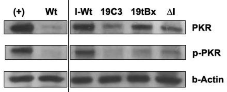
Figure 9.
Western blot analysis. Total proteins were extracted at 96 h post infection and immunoblotted for PKR, phospho-PKR, and β-actin. While untransformed cells infected with HCV (I-Wt) showed relatively high levels of both PKR and phospho-PKR, there was no significant difference evident in the concentration of PKR or phospho-PKR between cells transformed with 19C3, I19tBx, and ΔI group I introns. (+): IFN-induced positive control wild-type cells; Wt: untransformed, uninfected wild-type cells; I-Wt: HCV-infected untransformed wild-type cells, ΔI: intron lacking a trans-splicing domain.

