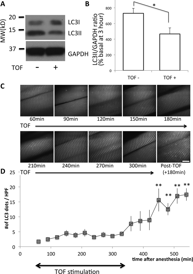
Figure 6. Electrical stimulation of the muscle reduces the amount of autophagy.
(A) Tibialis anterior muscles BL6 mice with (‘+’) or without (‘−‘) train-of-four (TOF) stimulation (continuous with 12-s interval) were harvested at 3 h after pentobarbital anesthesia and Western blots were performed for LC3 and GAPDH (as the loading control). Bars to the left of the figure represent the molecular size in kilodalton, (‘MW (kD)’). (B) Densitometric quantification of six different mice is shown as the bar graph. Relative ratio of LCII/GAPDH as compared to the basal value of freshly harvested tissue was plotted in percentage with the standard error. As compared to samples without TOF stimulation (‘TOF-‘), those with TOF (‘TOF+’) showed significantly reduced amount of LC3I and LC3II (*p<0.05, paired Student t-test, n=6). (C) Under pentobarbital anesthesia, the sternomastoid muscle of a GFP-LC3 transgenic mouse was exposed and observed under the confocal microscope up to 540 min after anesthesia. During the first 300 min (from 60min to 360min), electrical TOF stimulation was continuously applied at 12-s intervals. At 360 min, the stimulation was stopped and the muscles were observed another 180 min. (D) The numbers of autophagosome dots per high power field (HPF) were plotted against the time after anesthesia. Time-dependent increase of autophagosome formation by pentobarbital was suppressed by muscle activity both in tibialis anterior and sternomastoid muscles. Average value from n=6 is shown with the standard error bar. **p<0.01 by repeated measure ANOVA with Dunnett’s post-hoc test (n=6) as compared to the start of measurement (60min). GAPDH = Glyceraldehyde 3-phosphate dehydrogenase; MW (kD) = molecular weight in kilodalton.

