Abstract
Implant by definition “means any object or material, such as an alloplastic substance or other tissue, which is partial or completely inserted into the body for therapeutic, diagnostic, prosthetic, or experimental purpose.” The placement of a dental implant in an extraction socket at the time of extraction or explantation is known as immediate implant placement whereas delayed placement of implant signifies the implant placement in edentulous areas where healing has completed with new bone formation after the loss of tooth/teeth. Recent idea goes by “why late when it can be done immediately.” There are several advantages of immediate placement of implants, and lots of studies have been done. In this article, the advantages and disadvantages of immediate versus delayed placement of implants have been reviewed.
KEY WORDS: Extraction socket, immediate placement, implants
For years, endosseous implants have been the choice of treatment for restoring missing teeth successfully. In 1965, Branemark placed the first endosteal titanium implant successfully. Original protocols required the placement of implants into healed edentulous ridges and implant placement signifies the placement of the implant in the healed extracted socket after a minimum of 5–6 months. In 1989, Lazzara placed implants at the time of tooth extraction. Over the past few years, numerous studies have confirmed the reliability of implants placed at the time of tooth extraction.
Immediate implant placement, defined as the placement of dental implant immediately into fresh extraction socket site after tooth extraction, has been considered a predictable and acceptable procedure (Schwartz et al., 2000). The advantage of immediate implant placement into the extraction sockets over the delayed placement of implants are there is no need to wait for 4–6 months after extraction for the bone to form and crestal bone loss is found to be less in immediately placed implants rather than delayed placed implants.
Hansson et al. in 1983 and Ericsson in 2000 have found out that the decreased surgical trauma of immediate placement type will decrease the risk of bone necrosis and permit bone remodeling process to occur, that is, the healing period is rapid and allows the woven bone to be transformed into lamellar bone. The natural socket being rich in periodontal cells and matrix makes the healing faster and more predictable. Small osseous defects, which are frequently found adjacent to implants placed at the time of tooth extraction, can be grafted with autogenous bone obtained from edentulous ridges or other intraoral sites. Clinicians have also used other materials and methods to augment edentulous ridges and small bony defects adjacent to dental implants.
Diagnosis, Treatment Planning, Indications and Contraindications
Diagnosis and proper investigations are the key factors for successful treatment outcome in the case of immediate implant placements in the extraction sockets. The investigations to be done includes intra oral periapical radiographs, orthopantamographs and cone-beam computerized tomography.[1] The most important step in treatment planning is determining the prognosis of the tooth in question. Reasons for tooth extraction may include insufficient crown to root ratio, remaining root length, periodontal attachment level, furcation involvement, periodontal health status of teeth adjacent to the proposed implant site, nonrestorable caries lesions, root fractures with large endodontic posts, root resorption, and questionable teeth in need of endodontic retreatment.[2] For a successful treatment outcome, the following criteria should be fulfilled in case of immediate implant placement: (1) the patient should not have any contraindications to treatment, such as systemic diseases (e.g. diabetes), and he should not be consuming any prescription medications or recreational drugs; (2) the buccal and lingual plate of the extraction socket must be present; (3) the teeth adjacent to the extraction socket must be free of overhanging or insufficient restoration margins; (4) the patient most preferably should not use nicotine; and (5) the interradicular septum should be wide and intact following the tooth extraction. Few practitioners prefer to delay the placement of implants once periapical infections are found in the teeth to be extracted and replaced. But the study shows that a postoperative 2 years follow-up of patients who underwent immediate placement of implants following extraction of teeth which did show signs of periodontal and endodontic infection.[3,4] There should be a clear idea about the indications and contraindications for immediate implant placement. Block and Kent, 1991 summarized[4] the indications as (1) traumatic loss of teeth with a small amount of bone loss, (2) tooth lost because of gross decay without purulent exudates or cellulites, (3) inability to complete endodontic therapy, (4) presence of severe periodontal bone loss without purulent exudates (5) adequate soft tissue health to obtain primary wound closure.
The contraindications are: (1) presence of purulent exudates at the time of extraction, (2) adjacent soft tissue cellulites and granulation tissue, (3) lack of an adequate bone apical to the socket, (4) adverse location of the mandibular neurovascular bundle, maxillary sinus and nasal cavity, (5) poor anatomical configuration of remaining bone.
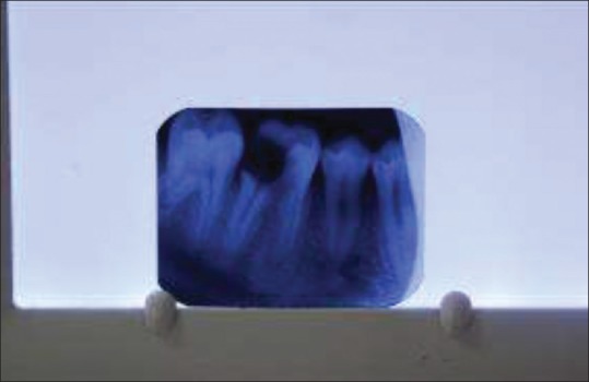
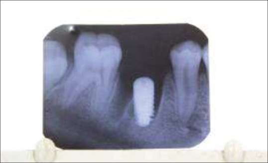
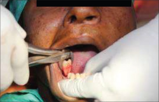
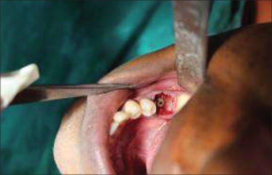
The esthetic zone is very important during treatment planning. The important things to be taken into consideration are: Scallop of periodontium, crestal bone level, smile line, morphology of gingival tissues, proposed inter implant distance, existing occlusal contact relation, and interproximal bone level.[5,6,7,8,9] Cone-beam computed tomography or radiographs should be evaluated for checking the availability of native bone and bone shape, quality, quantity, bone width, and bone height. It is recommended that a minimum of 4–5 mm of bone width should be available at the alveolar crest and at least 10 mm bone length should be available from the alveolar crest to a safe distance above the mandibular canal.[2] Sufficient distance must also be available to the maxillary sinus and the floor of the nose. A satisfactory esthetic result in the esthetic zone requires the interproximal bone height to be 5 mm or less, when measured from the contact point of the adjacent tooth. A minimum distance of 2.5–3 mm should be present between the implant and the adjacent teeth. Various surgical flap procedures can be used to gain access for tooth extraction.[10] With the experience, the surgeon can displace the marginal tissues buccal–lingually to gain access to the surgical site. The tooth must be extracted as atraumatically as possible. If any periapical infection was evident in the radiographs, then the extraction sockets must be curetted followed by immediate placement of the implants followed by fixation of the cover screw. The flap closure will be followed by sutures which may be resorbable or nonresorbable. In the case of any bony defects surrounding the implant surface, several types of bone grafts may be used either autogenous or alloplastic.[11] The immediate implant placement needs very minimal preparation since the extracted tooth socket preserves the anatomy of the tooth root which mimics the root form implants. The initial stability should be gained by placing the implant minimum 3 mm apical to the extraction site and 3 mm apical to the crestal bone.[12] The main factor determining the success of immediate placement is the initial stability of the implant. The extraction site must be evaluated to see whether it is suitable for immediate implant placement. Several publications have been there regarding the need of barrier membranes or bone grafts in the extraction sockets during placement of the immediate implants [Figures 1–4].[13]
Figure 1.

Preoperative X-ray
Figure 4.

Postoperative X-ray
Figure 2.

Atraumatic extraction
Figure 3.

Immediate implant placement
Discussion
Latest advancement in techniques and biomaterials has favored the indications for dental implant treatment. Teeth replacement using dental implants has proven to be a successful and predictable treatment procedure; different placement and loading protocols have evolved from the first protocols in order to achieve quicker and easier surgical treatment times.[14] Immediate placement of a dental implant in an extraction socket was initially described more than 30 years ago by Schulte and Heimke in 1976 The advantages of immediate implant placement techniques are reductions in the number of surgical interventions, a shorter treatment time, an ideal three-dimensional implant positioning, the presumptive preservation of alveolar bone at the side of the tooth extraction and soft tissue aesthetics. The common disadvantages of this technique are morphology of the side, the presence of periapical pathology, the absence of keratinized tissue, thin tissue biotype, and lack of complete soft tissue closure over the extraction socket.[15]
Over time, clinical experience has provided the criteria for immediate implant treatment success: Atraumatic tooth extraction, sterilization, and minimal invasive surgical approach, as well as implant primary stability.[16] The stages of extraction socket wound healing as described in the literature involve the osteophyllic, osteoconductive, and the osteoadaptive stages. The maximum blood supply for the cortical bone is from the periosteal blood supply. In 2000, Misch and Judy found out that if the buccal or facial cortical plate is lost during extraction it leads to reduced bone height and thickness for implant placement after the socket heals. In 2011, Khalid S. Hassan and Adel S. Alagl found out that there is a 25% decrease in the width of the alveolar bone during the 1st year following extraction of teeth and an average 4 mm decrease in height during the 1st year following multiple extractions. Even Carlson and Persson (1967) and Misch (1999) have observed a 40%-60% decrease in alveolar bone width after the first 2–3 years postextraction, and Christensen (1996) reported that there has been an annual bone resorption rate of at least 0.5–1% during the rest of a patient's life. There have been several evidence of immediate implant placement after tooth extraction helping to preserve the alveolar bone height and width with reduced marginal bone loss in previous studies. Osseointegration takes place with the implant surface while on the course of wound healing. Becker et al. found out that there has been 93.3% of 5-year success rate when the immediately placed implants are augmented with barrier membrane with an insignificant amount of crestal bone loss. In 2000, Misch and Judy found out that in case of delayed implant placements if the buccal or facial cortical plate is lost during extraction it leads to reduced bone height and thickness for implant placement after the socket heals thereby bone height and width are reduced forcing the operator to compromise with the size and width of the delayed implant to be placement. In a similar prospective study, A mean loss in facial crestal bone height of 0.8 mm after 6 months of submerged healing following immediate implant placement in 20 patients was reported by Covani and coworkers in which 38 implant sites included maxillary and mandibular anterior and premolar sites. 38% of the sites showed no change, 47% had between 0 mm and 1 mm of loss, and 15% had between 1 and 2 mm of loss but this amount of bone loss can be considered insignificant when compared to the bone loss after extraction of teeth without any immediate implant placement.
Very minimal preparation is needed while placing the immediate implants since the extracted tooth socket preserves the anatomy of the tooth root which mimics the root form implants. To gain the initial stability, the implant should be placed minimum 3 mm apical to the extraction site and 3 mm apical to the crestal bone. The initial stability of the implant is a main factor determining the success of immediate placement. The extraction site must be evaluated to see whether it is suitable for immediate implant placement. The stability of the implant may be checked with resonance frequency analysis. Several publications have been there regarding the need of barrier membranes or bone grafts in the extraction sockets during placement of the immediate implants.[17] Studies have revealed that crestal bone loss is evident in both delayed and immediate implant placements. But in case of immediate implant placement, the crestal bone loss was found to be less. The immediate implant placements with bone grafts to cover the gap between the socket walls and the implants showed better results with minimal crestal bone loss. Several methods of bone grafts were used, but the author himself have experienced that autogenous bone grafts obtained from patient's interradicular septal bone, interdental bone, and buccal cortical plate. The reason for choosing autogenous bone grafts was autogenous bone grafts have osteogenic as well as osteoinductive properties as well as it is obtained from patients own body and economic and reliable. The results were satisfactory with an insignificant amount of crestal bone loss surrounding the implants. Despite many articles previously described limited marginal bone level or gain in immediate implant therapy, caution is needed because few of these studies report radiographic outcomes.
Conclusion
Even though several studies prove the advantages of immediate implant placement over delayed implant placement case selection, proper diagnosis, treatment planning, and initial stability are very important factors for the success of an immediate implant. Several studies even show that there is not much of marginal bone loss difference between immediate and delayed implant placement. Nevertheless, beyond doubt, immediate implant placement saves time and needs less invasive surgical procedures with considerably very good esthetic outcome when final restorations are placed. Author's personal opinion is immediate implant placement in the extracted tooth socket is a well-accepted and practiced treatment worldwide which gives excellent success rates.
Footnotes
Source of Support: Nil
Conflict of Interest: None declared.
References
- 1.Becker W, Goldstein M. Immediate implant placement: Treatment planning and surgical steps for successful outcome. Periodontol 2000. 2008;47:79–89. doi: 10.1111/j.1600-0757.2007.00242.x. [DOI] [PubMed] [Google Scholar]
- 2.Becker W, Becker BE, Ricci A, Bahat O, Rosenberg E, Rose LF, et al. A prospective multicenter clinical trial comparing one- and two-stage titanium screw-shaped fixtures with one-stage plasma-sprayed solid-screw fixtures. Clin Implant Dent Relat Res. 2000;2:159–65. doi: 10.1111/j.1708-8208.2000.tb00007.x. [DOI] [PubMed] [Google Scholar]
- 3.Novaes AB, Jr, Novaes AB. Immediate implants placed into infected sites: A clinical report. Int J Oral Maxillofac Implants. 1995;10:609–13. [PubMed] [Google Scholar]
- 4.Villa R, Rangert B. Early loading of interforaminal implants immediately installed after extraction of teeth presenting endodontic and periodontal lesions. Clin Implant Dent Relat Res. 2005;7(Suppl 1):S28–35. doi: 10.1111/j.1708-8208.2005.tb00072.x. [DOI] [PubMed] [Google Scholar]
- 5.Ochsenbein C, Ross S. A reevaluation of osseous surgery. Dent Clin North Am. 1969;13:87–102. [PubMed] [Google Scholar]
- 6.Spear FM, Mathews DM, Kokich VG. Interdisciplinary management of single-tooth implants. Semin Orthod. 1997;3:45–72. doi: 10.1016/s1073-8746(97)80039-4. [DOI] [PubMed] [Google Scholar]
- 7.Becker W, Ochsenbein C, Tibbetts L, Becker BE. Alveolar bone anatomic profiles as measured from dry skulls. Clinical ramifications. J Clin Periodontol. 1997;24:727–31. doi: 10.1111/j.1600-051x.1997.tb00189.x. [DOI] [PubMed] [Google Scholar]
- 8.Kan JY, Rungcharassaeng K, Umezu K, Kois JC. Dimensions of peri-implant mucosa: An evaluation of maxillary anterior single implants in humans. J Periodontol. 2003;74:557–62. doi: 10.1902/jop.2003.74.4.557. [DOI] [PubMed] [Google Scholar]
- 9.Kois JC. Predictable single-tooth peri-implant esthetics: Five diagnostic keys. Compend Contin Educ Dent. 2004;25:895–905. quiz 905. [PubMed] [Google Scholar]
- 10.Becker W, Becker BE. Flap designs for minimization of recession adjacent to maxillary anterior implant sites: A clinical study. Int J Oral Maxillofac Implants. 1996;11:46–54. [PubMed] [Google Scholar]
- 11.Becker W. Treatment of small defects adjacent to oral implants with various biomaterials. Periodontol 2000. 2003;33:26–35. doi: 10.1046/j.0906-6713.2003.03303.x. [DOI] [PubMed] [Google Scholar]
- 12.Schropp L, Kostopoulos L, Wenzel A. Bone healing following immediate versus delayed placement of titanium implants into extraction sockets: A prospective clinical study. Int J Oral Maxillofac Implants. 2003;18:189–99. [PubMed] [Google Scholar]
- 13.Langer B, Sullivan DY. Osseointegration: Its impact on the interrelationship of periodontics and restorative dentistry. Part 3. Periodontal prosthesis redefined. J Periodontics Restorative Dent. 1989;9:240–61. [PubMed] [Google Scholar]
- 14.Esposito M, Grusovin MG, Coulthard P, Worthington HV. The efficacy of various bone augmentation procedures for dental implants: A Cochrane systematic review of randomized controlled clinical trials. Int J Oral Maxillofac Implants. 2006;21:696–710. [PubMed] [Google Scholar]
- 15.Chen ST, Wilson TG, Jr, Hammerle CH. Immediate or early placement of im-plants following tooth extraction: Review of biologic basis, clinical procedures, and out-comes. Int J Oral Maxillofac Implants. 2004;19(Suppl):12–25. [PubMed] [Google Scholar]
- 16.Siegenthaler DW, Jung RE, Holderegger C, Roos M, Hämmerle CH. Replacement of teeth exhibiting periapical pathology by immediate implants: A prospective, controlled clinical trial. Clin Oral Implants Res. 2007;18:727–37. doi: 10.1111/j.1600-0501.2007.01411.x. [DOI] [PubMed] [Google Scholar]
- 17.Ribeiro FS, Pontes AE, Marcantonio E, Piattelli A, Neto RJ, Marcantonio E., Jr Success rate of immediate nonfunctional loaded single-tooth implants: Immediate versus delayed implantation. Implant Dent. 2008;17:109–17. doi: 10.1097/ID.0b013e318166cb84. [DOI] [PubMed] [Google Scholar]


