Abstract
Lupus vulgaris (LV) is a common form of cutaneous tuberculosis in India, mostly involving the lower half of the body. Facial involvement is uncommon. Untreated disease may lead to significant morbidity due to atrophic scarring, mutilation, and deformity. We report a case of multi-focal LV in a 10-year-old boy affecting the nose and cheek that resulted in perforation of the nasal septum, a rarely reported complication.
Keywords: Lupus vulgaris, nasal perforation, childhood
INTRODUCTION
Head and neck region is frequently affected by granulomatous diseases that may be infective or noninfective in origin. Tuberculosis (TB) is the major infective cause of such disorders in India influenced by poverty, overcrowding, malnutrition and an upsurge in HIV. Cutaneous TB, an extra-pulmonary form, accounts for 1.5% cases of all extra-pulmonary TB. It accounts for about 0.1–0.9% of the total dermatology out-patients with the childhood prevalence varying from 18.7% to 53.9% in different studies.[1] Lupus vulgaris (LV), the most common clinical variant of cutaneous TB, commonly involves the lower extremities and the gluteal region in children of Indian subcontinent whereas involvement of the head and neck region is more common in Western countries. In the current era of extensive use of the anti-tubercular chemotherapy, LV involving the nasal region and leading to complications like nasal perforation is very rare.[2]
CASE REPORT
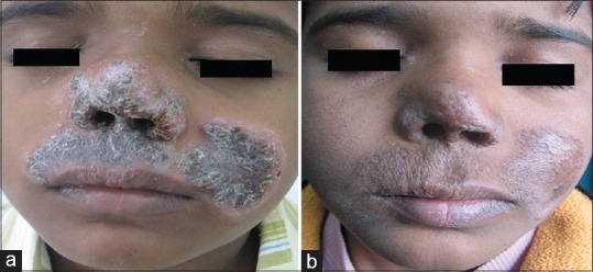
A 10-year-old boy presented with slowly progressive, reddish-brown scaly plaques involving the upper lip, nose, left cheek and thigh of one year duration. There was no preceding history of trauma. He denied having fever, weight loss or cough. Personal and family history of TB was absent. He complained of mild difficulty in nasal breathing since six months. Physical examination was normal, and he had been bacillus Calmette-Guérin vaccinated at birth. Cutaneous examination revealed multiple erythematous to hyperpigmented, thick, crusted plaques over the face extending from the left cheek to the upper lip, philtrum of nose, nasal alae, columella, nasal tip and mucosal aspect of the nose [Figure 1a]. Intranasal examination showed extension of thickly crusted plaque with consequent narrowing of the nasal cavity. Another plaque of similar morphology measuring approximately 4 cm × 4 cm in size was present over the left cheek [Figure 1a]; another on the left thigh revealed apple jelly nodules under diascopy. Patient had matted, and nontender left submandibular lymphadenopathy. Clinically, the differential diagnoses of cutaneous leishmaniasis, leprosy, Wegener's granulomatosis and midline nasal granuloma were considered, and patient was investigated.
Figure 1.

(a) Multiple papulonodular lesions coalescing to from an erythematous to hyperpigmented plaque over face. (b) Posttreatment-resolution of lesions with scarring after 8 weeks of anti-tuberculosis therapy
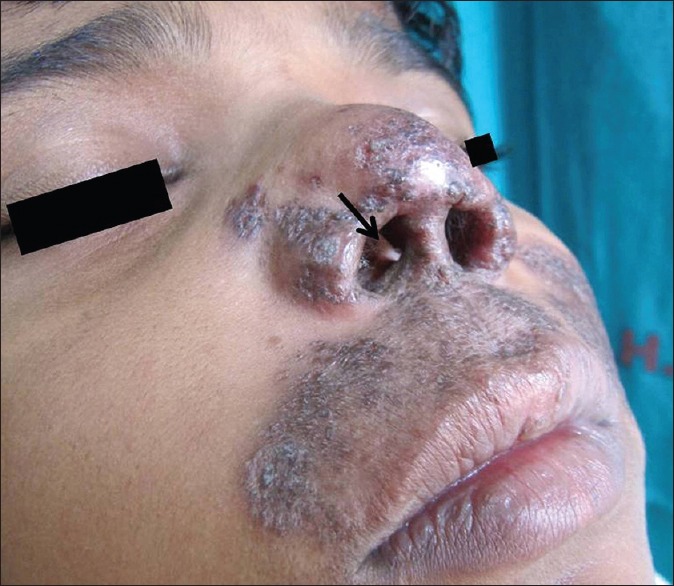
Hematological investigations were normal except for a raised erythrocyte sedimentation rate of 39 mm in the first hour. Mantoux test was positive (15 mm with central vesiculation). Chest radiograph and abdominal ultrasound were normal, and serology for HIV was nonreactive. Fine-needle aspiration cytology from the submandibular lymph nodes demonstrated tuberculoid granulomas with acid fast bacilli (AFB). Histopathology from the cutaneous plaques was consistent with LV with presence of noncaseating tuberculoid granuloma and Langhan's giant cells. Zeihl–Neelsen stain was negative for AFB [Figure 2].
Figure 2.

A large oval perforation (black arrow) in the nasal septum
Patient was started on category I anti-tubercular therapy (ATT) with initial two months of intensive phase with four drugs in standard doses (isoniazid, rifampicin, ethambutol and pyrazinamide) followed by four months of continuation phase with two drugs, isoniazid and rifampicin (2 HRZE/4HR). After eight weeks, he showed remarkable improvement and significant resolution of his lesions with scarring [Figure 1b]. At this point a large, oval perforation in the nasal septum was observed with positive trans-illumination [Figure 2]. Patient has been assigned for septoplasty.
DISCUSSION
Lupus vulgaris is a chronic, indolent paucibacillary form of cutaneous TB seen in patients with moderate to high immunity that classically presents as papulonodular lesions coalescing to form plaques with areas of progression as well as areas of scarring. The destructive nature of the disease with associated scarring adds to the morbidity. The clinical variants of the disease include classic, hypertrophic, ulcerative, atrophic and mutilating type. Hypertrophic type is the most common, whereas ulcerative and atrophic forms are least common in children.[1]
Involvement of the nose is rare and has been reported on limited occasions.[2] In addition to hematogenous or lymphatic spread from a distant focus, direct inoculation due to trauma to the nose caused by habitual picking forms an important source of infection. Sometimes the lesion may extend from surrounding skin to involve the nasal mucosa leading to nasal obstruction and discharge. Extension to involve anterior nasal cartilage of the nasal septum and anterior end of turbinates has been reported.[3] The resultant atrophic scarring following ATT may lead to extensive deformity and mutilation along with fibrotic contractures.[4]
The diagnosis of LV requires a strong correlation of clinical and laboratory findings. Epitheloid granulomas and Langhan's giant cells with or without caseous necrosis are the most common features on histopathology. Being a paucibacillary disease, the detection of AFB by conventional culture is difficult and identification by polymerase chain reaction is recommended.[1]
A retrospective review of 76 patients with nasal septal perforation reported infectious etiology to be the least common (3%) cause.[5] Cutaneous TB involving nose being a rare entity requires a high index of suspicion for diagnosis, especially in a country endemic for TB. Early diagnosis of LV is of utmost importance owing to the morbidity associated with scarring and mutilation which may result in gross disfigurement.
Footnotes
Source of Support: Nil
Conflict of Interest: None declared
REFERENCES
- 1.Singal A, Sonthalia S. Cutaneous tuberculosis in children: The Indian perspective. Indian J Dermatol Venereol Leprol. 2010;76:494–503. doi: 10.4103/0378-6323.69060. [DOI] [PubMed] [Google Scholar]
- 2.Garg A, Wadhera R, Gulati SP, Singh J. Lupus vulgaris of external nose with septal perforation – A rarity in antibiotic era. Indian J Tuberc. 2010;57:157–9. [PubMed] [Google Scholar]
- 3.Matsumoto FY, Clivati Brandt HR, Costa Martins JE, Rivitti EA, Romiti R. Nasoseptal perforation secondary to lupus vulgaris. J Dermatol. 2007;34:493–4. doi: 10.1111/j.1346-8138.2007.00318.x. [DOI] [PubMed] [Google Scholar]
- 4.Varshney S, Gupta P, Bist SS, Singh RK, Gupta N. Lupus vulgaris of nose. J Med Educ Res. 2009;11:91–3. [Google Scholar]
- 5.Diamantopoulos II, Jones NS. The investigation of nasal septal perforations and ulcers. J Laryngol Otol. 2001;115:541–4. doi: 10.1258/0022215011908441. [DOI] [PubMed] [Google Scholar]


