Abstract
Combined tumors of malignant melanoma (MM) and squamous cell carcinoma (SCC) are extremely rare and have unknown biological potential. Different theories of their development, including collision and dual/divergent differentiation, are proposed. Although some observations suggest an indolent course for such tumors, a case of MM metastasis was reported in a patient who initially presented with a combined MM-SCC tumor. Re-excisions of such tumors may show MM in situ and should be treated accordingly. Herein we present another case of a combined MM-SCC tumor in a 78 male patient with lentigo maligna seen after complete re-excision of the tumor.
Keywords: Melanoma, skin neoplasms, squamous cell carcinoma
INTRODUCTION
Collision tumors and tumors with divergent differentiation are rarely seen by dermatopathologists. In collision tumors, two morphologically distinct neoplasms arise in close proximity. In contrast, tumors with divergent differentiation are biphasic in nature with distinct cell types mingling within the same stroma. Combined tumors of malignant melanoma (MM) and squamous cell carcinoma (SCC) or basal cell carcinoma are sometimes referred to as “basomelanocytic” and “squamomelanocytic” tumors, respectively. Herein, we present a unique, biphasic tumor with features of MM and SCC in an elderly, male patient.
CASE REPORT
A 78-year-old male patient presented with a keratotic, cystic nodule of 1-month duration on the left forearm. A shave biopsy was performed and upon histopathological examination, a well-circumscribed, dermally based tumor was noted [Figure 1]. The tumor was pigmented with atypical keratinization and showed two morphologically distinct cell types [Figures 2 and 3]. The first was composed of large, eosinophilic cells that showed signs of keratinization [Figure 4]. The second population was composed of small, pigmented, spindle and epithelioid cells in cords and nests [Figure 4]. Both cell types showed prominent degrees of atypia - including irregular and enlarged nuclei; nuclear hyperchromasia; multiple mitotic figures; and necrotic cells. The squamous and melanocytic components were admixed within the same tumor stroma. No melanoma in situ (MIS) was seen on primary excision.
Figure 1.
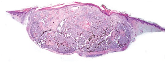
Well-circumscribed dermal tumor on sun damaged skin. H and E stained sections, ×20
Figure 2.
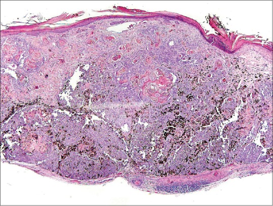
Tumor with pigment and keratin pearls. H and E stained sections, ×40
Figure 3.
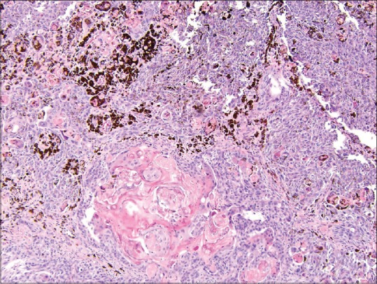
Tumor with pigment and atypical keratinization. H and E stained sections, ×100
Figure 4.
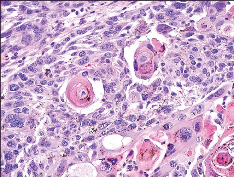
Two populations with admixed cells: Larger keratinizing cells and smaller spindled and epithelioid cells. H and E stained sections, ×400
Cytokeratin immunohistochemical stains AE1/3 (pan-cytokeratin), CK903, and CAM5.2 labeled the pleomorphic keratinocytes, whereas S-100, Melan-A, and HMB45 stained the atypical melanocytes [Figures 5 and 6]. Dual staining of both melanocytic and cytokeratin staining was not appreciated within individual tumor cells. The presence of markers for both types of cells indicated a diagnosis of squamomelanocytic tumor (melanocarcinoma).
Figure 5.
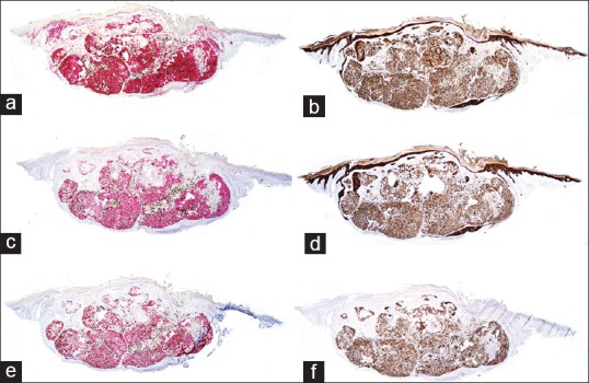
Immunohistochemical profile of the tumor. (a) Positive staining with S100, ×20. (b) Positive staining with pan-cytokeratin, ×20. (c) Positive staining with HMB45, ×20. (d) Positive staining with CAM 5.2, ×20. (e) Positive staining with MelanA, ×20. (f) Positive staining with CK903, ×20
Figure 6.
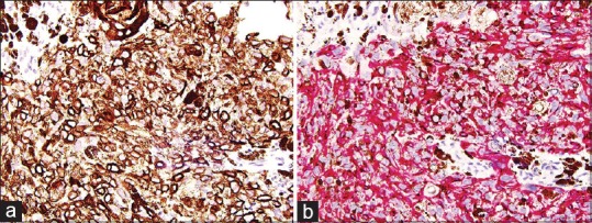
(a) CK903 staining large epithelioid cells. CK903 stained sections, ×400. (b) MelanA staining spindle cells. MelanA stained sections, ×400
Upon complete re-excision, the specimen showed residual SCC [Figure 7a] with adjacent single melanocytic proliferation of irregularly spaced and scattered melanocytes that was interpreted as MIS [Figure 7b].
Figure 7.
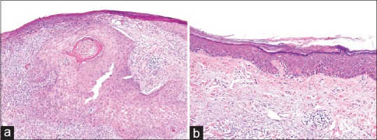
(a) Residual squamous cell carcinoma seen in the re-excision specimen, H and E stained sections, ×100. (b) Single cell melanocytic proliferation with irregularly spaced and scattered melanocytes, which may be interpreted as MIS, H and E stained sections, ×200
DISCUSSION
A variety of combined tumors involving keratinocytes and melanocytes have been described in dermatopathology.[1] These tumors have been given the designation of “basomelanocytic” and “squamomelanocytic” tumors depending on the composition. The term “melanocarcinoma” has also been used to describe these tumors. However, that term has received criticism as it was originally used as a term for melanoma.
Several theories on the development of these tumors have been proposed. They may be broken down into three distinct categories: Collision between two adjacent neoplastic process, dual differentiation of a single neoplastic cell line, or divergent differentiation of pluripotent stem cells.
The first category would be explained by colonization of a squamous carcinoma by an adjacent melanoma. A variant of this hypothesis, and one favored by Miteva et al., suggests that paracrine stimulation of cells through a tumor cytokine milieu could “induce” a secondary neoplasm within the confines of a primary tumor.[2]
Dual differentiation of a single cell line would result in a monoclonal cell population expressing both melanocytic as well as keratinocytic cell markers. This would explain the aberrant expression cytokeratin seen in <10% of MM cells.[3] In addition, Rosen reported a case of a squamomelanocytic tumor in which cells expressed both S100 and keratin.[4] In his study, he performed an ultra-structural analysis, which showed the presence of both tonofilaments and melanosomes in some tumor cells. We did not observe dual staining in our case. Dual staining with many of the melanocytic and keratinocytic markers can be quite difficult to interpret due to overlapping cytoplasmic and nuclear staining patterns.
We favor the mechanism of divergent differentiation of pluripotent stem cells. The resultant tumor would therefore represent a variant of a single tumor such as a MM with divergent staining patterns. Pool et al. was the first to describe a series of four squamomelanocytic tumors with melanocytes and keratinocytes intermingled together.[5] They were well-circumscribed nodules: One connected with the epidermis and the other three were dermal tumors. Lentigo maligna was also present in one case. The tumor cells in the Pool et al. series showed divergent differentiation with immunostains. Similar findings were reported by Satter et al.[1] The ductal structures in squamomelanocytic tumor have also been described by several authors.[6,7] A case report by Pouryazdanparast et al. showed residual MIS at wide local excision.[8] Amerio et al. described a case of squamomelanocytic tumor, which was treated as MM with the depth 4.3 mm, followed by excision and sentinel lymph node biopsy.[9,10] A metastasis of MM was seen in the lymph node.
The rarity of the squamomelanocytic tumor limits what is known about its incidence and prognosis. Thus, it has often been referred to as a “tumor having the uncertain biological potential.[11] It was considered to have a more indolent course by some authors who observed their patients in the ensuing 9 years post-diagnosis.[5] This may be because the melanoma remains confined to the epithelium of the invasive carcinoma. Like prior reported cases, a MM in situ was discovered in our patient's re-excision. Until more studies confirm the origin of squamomelanocytic tumors and elucidate its origin and clinical behavior, such tumors should be treated and measured as MMs and the prognosis regarded as uncertain. Multiple step sections and thorough examination of the re-excision specimen should be performed to ensure the complete elimination of the tumor.
Footnotes
Source of Support: Nil
Conflict of Interest: None declared
REFERENCES
- 1.Satter EK, Metcalf J, Lountzis N, Elston DM. Tumors composed of malignant epithelial and melanocytic populations: A case series and review of the literature. J Cutan Pathol. 2009;36:211–9. doi: 10.1111/j.1600-0560.2008.01000.x. [DOI] [PubMed] [Google Scholar]
- 2.Miteva M, Herschthal D, Ricotti C, Kerl H, Romanelli P. A rare case of a cutaneous squamomelanocytic tumor: Revisiting the histogenesis of combined neoplasms. Am J Dermatopathol. 2009;31:599–603. doi: 10.1097/DAD.0b013e3181a88116. [DOI] [PubMed] [Google Scholar]
- 3.Ben-Izhak O, Stark P, Levy R, Bergman R, Lichtig C. Epithelial markers in malignant melanoma. A study of primary lesions and their metastases. Am J Dermatopathol. 1994;16:241–6. doi: 10.1097/00000372-199406000-00003. [DOI] [PubMed] [Google Scholar]
- 4.Rosen LB, Williams WD, Benson J, Rywlin AM. A malignant neoplasm with features of both squamous cell carcinoma and malignant melanoma. Am J Dermatopathol. 1984;6(Suppl):213–9. [PubMed] [Google Scholar]
- 5.Pool SE, Manieei F, Clark WH, Jr, Harrist TJ. Dermal squamo-melanocytic tumor: A unique biphenotypic neoplasm of uncertain biological potential. Hum Pathol. 1999;30:525–9. doi: 10.1016/s0046-8177(99)90195-8. [DOI] [PubMed] [Google Scholar]
- 6.Rongioletti F, Baldari M, Carli C, Fiocca R. Squamomelanocytic tumor: A new case of a unique biphenotypic neoplasm of uncertain biological potential. J Cutan Pathol. 2009;36:477–81. doi: 10.1111/j.1600-0560.2008.01061.x. [DOI] [PubMed] [Google Scholar]
- 7.Wen YH, Giashuddin S, Shapiro RL, Velazquez E, Melamed J. Unusual occurrence of a melanoma with intermixed epithelial component: A true melanocarcinoma?: Case report and review of epithelial differentiation in melanoma by light microscopy and immunohistochemistry. Am J Dermatopathol. 2007;29:395–9. doi: 10.1097/DAD.0b013e31812f5235. [DOI] [PubMed] [Google Scholar]
- 8.Pouryazdanparast P, Yu L, Johnson T, Fullen D. An unusual squamo-melanocytic tumor of uncertain biologic behavior: A variant of melanoma? Am J Dermatopathol. 2009;31:457–61. doi: 10.1097/DAD.0b013e318182c7dc. [DOI] [PubMed] [Google Scholar]
- 9.Amerio P, Carbone A, Auriemma M, Tracanna M, Di Rollo D, Angelucci D. Metastasizing dermal squamomelanocytic tumour: More evidences. J Eur Acad Dermatol Venereol. 2011;25:734–5. doi: 10.1111/j.1468-3083.2011.03999.x. [DOI] [PubMed] [Google Scholar]
- 10.Amerio P, Carbone A, Auriemma M, Tracanna M, Di Rollo D, Angelucci D. Metastasizing dermal squamomelanocytic tumour. J Eur Acad Dermatol Venereol. 2011;25:489–91. doi: 10.1111/j.1468-3083.2010.03742.x. [DOI] [PubMed] [Google Scholar]
- 11.Leonard N, Wilson N, Calonje JE. Squamomelanocytic tumor: An unusual and distinctive entity of uncertain biological potential. Am J Dermatopathol. 2009;31:495–8. doi: 10.1097/DAD.0b013e31819b4077. [DOI] [PubMed] [Google Scholar]


