Dermoscopy of normal skin reveals normal reticular pattern of pigment network which consists of homogeneous pigmented lines corresponding to rete network and pale areas in between these lines. This normal reticulate pigmentary network is reversed in some cases of evolving lesions of vitiligo.
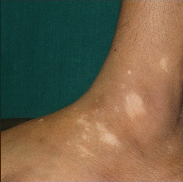
Dermoscopy was done on evolving lesions of vitiligo [Figure 1] in 2 of our patients with established vitiligo. A triple light source dermoscope (Ultracam TLS; Dermaindia, Tamil Nadu, India) having white light, polarised light and ultraviolet light was used.
Figure 1.

Evolving lesions of vitiligo: Clinical image showing evolving lesions of vitiligo
Histopathological examination was done for correlation.
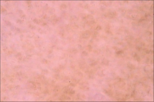
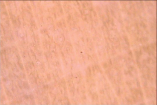
On polarized light dermoscopy of the evolving lesion of vitiligo, there was net like pigmentary network formed by homogeneous white lines and pigmented areas in between these lines [Figure 2] which was opposite to that of normal reticulate pigmentary network [Figure 3] appearing as reversed pigmentary network.
Figure 2.

Evolving lesions of vitiligo: Reversed pigmentary network
Figure 3.

Evolving lesions of vitiligo: Normal reticulate pigmentary network
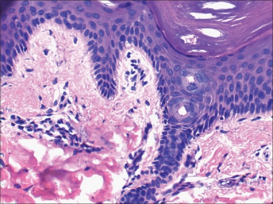
Histopathological examination of lesions which showed reversed pigmentary pattern was done which revealed patchy reduction of melanocytes with sparse perivascular and junctional lymphocytic infiltrate compatible with the diagnosis of vitiligo [Figure 4].
Figure 4.

Evolving lesions of vitiligo: Reduced melanocytes with sparse perivascular lymphocytic infiltrate (H and E, ×40)
Normal reticulate pattern of pigmentation seen over normal skin corresponds to the pigmentation of the keratinocytes along the rete ridges while the pale area in between corresponds to the papillary dermis.[1] In vitiligo there is gradual loss of melanocytes and melanin due to which light directly passes into the dermis without being reflected by the melanocytes and melanin. This leads to a window through which light passes into the dermis and is reflected by dermal collagen. In initial stages of evolving vitiligo this leads to area of relative hyperpigmentation produced by the pale area corresponding to papillary dermis in normal reticulate pattern of pigmentation. This leads to the appearance of “reversed pigmentary network pattern” in evolving vitiligo.
The dermoscopic finding of “reversed pigmentary network pattern” has not been mentioned previously in vitiligo. Reversed pigmentary network pattern has been described previously in case of melanoma and melanocytic nevi.[2] The fact that “reversed pigmentary network pattern” exists in vitiligo may help us to identify vilitigo at an early stage.
REFERENCES
- 1.Haldar SS, Nischal KC, Khopkar US. Dermoscopy: Applications and patterns in diseases of the brown skin. In: Uday K, editor. Dermoscopy and Trichoscopy in Diseases of the Brown Skin: Atlas and Short Text. New Delhi, India: Jaypee Brothers Medical Publishers; 2012. p. 14. [Google Scholar]
- 2.Basak PY, Hofmann-Wellenhof R, Massone C. Three cases of reverse pigment network on dermatoscopy with three distinctive histopathologic diagnoses. Dermatol Surg. 2013;39:818–20. doi: 10.1111/dsu.12186. [DOI] [PubMed] [Google Scholar]


