Abstract
Sepsis is a severe illness in which the bloodstream is overwhelmed by bacteria. Despite effective antibiotic treatment, the mortality of septic shock remains high. In this study, we examined a potential usage of paeoniflorin, anti-inflammatory component for the treatment of sepsis. We established an inflammatory cell line by stimulating human THP-1 cell line with bacterial lipoprotein (BLP), which resulted in an activation of nuclear factor κB (NF-κB) p65 dependent-signal pathway, and in consequence, an increase in tumor necrosis factor α (TNF-α) and interleukin (IL)-6 expression. With this model, we studied the effect of paeoniflorin on the expression of NF-κB and Toll-like receptor 2 (TLR2) mediated signal transduction. Our data indicated that paeoniflorin directly inhibited activation of NF-κB p65, thereby reduced the expression of TNF-α and IL-6 in the BLP stimulated THP-1 cells. Paeoniflorin was also found to inhibit IκB phosphorylation and degradation. However, no significant differences in TLR2 and myeloid differentiation factor 88 (MyD88) expression were observed; therefore, these signaling molecules may not have much anti-inflammatory effect in our cellular model. As such, our current study provided a molecular base for the potential use of paeoniflorin in therapeutic treatment of sepsis induced by bacterial lipoprotein.
Keywords: paeoniflorin, bacterial lipoprotein, Toll-like receptor, TnF-α
Introduction
Sepsis arises from complex interactions between the infecting microorganism and host-immune inflammation [1]. Bacterial pathogens and their products trigger the inflammatory response by transcriptional activation of inflammatory genes, leading to the release of large numbers of inflammatory mediators. Although these mediators are important for host defense against invading bacteria, they damage cells and tissues. When their production is uncontrolled and/or excessive, mediators may cause multiple organ dysfunction syndrome (MODS). Despite significant advances in antibiotic and supportive therapies, sepsis-induced MODS remains the most common cause of death in critically ill patients admitted to intensive care units. Thus, MODS represents a significant challenge to physicians worldwide [2].
Bacterial lipoprotein (BLP) from the cell wall of both Gram-positive and Gram-negative bacteria, characterized by a unique NH2-terminal lipoamino acid, N-acyl-S-diacylglycerol cysteine, is best known as an activating component for monocytes and macrophages, inducing the production of inflammatory cytokines [3]. Bacterial cell wall components are recognized by pattern recognition receptors on immune cells, especially, the Toll-like receptor (TLR) family [4]. Human TLR2 transmits intracellular signals in response to BLP, which activates downstream signaling molecules, including myeloid differentiation factor 88 (MyD88) and nuclear factor (NF)-κB. Nuclear factor κB is a key transcription factor for regulating the immune response and it mediates pro-inflammatory cytokine production [5].
Paeoniflorin (MW 480.45 Da), a characteristic and chief bioactive component from the Paeonia lactiflora pall root, has been reported to have many pharmacological effects including antihyperglycemic [6], anti-inflammatory [7], hepatoprotective [8], cognition-enhancing [9], neuroprotective [10, 11], and anti-cancer [12] properties. Recent studies [13] suggest that paeoniflorin reduces mortality in a rat model of cecal ligation and puncture and lipopolysaccharide (LPS) administration (intraperitoneal injection – ip). It also reduces cytokines and myeloperoxidase in the lung, liver and small intestine. Paeoniflorin's anti-inflammatory activity may inhibit NF-κB pathway activation by inhibiting IκB kinase activity.
In the present study, we have investigated the effects of paeoniflorin on NF-κB and TLR2 signal transduction pathways and apoptosis in BLP-induced human THP-1 cells.
Material and methods
Reagents and antibodies
Paeoniflorin was purchased from the National Institute for the Control of Pharmaceutical and Biological Products (Nanjing, China) with a purity of 99% as determined by high-performance liquid chromatography (HPLC). It was dissolved in RPMI 1640 medium, and then diluted as needed in cell culture medium. Bacterial lipoprotein, a synthetic BLP (Pam3Cys-Ser-(Lys) 4∙3HCl) was purchased from Alexis Biochemicals (San Diego, CA). Polyclonal antibodies (pAb) for TLR2 NF-κB p65 and IκB, and histone H3 (3H1) rabbit monoclonal antibody were obtained from Cell Signaling Technology (Beverly, MA). Anti-MyD88 pAb was purchased from eBioscience (San Diego, CA). β-Actin antibody was obtained from Boster (Wuhan, China).
Cell lines and culture
The human monocyte cell line, THP-1 (the National Institute of Cells, Shanghai, China) was maintained in culture in RPMI 1640 medium supplemented with 10% heat inactivated fetal bovine serum, penicillin (100 units/ml), streptomycin sulfate (100 μg/ml), and glutamine (2.0 M) at 37°C, in a humidified 5% CO2 atmosphere.
Cytokine analysis
THP-1 cells (2 × 10⁵ cells/well) were incubated, in 24-well plates. Control cells were treated with incomplete RPMI 1640 medium. Treated cells were given 1,000 ng/ml of BLP for 1, 2, 4, 6, 8, 12, 24, 36 and 48 h. Paeoniflorin at various concentrations (10−8 to 10−4 M) incubated with 1,000 ng/ml BLP was allowed to stimulate THP-1 cells for 4 and 48 h (n = 4, performed in duplicate). After incubation, the cell suspension was collected and immediately centrifuged at 4°C for 10 min at 1,500 rpm. The supernatant was collected and stored at –80°C for tumor necrosis factor α (TNF-α) and IL-6 measurement using a commercially available ELISA (Sizhengbo Biology Technology Company, Beijing, China) according to the manufacturer's instructions.
Western blot
THP-1 cells (2 × 10⁶) were cultured with 100 ng/ml BLP or 10−4 M paeoniflorin for 20 min, or they were not treated. After washing twice with cold PBS, cells were lysed in lysis buffer containing 1.0% Nonidet P-40, 150 mM NaCl, 10 mM Tris-Cl (pH 7.5), and 1 mM EDTA. The supernatant was transferred (cytoplasmic fraction) into a pre-chilled microcentrifuge tube and stored at –80°C until use. The nuclear pellet was resuspended in lysis buffer (DTT, lysis buffer, protease inhibitor cocktail). Then, the supernatant (nuclear fraction) was prepared as described previously. Protein concentrations of the extracts were measured using a Bicinchoninic Acid (BCA) Protein Assay. Equal amounts of protein were subjected to SDSPAGE, and then transferred onto immobilon-P membranes (Millipore, Bedford, MA). The membranes were blocked for 1 h at room temperature with 5% nonfat milk and probed overnight (maintained at 4°C) with the appropriate primary antibodies under conditions recommended by the manufacturer (anti-TLR2, anti-MyD88, anti-NF-κB p65, anti-IκB, phosphorylated specific anti-IκB), respectively. Blots were then incubated with the appropriate horseradish peroxidase-conjugated secondary antibodies at room temperature for 2 h, developed with SuperSignal Chemiluminescent substrate (Pierce Chemical, Rockford, IL) and exposed to X-Omat BT films (Kodak). Quantification of the developed target bands was determined by densitometric analysis.
Flow cytometric analysis
To assay the effect of paeoniflorin on cell apoptosis, THP-1 cells were exposed to different drugs for 24 h. Then, cultured cells were trypsinized and 1 × 10⁶ cells were collected by centrifugation. Cells were washed with PBS and centrifuged at 1,000 rpm for 5 min to obtain a cell pellet. The supernatant was discarded and the cell pellet was resuspended in stain containing Annexin V-FITC and propidium iodide (PI). This was mixed well and incubated in the dark for 10-15 min at 15-25°C. Cells were analyzed by flow cytometry (excitation = 488 nm; emission = 530 nm) for Annexin V-FITC detection (FL1 channel). A filter (> 600 nm) was used for PI detection (FL2 channel). Electronic compensation of the instrument was required to exclude overlapping of the two emission spectra.
Statistical analysis
All values were expressed as mean ± SD. Groups were compared by one-way ANOVA. Inter-group differences were compared using an Independent-Samples t-test. P < 0.05 was considered to be statistically significant.
Results
Effects of BLP on TNF-α and IL-6 production
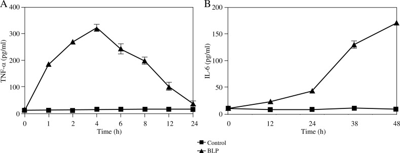
We first demonstrated that BLP induced a rapid release of TNF-α from THP-1 cells after 1 h incubation with BLP. The peak release was at 4 h, and then TNF-α production decreased slowly within 24 h. Unstimulated THP-1 cells had less spontaneous TNF-α release throughout the experiments (Fig. 1A). Also, IL-6 released over a different time course. It was slow initially, but steadily increased with BLP treatment duration (Fig. 1B). Thus, our data indicated that BLP-induced THP-1 cells produce high concentrations of TNF-α and IL-6.
Fig. 1.
Time course of BLP-induced TNF-α and IL-6 release, following stimulation with 1000 ng/ml of BLP. A) THP-1 cells, 2 × 10⁵ were incubated with control and 1000 ng/ml of BLP for different periods of time (0, 1, 2, 4, 6, 8, 12 and 24 h). The level of TNF-α was determined with the enzyme-link immunosorbent assay kits and plotted vs. time (hour). B) THP-1 cells, 2 × 10⁵ were incubated with control and 1000 ng/ml of BLP for different periods of time (0, 12, 24, 36 and 48 h). The level of IL-6 was plotted vs. time
Paeoniflorin inhibited TNF-α and IL-6 release in activated THP-1 cells
To investigate various concentrations of paeoniflorin on BLP-induced TNF-α and IL-6 release in THP-1 cells, cells were incubated with the compound (10−4, 10−5, 10−6, 10−7 and 10−8 M paeoniflorin), and TNF-α and IL-6 release was measured at 4 and 48 h, respectively. As seen in Table 1, paeoniflorin inhibited both TNF-α and IL-6 production stimulated by BLP treatment in a dose-dependent fashion.
Table 1.
The BLP group was incubated with BLP (1000 ng/ml) in incomplete medium while the control group was only incubated with medium. Treatment with 10−8 M of paeoniflorin resulted in a significant reduction in TNF-α release at 4 h compared to the BLP group (**p < 0.01). A similar trend was seen at IL-6 production. However, treatment with 10−7 M of paeoniflorin resulted in a significantly greater reduction in IL-6 release than 10−8 M at 48 h compared to the BLP group (*p < 0.05). Data are expressed as mean ± SD; n = 4 in each group (*p < 0.05) vs. BLP group (**p < 0.01) vs. BLP group
| Group | Paeoniflorin (mol/l) | TNF-α (pg/ml) | IL-6 (pg/ml) |
|---|---|---|---|
| Control | – | 13.04 ±1.40 | 10.76 ±4.72 |
| BLP | – | 336.99 ±11.44 | 185.64 ±8.58 |
| BLP + paeoniflorin | 10−8 | 251.55 ±16.05** | 175.00 ±2.45* |
| 10−7 | 202.51 ±17.56** | 168.06 ±1.69** | |
| 10−6 | 159.44 ±5.84** | 157.13 ±4.89** | |
| 10−5 | 144.18 ±6.00** | 133.00 ±1.56** | |
| 10−4 | 113.35 ±5.83** | 115.31 ±2.15** |
BLP – bacterial lipoprotein, TNF-α – tumor necrosis factor α, IL-6 – interleukin 6
Paeoniflorin pretreatment affects the BLP-mediated signaling pathway
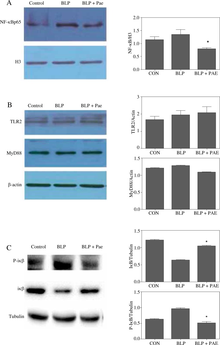
The NF-κB pathway plays a critical role in cytokine secretion, and NF-κB is present in the cytosol in an inactive state. It forms compounds with inhibitory IκB protein. And NF-κB activators initiate various signal transduction pathways that ultimately rapidly phosphorylate IκB protein. IκB degradation exposes the nuclear localization sequences on NF-κB p65, leading to NF-κB translocation to the nucleus. To measure these proteins in our current system, we treated THP-1 cells with BLP. Then, as early as 20 min after exposure to BLP (100 ng/ml), NF-κB was observed to increase as evidenced by Western blot, and this activation was partially inhibited by paeoniflorin (1 × 10⁻⁴ M) (Fig. 2A). Also, THP-1 cells exposed to paeoniflorin had decreased IκB phosphorylation (Fig. 2C), suggesting that paeoniflorin could prevent IκB phosphorylation and subsequent degradation, blocking nuclear translocation of NF-κB.
Fig. 2.
Effect of paeoniflorin on expression of NF-κB p65 in nuclei, phosphorylation of IκB and TLR2, MyD88 in cytoplasm of THP-1 cells. (A) BLP induced the activation of NF-κB in THP-1 cells. Pae inhibited the phosphorylation of IκB (C) and NF-κB protein (A). (B) No change of expression of TLR2 and MyD88 in response to BLP and Pae treatment. THP-1 cells were incubated with medium, BLP (100 ng/ml) and BLP (100 ng/ml) + Pae (10-4 M) for 20 min and analyzed by western blotting for NF-κB, TLR2, MyD88 and P-IκB (*p < 0.05, significant decrease compared with BLP group)
Western blot revealed that TLR2 and MyD88 expression did not change with either BLP or paeoniflorin treatment of cells (Fig. 2B). Thus, neither of these molecules changed under our experimental conditions.
BLP induces apoptosis in THP-1 cells
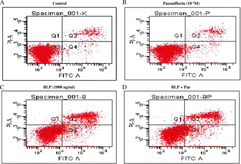
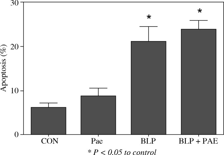
As shown in Fig. 3, BLP promoted apoptosis of THP-1 cells (6.1% in control; 20.9% in BLP-treated cells; p < 0.05). However, paeoniflorin (10−4 M and 24 h) did not induce apoptosis in THP-1 cells (control = 6.1%; paeoniflorin-treated cells = 8.6%; p > 0.05). Finally, co-stimulation of THP-1 cells with both 1,000 ng/ml BLP and 10−4 M paeoniflorin did not increase apoptosis compared to BLP alone (20.9% for BLP-treated cells; 23.8% for cells co-stimulated with BLP and paeoniflorin; p > 0.05) (Fig. 4).
Fig. 3.
Effects of BLP and paeoniflorin on apoptosis of THP-1 cells. Control cells were treated with incomplete RPMI- 1640 medium (A). THP-1 cells were treated for 24 h with paeoniflorin 10–4 M (B), with 1,000 ng/ml BLP (C), or with BLP 1000 ng/ml + Pae 10–4 M (D). The cells were harvested for quantification of apoptosis by flow cytometry measured as the percentage of Annexin V-FITC positive cells
Fig. 4.
Bacterial lipoprotein at doses of 1000 ng/ml at 24 h induced apoptosis which was significantly increased compared with control (*p < 0.05, significant increase compared with control). Paeoniflorin did not result in apoptosis in THP-1 cells (p > 0.05)
Discussion
Septic shock is a serious problem usually encountered in the hospital intensive care unit. Despite effective antibiotics, the mortality of septic shock remains high [14]. We investigated the therapeutic potential of paeoniflorin in sepsis induced by bacterial lipoprotein, and studied its potential mechanism of action in a cultured human monocyte cell line.
Both experimental and clinical studies indicate that harmful tissue events, including infections, are sensed by macrophages and monocytes, which in turn secrete pro-inflammatory cytokines, such as TNF-α, IL-1β, and IL-6, and anti-inflammatory cytokines IL-4 and IL-10 [15, 16]. Appropriate concentrations of cytokines are essential for cell-mediated microbicidal activity but excessive production can promote an uncontrolled inflammatory response, multiple organ failure, and ultimately death [15]. Reducing inflammation is important for critical care medicine. Thus, we evaluated paeoniflorin's effect on production of inflammatory mediators in bacterial lipoprotein-induced THP-1 cells to understand its anti-inflammatory mechanism. We conclude that BLP stimulation induces release of TNF-α and IL-6 in THP-1 cells, because these were significantly greater than in control cells. Also, TNF-α was secreted prior to IL-6. Thus, BLP can stimulate the production of pro-inflammatory factors to induce inflammation and paeoniflorin dose-dependently inhibited BLP-induced production of TNF-α and IL-6 by inhibiting phosphorylation and degradation of IκB and subsequent down-regulation of NF-κB p65 protein.
The innate immune system is the first line of defense to protect the host from invading microbial pathogens. Tolllike receptors expressed on antigen presenting cells serve central roles in the induction of the innate immune response as well as in a subsequent development of adaptive immune responses. To date, 11 human TLRs have been identified. Toll-like receptors are responsible for recognition of PAMPs (pathogen-associated molecular patterns), which are required for downstream signaling [4]. An adaptor molecule, MyD88, is involved in the signaling pathway via TLR2 so we speculated that TLR2 and MyD88 expression could be affected, as observed with NF-κB p65. However, TLR2 and MyD88 expression was not changed by BLP and paeoniflorin in our current study. However, signaling alterations within this pathway may still be important because protein phosphorylation or degradation could be altered after treatment with either of these compounds.
The role of apoptosis in sepsis and its clinical sequelae of systemic inflammatory response syndrome and MODS remain elusive [17]. Previous studies suggest that BLP and LPS can induce apoptosis in human monocytes, which for BLP are TLR-2-dependent and augmented by CD14 and PMA [3]. In Antonios O's research [18], TLR2 signals for apoptosis through MyD88 via a pathway involving Fas-associated death domain protein (FADD) and caspase 8. Inhibition of NF-κB activation downstream of TLRs augments the apoptosis response. These data indicated that TLR2 is a novel “death receptor” that engages the apoptosis machinery without a conventional cytoplasmic death domain. In our study, BLP at 1000 ng/ml and 24 h induced apoptosis, which agrees with previous studies. However, paeoniflorin at a dose of 10−4 M did not protect against BLP-induced apoptosis in THP-1 cells.
Acknowledgements
We thank LetPub for its linguistic assistance during the preparation of this manuscript.
The authors declare no conflict of interest.
References
- 1.Hotchkiss RS, Karl IE. The pathophysiology and treatment of sepsis. N Engl J Med. 2003;348:138–150. doi: 10.1056/NEJMra021333. [DOI] [PubMed] [Google Scholar]
- 2.Riedemann NC, Guo RF, Ward PA. The enigma of sepsis. J Clin Invest. 2003;112:460–467. doi: 10.1172/JCI19523. [DOI] [PMC free article] [PubMed] [Google Scholar]
- 3.Aliprantis AO, Yang RB, Mark MR, et al. Cell activation and apoptosis by bacterial lipoproteins through Toll-like receptor-2. Science. 1999;285:736–739. doi: 10.1126/science.285.5428.736. [DOI] [PubMed] [Google Scholar]
- 4.Akira S, Uematsu S, Takeuchi O. Pathogen recognition and innate immunity. Cell. 2006;124:783–801. doi: 10.1016/j.cell.2006.02.015. [DOI] [PubMed] [Google Scholar]
- 5.Hayden MS, West AP, Ghosh S. NF-κB and the immune response. Oncogene. 2006;25:6758–6780. doi: 10.1038/sj.onc.1209943. [DOI] [PubMed] [Google Scholar]
- 6.Hsu FL, Lai CW, Cheng JT. Antihyperglycemic effects of paeoniflorin and 8-debenzoylpaeoniflorin, glucosides from the root of Paeonia lactiflora. Planta Med. 1997;63:323–325. doi: 10.1055/s-2006-957692. [DOI] [PubMed] [Google Scholar]
- 7.Zheng YQ, Wei W, Zhu L, Liu JX. Effects and mechanisms of paeoniflorin, a bioactive glucoside from paeony root, on adjuvant arthritis in rats. Inflamm Res. 2007;56:182–188. doi: 10.1007/s00011-006-6002-5. [DOI] [PubMed] [Google Scholar]
- 8.Chang YQ, Zhao ZF, Liu MS. Effects of Paeoniflorin on the Expression of HMGB-1 in RAW264.7 Macrophages induced by LPS. J Chang Zhi Medical College. 2009;23:81–84. [Google Scholar]
- 9.Li Q, Zhong SZ, Ma SP. Paeoniflorin ameliorates cognitive impairment and neurodegeneration in senescent mice induced by D-galactose and sodium mitrite. Pharm Clin Res. 2009;17:172–176. [Google Scholar]
- 10.Liu J, Jin DZ, Xiao L, Zhu XZ. Paeoniflorin attenuates chronic cerebral hypoperfusion-induced learning dysfunction and brain damage in rats. Brain Res. 2006;1089:162–170. doi: 10.1016/j.brainres.2006.02.115. [DOI] [PubMed] [Google Scholar]
- 11.Sun R, Lv LL, Liu GQ. Effects of paeoniflorin on cerebral ischemia in Mongolia gerbils. Zhongguo Zhang Yao Za Zhi. 2006;31:832–835. [PubMed] [Google Scholar]
- 12.Wu H, Li W, Wang T, et al. Paeoniflorin suppress NF-κB activation through modulation of IκBα and enhance 5-fluorouracil-induced apoptosis in human gastric carcinoma cells. Biomed Pharmacother. 2008;62:659–666. doi: 10.1016/j.biopha.2008.08.002. [DOI] [PubMed] [Google Scholar]
- 13.Jiang WL, Chen XG, Zhu HB, et al. Paeoniflorin Inhibits Systemic Inflammatory and Improves Survival in Experimental Sepsis. Basic Clin Pharmacol Toxicol. 2009;105:64–71. doi: 10.1111/j.1742-7843.2009.00415.x. [DOI] [PubMed] [Google Scholar]
- 14.Glauser MP, Zanetti G, Baumgartner JD, Cohen J. Septic shock: pathogenesis. Lancet. 1991;338:732–736. doi: 10.1016/0140-6736(91)91452-z. [DOI] [PubMed] [Google Scholar]
- 15.Wang JH, Doyle M, Manning BJ, et al. Bacterial lipoprotein induces endotoxin-independent tolerance to septic shock. J Immunol. 2003;170:14–18. doi: 10.4049/jimmunol.170.1.14. [DOI] [PubMed] [Google Scholar]
- 16.Jaworek Jl, Konturek SJ, Macko M, et al. Endotoxemia in newborn rats attenuates acute pancreatitis at adult age. J Physiol Pharmacol. 2007;58:131–147. [PubMed] [Google Scholar]
- 17.Papathanassoglou ED, Moynihan JA, Ackerman MH. Does programmed cell death (apoptosis) play role in the development of multiple organ dysfunctions in critically ill patients: A review and a theoretical framework. Crit Care Med. 2000;28:537–549. doi: 10.1097/00003246-200002000-00042. [DOI] [PubMed] [Google Scholar]
- 18.Aliprantis AO, Yang RB, Weiss DS, et al. The apoptotic signaling pathway activated by Toll-like receptor-2. EMBO J. 2000;19:3325–3336. doi: 10.1093/emboj/19.13.3325. [DOI] [PMC free article] [PubMed] [Google Scholar]


