
Figure 2. Combinatorial expression of TFs in Lin B.
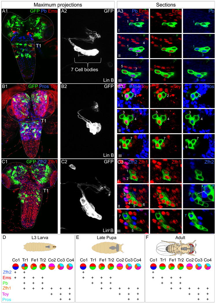
(A-C): L3 CNSs with Lin B MARCM clones in T1 (boxed) expressing mCD8::GFP under the control of VGlut-Gal4. (A1, B1, C1) Ventral views of maximal confocal projections immunostained with anti-Pb (blue) and anti-Ems (red) (A1), anti-Toy (blue) and anti-Pb (red) (B1) and anti-Pros (blue) and anti-Toy (red) (C1). (A2, B2, C2) show magnifications of the GFP+ clones in A1, B1, C1, respectively. (A3, B3, C3) show confocal sections of each clone from ventral (I) to dorsal (III). Note: One of the two Zfh2+ cells consistently expresses lower levels of this TF, as illustrated by the inset in C3 that shows a higher magnification and intensity of this cell and a non-expressing cell for comparison. Pros levels are also consistently higher in one of the two Pros-expressing cells (B3).
(D-F): Summary of the post-mitotic TF combinatorial code in Lin B in the L3 CNS (D), late pupa (E), and adult (F); (see also Figure S2, S3 and S4). Note that although the current code does not discriminate between Fe1 and Tr2 or Co3 and Co4, differing TF levels (e.g. Pros in Co3 and Co4) may contribute to their distinct identities.