A 50-year-old man presented with amoxicillin-induced cholestatic liver injury with resultant dialysis-dependent acute kidney injury. Total bilirubin ranged from 30 to 33 mg/dL, AST/ALT 200 to 400 U/L, and alkaline phosphatase from 3,000 to 4,000 U/L.
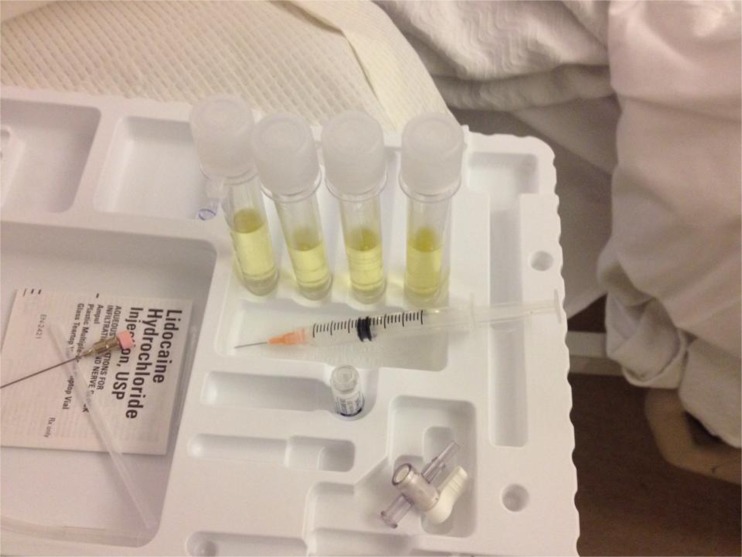
Because of fever and delirium, lumbar puncture (LP) was performed on hospital day 30 after normal head imaging. Moderate xanthochromia was immediately visualized after a non-traumatic tap. Analysis revealed RBC 1/cu mm, WBC 1/cu mm, minimally elevated protein (50 mg/dL), and normal glucose (82 mg/dL).
Xanthochromia, “blond color” in Greek,1 is caused by pigment in the cerebral spinal fluid (CSF). Xanthochromia is classically associated with subarachnoid hemorrhage (SAH): red blood cells lyse within hours and are metabolized from oxyhemoglobin (pink) to bilirubin (yellow). Xanthochromia is present in >90 % of patients with a SAH within 12 h of onset,1–3 but can also occur with increased CSF protein (≥150 mg/dL) due to bilirubin binding, traumatic LP with delayed analysis, and serum hyperbilirubinemia (>10–15 mg/dL), which was the most likely cause in this case.1,2,4 Literature suggests an association between CSF and serum bilirubin levels but not the duration of hyperbilirubinemia.5 Spectrophotometry can be used to identify bilirubin with or without oxyhemoglobin.3,6,7 However, xanthochromia should continue to raise concern for SAH in appropriate clinical scenarios (Fig. 1).
Fig. 1.
CSF vials after lumbar puncture, with the hallmark blond color of xanthochromia.
Acknowledgments
Conflict of interest
The authors each declare that they have no conflicts of interest.
References
- 1.Chalmers AH. Comments on the Proposed National Guidelines for Analysis of Cerebrospinal Fluid for Bilirubin in Suspected Subarachnoid Haemorrhage. Produced by a working group of UK NEQAS for Immunochemistry and in conjunction with the National Audit Group of the Association of Clinical Biochemists. Clin Biochem Rev. 2003;24(4):131–133. [Google Scholar]
- 2.Seehusen DA, Reeves MM, Fomin DA. Cerebrospinal fluid analysis. Am Fam Physician. 2003;68(6):1103–1108. [PubMed] [Google Scholar]
- 3.Williams A. Xanthochromia in the cerebrospinal fluid. Pract Neurol. 2004;4:174–175. doi: 10.1111/j.1474-7766.2004.11-213.x. [DOI] [Google Scholar]
- 4.Greenlee JE, Carroll KC. Cerebrospinal fluid in central nervous system infections. In: Scheld WM, Whitley RJ, Marra CM, editors. Infections of the Central Nervous System. 3. Philadelphia: Lippincott Williams & Wilkins; 2004. p. 5. [Google Scholar]
- 5.Rosenberg DG, Galambos JT. Yellow spinal fluid. Diagnostic significance of cerebrospinal fluid in jaundiced patients. Am J Dig Dis. 1960;5:32–48. doi: 10.1007/BF02233023. [DOI] [PubMed] [Google Scholar]
- 6.Petzold A, Keir G, Sharpe LT. Spectrophotometry for xanthochromia. N Engl J Med. 2004;351(16):1695–1696. doi: 10.1056/NEJM200410143511627. [DOI] [PubMed] [Google Scholar]
- 7.Chu K, Hann A, Greenslade J, Williams J, Brown A. Spectrophotometry or visual inspection to most reliably detect xanthochromia in subarachnoid hemorrhage: systematic review. Ann Emerg Med. 2014;64(3):256–264.e5. doi: 10.1016/j.annemergmed.2014.01.023. [DOI] [PubMed] [Google Scholar]


