Abstract
Objective: To compare the efficacy of volar and dorsal plate fixation for unstable dorsal distal radius fractures. Methods: Forty-seven cases were selected from patients undergoing surgical reduction and internal fixation treatment in our hospital from August 2006 to October 2010, with 21 males and 26 females, aged 39-73 years old. Patients were divided into two groups: volar plate fixation group (Group A) which has 32 cases, including 27 cases with locking plate, 5 cases with ordinary T plate, and 4 cases combined with dorsal Kirschner wire fixation; dorsal plate fixation group (Group B) which has 15 cases, including 7 cases with locking plate. The efficacy of the two fixation methods were compared in terms of postoperative wrist function, X-ray score, and postoperative complications. Results: Compared with those of preoperative groups, the volar tilt, ulnar deviation and radial styloid height in both group A and B were significantly improved one week after surgery as shown by X-ray imaging. Comparison of X-ray images one week after surgery with those of six months after surgery showed no significant changes in volar tilt, ulnar deviation or radial styloid height. 87.5% of patients in group A and 86.7% of patients in group B got “excellent” in their wrist function assessment, and there was no significant difference between the two groups (X2=0.825, P=1.000). But patients in group A hax significantly lower incidence rate of postoperative complications than group B (X2=4.150, P=0.042). Conclusion: For unstable distal radius fractures with dorsal displacement, volar plate fixation can achieve satisfactory reduction results, and cause less tendon damage or other complications than dorsal plate fixation.
Keywords: Distal radius fracture, unstable, internal fixation, volar plate fixation, dorsal plate fixation
Introduction
Unstable distal radius fractures are mostly found in elderly patients of osteoporosis or young patients of high-energy injury. The dorsal displacement fracture is the most common one among the unstable wrist fractures. From August 2006 to October 2010, we did volar or dorsal plate fixation in forty-seven patients with dorsal unstable distal radius fractures and the outcomes were satisfactory. Through retrospective comparative analysis, we evaluated the efficacy of the two different fixation approaches on dorsal distal radius fractures.
Materials and methods
Clinical data and grouping
From August 2006 to October 2010, forty-seven patients were treated with open reduction and plate fixation and followed up after surgery. There were twenty-one male and twenty-six female, aging from 39 to 73 years old, and they all have closed fracture. They were divided into two groups. In group A, the patients were treated with volar plate fixation. Among the thirty-two patients, twenty-seven patients had locking plate, five patients had T plate, and four patients had combined dorsal Kirschner wire. In group B, fifteen patients were treated with dorsal plate fixation, including seven cases of locking plate. The follow-up is 6-18 months postoperative and average time is 13.5 months.
Diagnosis and inclusive and exclusion criteria
(1) Diagnostic criteria: a. history: confirmed palm stays; b. symptoms and signs: wrist fork or spear-like deformity, swelling, tenderness, and bone rubbing; c. X-ray: confirms the diagnosis.
(2) Inclusion criteria: a. X-ray: distal radius fractures with dorsal cortical bone crushing, articular surface displacement greater than 2 mm, palmar angle tilted more than 25 degrees, dorsal distal radial shortening greater than 5 mm, unstable reduction and easy recurrence of dorsal displacement [1]; b. age: 35-80 years old; c. treatment: open reduction and plate fixation.
(3) Exclusion criteria: a. patients older than 80 years old or young than 35 years old; b. treatment is not open reduction and plate fixation.
Surgical approaches
The patients were given brachial plexus anesthesia with arms at supine position, and given inflated tourniquet at the roots of upper arm.
In the volar plate fixation group (group A, 32 cases): at volar side of distal radius, 6 cm longitudinal incision was made between radial volar flexor tendon and the radial artery, and then skin, subcutaneous tissue and fascia were cut open layer by layer. The radial artery was dragged to the radial side, and the radial flexor carpi and median nerve were dragged to the ulnar side. Periosteum was stripped down to expose the fracture and the hematoma and soft tissue at the stump were cleared. After reduction, C-arm fluoroscopy was used to confirm palm inclination, ulnar deviation, articular surface flatness and radial length. Fine Kirschner was use for temporary fixation, and volar locking plate or T plate was used for final fixation. Bone defects were repaired with artificial or autologous bone grafts. For the articular and dorsal displacement, which have difficulty to maintain reduction, small incision dorsal reduction and one or two pieces of Kirschner wire were used and the end of wire was kept outside for early removal. The location and length of the screw plate were checked with fluoroscopy again, and the fine Kirschner wire was removed, pronator muscle was repaired and the fascia and skin were sutured layer by layer.
In the dorsal plate fixation group (group B, 15 cases): forearm was placed at pronation, and 6 cm longitudinal incision was made along radius from distal end to proximal end. Skin, subcutaneous tissue and fascia were cut open layer by layer. The cut was between the radial extensor digitorum longus and extensor capri radialis brevis. Periosteum was stripped down to expose the fracture and the hematoma and soft tissue at the stump were cleared. After reduction, C-arm fluoroscopy was used to confirm palm inclination, ulnar deviation, articular surface flatness and radial length. Fine Kirschner was use for temporary fixation, and volar locking plate or T plate was used for final fixation. For dorsal plate fixation, it usually needs remodeling to make the plate fit into the defects. Bone defects were repaired with artificial or autologous bone grafts. The location and length of the screw plate were checked with fluoroscopy again, and the fine Kirschner wire was removed, pronator muscle was repaired and the fascia and skin were sutured layer by layer.
After surgery, the patients were given dorsal plaster fixation at functional position for two weeks and then the plaster was removed and patients started to exercise wrist joint. Shoulder, elbow, fingers and metacarpophalangeal joints can start functional activity right after surgery.
Assessment parameters
Parameters: postoperative X-ray, postoperative complications and wrist function.
Methods: wrist joint X-ray was taken at one week, four week, eight week, twelve week, half year and one year after surgery. At the meantime, complications and wrist function were assessed.
Evaluation
X-ray: according to Lidstrom scoring criteria [2], the volar tilt, ulnar deviation, radial styloid height and articular surface flatness were evaluated.
Wrist joint function: according to Dienst wrist rating system [3], the following comparisons were made: a. X-ray score in same group at one week vs. half year postoperatively; b. X-ray score between groups at two weeks postoperatively; c. X-ray score between groups at half year postoperatively; d. complications.
Statistical analysis
SPSS11.0 statistical software was used for analysis, One-Way ANOVA was used to analyze numerical data between groups and chi-square test was used for categorical variables. P<0.05 is considered statistically significant.
Results
The patients’ age, gender, fracture type and other information are compared as shown in Table 1.
Table 1.
Comparison of clinical data between two groups
| Groups | Gender (cases) | Fractures type (cases) | |||||
|---|---|---|---|---|---|---|---|
|
| |||||||
| Male | Female | Age | A3 | B2 | C2 | C3 | |
| Group A | 15 | 17 | 61.3±0.79 | 4 | 3 | 12 | 13 |
| Group B | 6 | 9 | 59.8±0.81 | 1 | 2 | 5 | 7 |
| Test value | x2=0.195 | t=0.119 | x2=0.758 | ||||
| P value | 0.659 | 0.906 | 1.000 | ||||
X-ray scores
In both group A and B, the volar tilt, ulnar deviation, radial styloid height were significantly improved at one week after surgery. The difference was statistically significant (F=7.304, P=0.001). In either group A or B, there was no significant change in volar tilt, ulnar deviation and radial styloid height at one week vs. six months after surgery, as shown in Table 2.
Table 2.
Preoperative and postoperative X-ray measurement values in groups A and B
| Group | Parameters | Time points | F value | P value | ||
|---|---|---|---|---|---|---|
|
| ||||||
| Preoperative | 1 week postoperative | 6 months postoperative | ||||
| A | Volar tilt | -27.31±6.57* | 9.82±2.18 | 9.76±2.38 | 7.304 | 0.001 |
| Ulnar deviation | 5.17±2.70* | 19.04±3.76 | 18.51±2.83 | 4.275 | 0.016 | |
| radial styloid height | 2.35±3.84* | 18.23±0.59 | 18.04±1.31 | 6.088 | 0.003 | |
| B | Volar tilt | -27.50±6.32* | 9.87±1.97 | 9.83±2.01 | 8.214 | 0.001 |
| Ulnar deviation | 5.88±2.17* | 18.25±2.47 | 18.20±1.98 | 4.372 | 0.019 | |
| radial styloid height | 3.17±2.12* | 18.34±1.17 | 17.98±1.54 | 5.296 | 0.009 | |
Note: One-Way ANOVA analysis shows that there is significant difference in X-ray measurement values among different time points inside group A and B (P<0.01);
indicates that the value in preoperative is significantly different from both 1 week after surgery and 6 months after surgery.
Wrist function scores
In group A, the scores were graded as “excellent” in 17 cases, “good” in 11 cases, “fair” in 3 cases and “poor” in 1 case. In group B, the scores were graded as “excellent” in 8 cases, “good” in 5 cases, “fair” in 2 cases and “poor” in 1 case. The rate of “excellent” and “good” score in group A is 87.5%, and the rate in group B is 86.7%. There is no significant difference between two groups (x2=0.825, P=1.000), as shown in Table 3.
Table 3.
Wrist joint function assessment results six months after surgery
| Groups | Cases | Excellent | Good | Fair | Poor | Rate of Excellence |
|---|---|---|---|---|---|---|
| A | 32 | 17 | 11 | 3 | 1 | 87.5% |
| B | 15 | 8 | 5 | 2 | 1 | 86.7% |
Note: Comparison of two groups, X 2=0.825, P=1.000.
Postoperative complications
In group A, there were one case of median nerve traction injury, one case of wound infection, and two cases of tendon adhesion. In the group B, there were one case of incision infection, one case of tendon adhesion, and three cases of extensor pollicis longus tenderness. The patients with infection were given intravenous antibiotics and the swelling subsided. For the tendon adhesion, the adhesiolysis was performed when the internal fixation was removed. There was significant difference in occurrence of complications between two groups (x2=4.150, P=0.042).
Discussion
Distal radius fracture is one of the most common fractures in clinical. Most distal radial fractures can be cured by reduction and external plaster fixation [4]. For those unstable distal radius fractures with severe osteoporosis and high-energy injury, further internal fixation is required because of obvious dorsal displacement, dorsal bone crushing or difficulty of manual reduction or maintaining the reduction [5]. Currently, it is still controversial which internal fixation method is more beneficial. Some researchers believe that, for unstable dorsal radial fracture with dorsal displacement, simple volar fixation may cause decline of intraoperative fracture reduction angle and height. Murakami et al [6] found that there were less tissue damage in volar plate fixation and the plate could fit better than in the dorsal fixation.
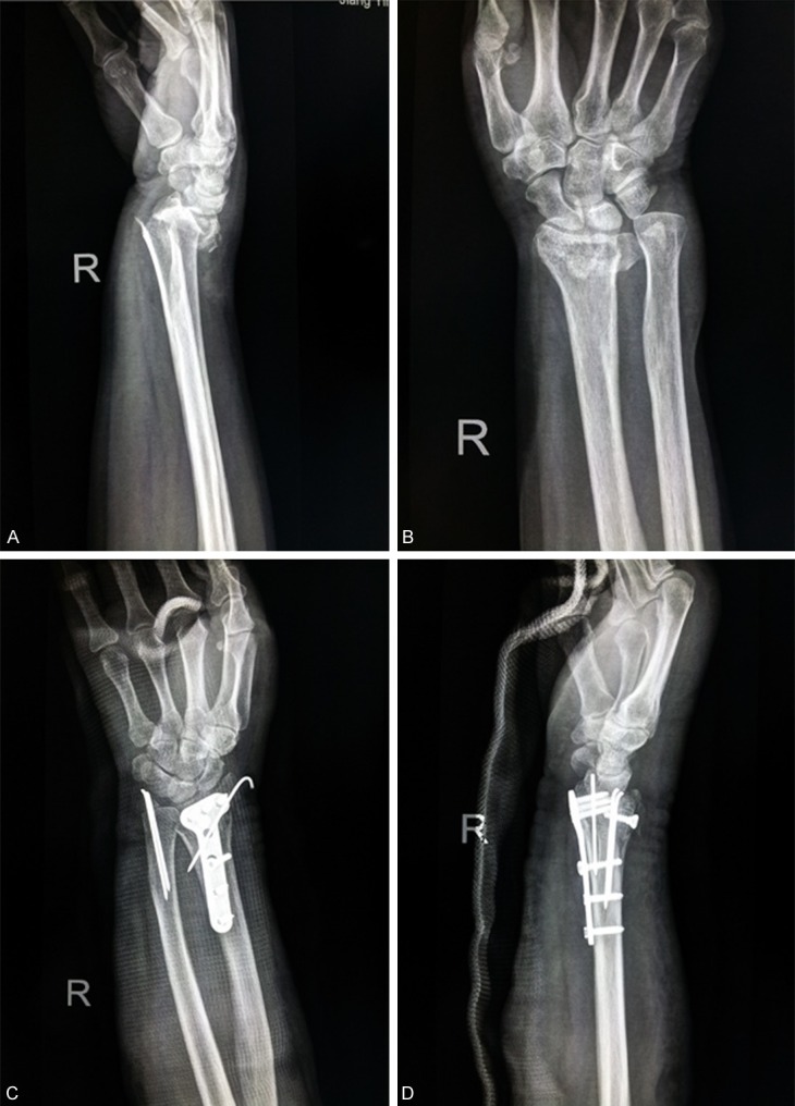
Our retrospective analysis found that simple volar plate fixation in unstable distal radial fracture could improve volar tilt, ulnar deviation, radial length and other parameters (Figure 1). In this study, thirty-two patients underwent volar plate fixation (combined with Kirschner pin fixation through dorsal small incision in four cases), postoperative X-ray showed improved volar tilt, ulnar deviation and radial length, half year follow-up showed no occurrence of fracture displacement. Since most distal radial fractures are associated with osteoporosis and the fractures are most likely comminuted, adequate bone should be grafted, and distal volar locking plate should be used as its distal 3-4 locking screws can form a stable “inner supporter” structure [7]. Since volar plate fits the anatomy well and does not need to bend over, and pronator muscles may overlie the volar plate, postoperative tendon irritation and wear are less likely to occur. For patients with severe comminuted dorsal bone fracture which is hard to reduce, volar plate fixation combined with Kirschner pin fixation through dorsal small incision can be as effective as dorsal plate, while avoiding the wearing of hallucis longus tendon caused by irritation of the dorsal plate.
Figure 1.

X-ray images of a typical case of unstable dorsal distal radius fracture before and after volar plate fixation combined with Kirschner pin fixation. A. Lateral view of unstable dorsal distal radius fracture before surgery; B. Frontal view of unstable dorsal distal radius fracture before surgery; C. Lateral view of unstable dorsal distal radius fracture after surgery; D. Frontal view of unstable dorsal distal radius fracture after surgery.
Statistical analysis showed that there is no significant difference in fracture reduction rate and fracture re-displacement rate between seventeen patients treated with dorsal plate fixation and volar plate fixation, but the incidence of long-term complications in dorsal plate fixation group is significantly higher than that of the volar fixation group. Studies showed that dorsal plate did not fit the local anatomy well and often required pre-bending, which caused deformation of the screw thread in the locking plate and made it hard to place the screw in. Dorsal plates are often placed underneath the extensor tendons, and there is little soft tissue between the plate and the tendons, so the tendons are prone to irritation, wear or breakage.
Some scholars have found that it did not significantly affect the therapeutic outcome whether the wrist exercised early or late postoperatively in patients with distal radial fractures. AAOS guidelines on distal radius fracture treatment suggest that patients do not need to exercise their wrists at the early stage even after a firm fracture fixation [8]. Therefore, we routinely immobilized the wrist joint with plaster for two weeks after surgery, but shoulder, elbow and interphalangeal joint were not immobilized, the joint function and activity were not affected postoperatively. And by the short-term postoperative plaster immobilization of the wrist, occurrence of early postoperative fracture displacement in patients with unstable distal radius fractures can be effectively prevented. This has also been shown by Rhee and colleagues who reported that volar locking plating of distal radial fractures was a reliable form of treatment without substantial late displacement [9].
It is noteworthy to point out that 87.5% of patients undergoing volar plate fixation got “excellent” in wrist function assessment in our study, while another study with a smaller sample size showed that only 30% of their patients got “excellent” in wrist function assessment, nevertheless compared with dorsal plate, they concluded that treatment of unstable distal radius fractures with a volar plate provided stable internal fixation and allowed early function and was associated with a low complication rate [10]. Another report showed that corrective osteotomies of distal radius malunions can be done with either volar or dorsal plate fixation, but it might result in some better flexion, if performed volarly [11], which also agrees with our conclusion.
In summary, for unstable distal radius fractures with dorsal displacement, volar plate fixation can achieve satisfactory reduction results, and cause less tendon damage or other complications than dorsal plate fixation.
Disclosure of conflict of interest
None.
References
- 1.Gong FL, Li J, Fan QB, Jiang Y, Xia HG, Fan JF. Three different methods for the treatment of unstable distal radial fracture comparative study. Chinese Journal of Bone and Joint Injury. 2011;2:842–3. [Google Scholar]
- 2.Lidstrom A. Fractures of the distal radius. A clinical and statistical study of end result. Acta Orthop Scand (Supply) 1959;41:1–118. [PubMed] [Google Scholar]
- 3.Dienst M, Wozasek GE, Seligsonp D. Dynamic external fixation for distal radiusfractums. Clin Orthop Relat Res. 1997;338:160–71. doi: 10.1097/00003086-199705000-00023. [DOI] [PubMed] [Google Scholar]
- 4.Liu Z. Distal radial fracture treatment methods reasonably choose. Chinese Traumatology. 2010;23:571–3. [Google Scholar]
- 5.Zhang XP. Distal radial fracture choice of treatment and thinking. Chinese Journal of Orthopaedics and Traumatology. 2011;24:887–889. [PubMed] [Google Scholar]
- 6.Murakami K, Abe Y, Takahashi K. Surgical treatment of unstable distal radius fractures with volar locking plates. J Orthop Sci. 2007;12:134. doi: 10.1007/s00776-006-1103-0. [DOI] [PubMed] [Google Scholar]
- 7.Li X, Gao W, Wang QG, Li HQ, Tao J, Li F, Huang JH, Wu XM, Wang JD, Wu XF, Zhou ZH, Gao KD, Lu HM. AO novel locking fixation system in the treatment of unstable distal radius fractures observed. Chinese Journal of Orthopaedic Trauma. 2010;12:6–11. [Google Scholar]
- 8.Yu X, Yu GR. Guide of the AAOS treatment of distal radius fractures. Chinese Journal of Orthopedics. 2010;18:45–9. [Google Scholar]
- 9.Rhee SH, Kim J, Lee YH, Gong HS, Lee HJ, Baek GH. Factors affecting late displacement following volar locking plate fixation for distalradial fractures in elderly female patients. Bone Joint J. 2013;95:396–400. doi: 10.1302/0301-620X.95B3.30514. [DOI] [PubMed] [Google Scholar]
- 10.Giannotti S, Alfieri P, Magistrelli L, Casella F, Palmeri L, Guido G. Volar fixation of distal radial fracture using compression plate: clinical and radiographic evaluation of 20 patients. Musculoskelet Surg. 2013;97:61–5. doi: 10.1007/s12306-012-0238-8. [DOI] [PubMed] [Google Scholar]
- 11.Rothenfluh E, Schweizer A, Nagy L. Opening wedge osteotomy for distal radius malunion: dorsal or palmar approach? J Wrist Surg. 2013;2:49–54. doi: 10.1055/s-0032-1326725. [DOI] [PMC free article] [PubMed] [Google Scholar]


