SUMMARY
GlideScope® is a recently developed videolaryngoscope that helps to achieve a good view of the laryngeal inlet and the vocal cords. Videolaryngoscopy has been proven effective in patients with unusual anatomical or pathological features, suggesting the possibility of a difficult endotracheal intubation. This device may also be useful for otorhinolaryngologists by facilitating access to the larynx and tongue base, especially in selected cases, where good visualisation of disease-altered structures is vital. According to the current literature, GlideScope® has been used for surgical procedures involving the tongue base, such as biopsies, foreign body removal and radiofrequency treatment of obstructive sleep apnoea. We believe that the use of this kind of videolaryngoscopy might be also indicated for laryngeal surgery as a valid alternative to the placement of a direct laryngoscope. This technique, especially in those cases with anatomical issues or important comorbidities, may be preferred to ambulatorial flexible or rigid laryngoscopy, and in planning surgical procedures in "difficult" patients due to the operating room setting comprising constant anaesthesiological support. In our experience, we performed five procedures involving the larynx with the GlideScope® in patients presenting unusual clinical characteristics that potentially compromised surgical outcome. No complications related to videolaryngoscopy were found. We recommend the use of GlideScope® for small surgical procedures involving the larynx in selected patients.
KEY WORDS: Videolaryngoscopy, Laryngeal Surgery, Difficult Intubation
RIASSUNTO
Il GlideScope® è un videolaringoscopio di recente sviluppo, che permette di visualizzare in maniera ottimale l'aditus ad laryngem ed il piano glottico. La videolaringoscopia con l'ausilio del GlideScope® è indicata in pazienti con caratteristiche anatomiche delle vie aereodigestive superiori (VADS) o con patologie che possano far prevedere una intubazione difficile. Questo strumento può essere di grande aiuto anche per lo specialista otorinolaringoiatra, in quanto l'accesso alla laringe ed alla base della lingua è facilitato, soprattutto in casi selezionati, nei quali una buona visualizzazione delle strutture interessate da patologia è di vitale importanza. In letteratura il GlideScope® è stato utilizzato per procedure chirurgiche sulla base linguale, come biopsie, rimozione di corpi estranei e trattamento con radiofrequenze delle apnee ostruttive del sonno. Crediamo che la videolaringoscopia possa essere indicata anche per la chirurgia della laringe, come valida alternativa all'utilizzo del laringoscopio convenzionale. Questa tecnica, soprattutto in caso di comorbidità importanti o di alterazioni anatomiche, è da preferire alla fibrolaringoscopia ambulatoriale, in quanto si esegue in ambiente operatorio e con supporto anestesiologico. La nostra esperienza consta di cinque casi di chirurgia laringea con l'utilizzo del GlideScope®, in pazienti con caratteristiche cliniche potenzialmente pregiudicanti il successo chirurgico. Non sono state riscontrate complicanze dovute all'utilizzo del videolaringoscopio. Raccomandiamo pertanto l'utilizzo del GlideScope® per procedure di piccola chirurgia laringea, in pazienti selezionati.
Introduction
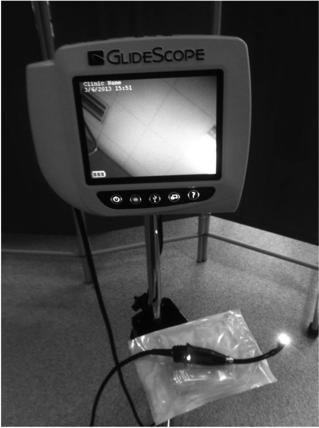
GlideScope® (Verathon Medical, Bothell, WA, USA) is a recently developed videolaryngoscope, which allows easier airway management in difficult conditions, such as neonatal intubation, morbidly obese patients or a restricted view of the laryngeal inlet. This device can be useful for several clinical situations due to its particular features: 1) multiple sizes are available; 2) it can be used even in preterm/small children; 3) the possibility to record the video images makes it suitable for academic purposes; and 4) the anti-fog system prevents poor visualisation due to secretions and the device can be operational in seconds, when needed (Fig. 1). While videolaryngoscopy has been proven effective in achieving a better view of the glottis, the success rate of intubation seems to be essentially the same as conventional direct
Fig. 1.
The GlideScope videolaryngoscope.
laryngoscopy (DL): for this reason, at present Glide- Scope® is recommended with strong evidence only as a rescue choice after a failed intubation attempt using DL 1. Nonetheless, GlideScope® can be helpful not only for the anaesthesiologist, but also for other scenarios of upper airway management. For example, a perfect view of the vocal cords is vital in laryngeal surgery, and it is frequent for the ENT specialist to experience technical issues due to the misplacement of the laryngoscope, due to particular anatomical or pathological features (i.e. traumatic injuries, cervical spine immobilisation, etc.). The use of the GlideScope® can help in managing these issues and facilitate the execution of several procedures in addition to simple endotracheal tube placement. According to the current scientific literature, GlideScope® has been used for examination and biopsies of the tongue base, removal of foreign bodies, and radiofrequency treatment of obstructive sleep apnoea 2. The aim of this study was to explore selected laryngeal and hypopharyngeal procedures as other conditions in which GlideScope® may help achieving an optimal view, thus providing greater ease for surgeons. We believe that the GlideScope®, for certain surgical procedures, may be preferred to surgical procedures performed with fiberoptic laryngoscopy in those patients regarded as 'complex' for anatomical issues or relevant comorbidities. The procedure is executed in an operating theatre, which may be important in case of complications: furthermore, using GlideScope®, intubation is not generally required, thus making the procedure faster and less invasive for the patient. In the recent medical literature, no data were found about GlideScope®-guided surgical procedures involving the larynx. In our department, we performed laryngeal surgery with the use of GlideScope® on five patients.
Clinical technique
Case 1
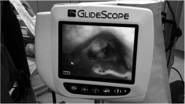
An 84-year-old man came to our observation with a long history of dysphonia. Fibre optic laryngoscopy revealed a 5 mm mass involving the middle third of the left vocal cord. The patient was scheduled for a surgical biopsy but, due to severe cardiovascular and respiratory comorbidities, general anaesthesia was not possible. Under Glide- Scope® view, the procedure was rapidly executed in an operating theatre setting (Fig. 2), under mild sedation, and the patient did not need further hospitalisation. Bleeding was minimal and no respiratory problems occurred.
Fig. 2.
The GlideScope device helped in achieving correct visualisation of a laryngeal mass, under mild sedation, without needing to place a direct laryngoscope (this picture refers to case 1).
Case 2
A 21-year-old woman with Down syndrome was admitted to our department with increasing dysphonia. Indirect laryngoscopy was impossible due to the characteristic macroglossy, while with fibre optic laryngoscopy visualisation of the vocal cords was partial (Cormack-Lehane grade II) and spoiled by vivid reflexes. A large cyst involving the anterior third of the right vocal cord was visualised, and surgery was scheduled. Intubation proved hard to achieve, so GlideScope® videolaryngoscopy was used. With the device still in, we proceeded with the removal of the cyst, thus sparing all the time and the difficulties associated with direct laryngoscope positioning. The procedure was brief and with no immediate complication. The patient was hospitalised and went home the following day.
Case 3
A 44-year-old man with pathological obesity (BMI = 38) underwent fibre optic laryngoscopy in our department for complaints of dysphagia. A medial epiglottic cyst measuring about 1 cm was found. Microlaryngoscopy surgery was programmed, but DL with the Macintosh blade was especially difficult to perform due to the patient's physical structure. The GlideScope® was used and a good view of the aditus ad laryngem was obtained. The cyst was successfully removed in a short period of time.
Case 4
A 28-year-old woman came to our observation with a history of dysphonia. Indirect and fibre optic laryngoscopy showed the presence, in the middle third of the right vocal cord, of two small, exophytic lesions, compatible with a diagnosis of laryngeal papillomatosis. Due to the possibility of respiratory obstruction, the patient was scheduled for surgical removal of the lesions. The patient presented severe prognathism and a very short interincisive distance (1.9 cm), and thus the intubation procedure required several attempts, and was achieved with the use of the GlideScope ®. We decided to maintain the inserted device and an excisional biopsy of both lesions was performed. The procedure ended with consistent time savings. Histology confirmed clinical diagnosis, and the patient was discharged without hospitalisation.
Case 5
A 74-year-old man with a long history of smoking and increasing dysphonia came to our observation. Fibre optic laryngoscopy revealed a bulky, 2 cm mass in the left pyriform sinus which imposed surgical biopsy. The patient suffered from a severe form of congestive heart failure and preoperative risk was assessed as high (ASA class 4). We decided to perform a biopsy under mild sedation with the use of GlideScope® videolaryngoscopy. The procedure was brief and without complications. Histology revealed a squamous cell carcinoma, for which radiotherapy treatment was programmed.
Discussion
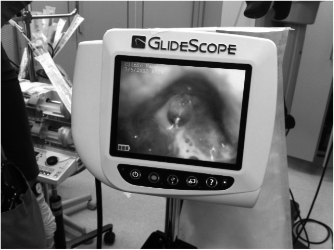
The GlideScope® videolaryngoscopy device is frequently helpful in airway management, especially in achieving a better view of the glottis in difficult intubations. It is currently used as a primary or a rescue device for several kinds of patients, from paediatric cases to those with cervical spine immobilisation. Moreover, the numerous features in common with direct laryngoscopes make it easy to use without any special training 3. Furthermore, the chance of registering the procedure with good video quality should be regarded as useful for both academic teaching in university hospitals and forensic issues (Fig. 3). Few complications related to the use of GlideScope® have been described, the majority of which concern difficulties in placing the endo-tracheal tube for ventilation, due to the presence of potential blind spots. However, less intensity is needed to expose the glottis in comparison to direct laryngoscopy, and the GlideScope® is considered safe 4. We encountered no particular anatomical difficulty while performing these procedures, and we always obtained a good view of the anatomical district. Surgery using the GlideScope® can be performed with one hand, while the other hand holds the instrument, or with both hands, with a second operator holding the device: both operators can see the images on the screen, which can be useful in an academic setting for teaching purposes. No evidence of the use of a GlideScope® in laryngeal surgery seems to have been published in the medical literature: this may be due to the relatively recent introduction of this device, which may not yet be available in all institutions. In our experience, GlideScope® videolaryngoscopy has considerably shortened the duration of surgical procedures because the operation can start immediately, without the time needed for intubation. Moreover, while performing a biopsy, GlideScope® allows a better view and no need for general anaesthesia, with the possibility of easier access to some delicate regions like the piriform sinuses and better patient compliance. We performed upper aero-digestive tract surgery (excisional biopsies) on five patients, all of whom were difficult to intubate for either anatomical or pathological reasons. We managed to achieve an optimal view of the glottis (Cormack and Lehane grade I) in all procedures we performed. For all procedures, we used the instrumentation commonly needed for microlaryngoscopy with the addition of some instruments from our endoscopic sinus surgery set (due to the fact that their curvature is more suitable). Thus, we think that it would be helpful to have a new set of instruments specifically designed for GlideScope®-guided surgery, and have started the designing process. The outcome was generally satisfying; no particular complication related to use of the GlideScope ® was encountered. We believe that GlideScope® videolaryngoscopy should be considered for lesions suitable for biopsy, diagnosed with traditional fibre optic endoscopy, for which surgery in direct microlaryngoscopy or in fiberoptic laryngoscopy is not optimal due to the patients' conditions. GlideScope® allows a complete acquisition of clinical data, with a reduced risk in these types of patients. Nevertheless, we believe that direct suspension microlaryngoscopy should remain the gold standard for the treatment of laryngeal lesions; GlideScope® can be considered as an alternative to local ambulatory surgical procedures, once a biopsy is necessary, to perform surgery in presence of unfavourable anatomy or high risk associated with endotracheal intubation.
Fig. 3.
Videolaryngoscopy offers the possibility to record the procedure on the monitor, which can be of great interest for both academic and medicolegal purposes.
Conclusions
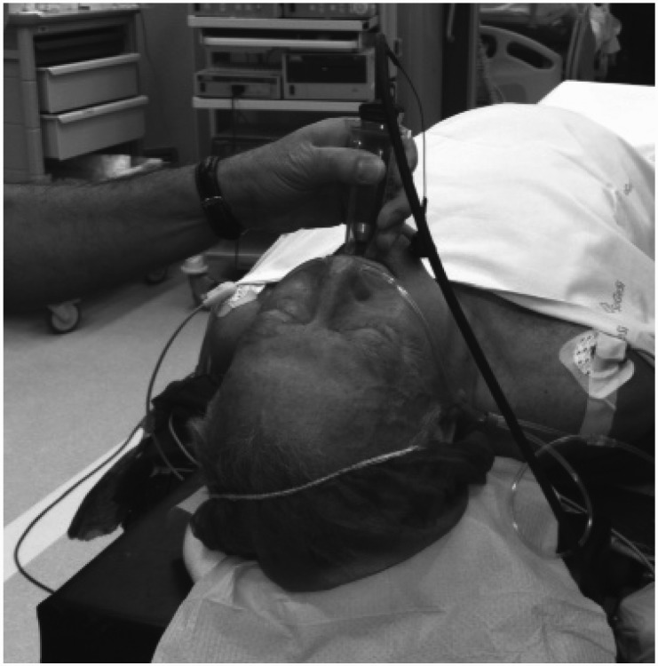
GlideScope® videolaryngoscopy offers several opportunities in many fields other than airway management for difficult intubations. A good exposure of upper aero-digestive tract, the possibility to register images and the short learning curve make it an interesting device to use in academic settings. Recently, tongue base surgery and foreign body removal were attempted under GlideScope® view, and seemed to show positive results. We believe that the advantages this technology may bring in upper airway surgery are numerous; for this reason, we tested a new potential use of GlideScope®, by performing laryngeal surgery in selected cases. The results were good, with no additional complication and a consistent time savings. Moreover, thanks to the possibility of achieving an optimal view of the laryngeal inlet, we could perform a relatively easy procedure even in patients with complicated anatomical features. We believe that this technique should be preferred over ambulatorial procedures, due to the easier handling of unforeseen complications such as bleeding or respiratory problems needing intubation (Fig. 4). We suggest limited use of GlideScope® videolaryngoscopy for selected patients who must undergo a surgical procedure for diagnostic and/or therapeutical reasons and present a high risk of intubation or adverse anatomical features.
Fig. 4.
GlideScope-guided procedures are executed in an operating theatre setting, with the help of an anaesthesiologist. We believe this makes this technique preferable over ambulatorial fibre optic laryngoscopy, especially in patients with relevant comorbid conditions.
References
- 1.Healy DW, Maties O, Hovord D, et al. A systematic review of the role of videolaryngoscopy in successful orotracheal intubation. BMC Anesthesiol. 2012;12:32–32. doi: 10.1186/1471-2253-12-32. [DOI] [PMC free article] [PubMed] [Google Scholar]
- 2.Shenoy PK, Aldea M. The use of GlideScope for biopsies of the tongue base. J Laryngol Otol. 2012;7:1–2. doi: 10.1017/S002221511200271X. [DOI] [PubMed] [Google Scholar]
- 3.Xue FS, Zhang GH, Liu J, et al. The clinical assessment of Glidescope in orotracheal intubation under general anesthesia. Minerva Anestesiol. 2007;73:451–457. [PubMed] [Google Scholar]
- 4.Niforopoulou P, Pantazopoulos I, Demestiha T, et al. Video-laryngoscopes in the adult airway management: a topical review of the literature. Acta Anaesthesiol Scand. 2010;54:1050–1061. doi: 10.1111/j.1399-6576.2010.02285.x. [DOI] [PubMed] [Google Scholar]


