

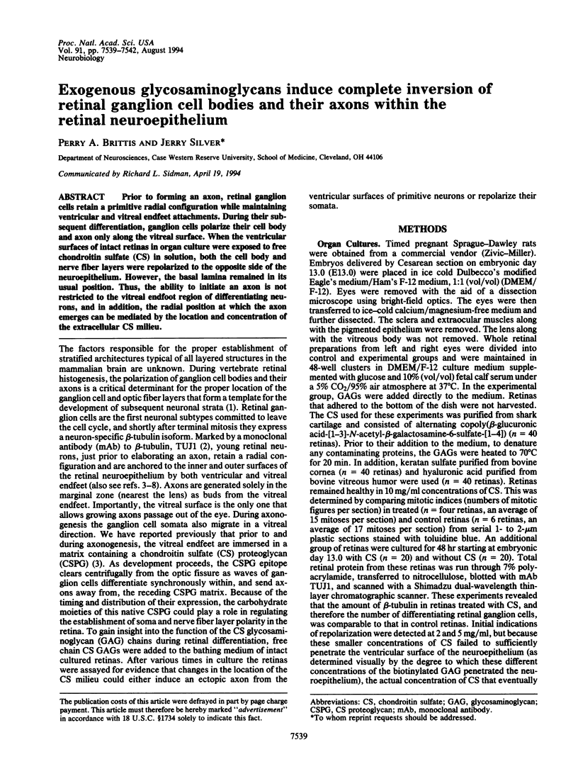
Abstract
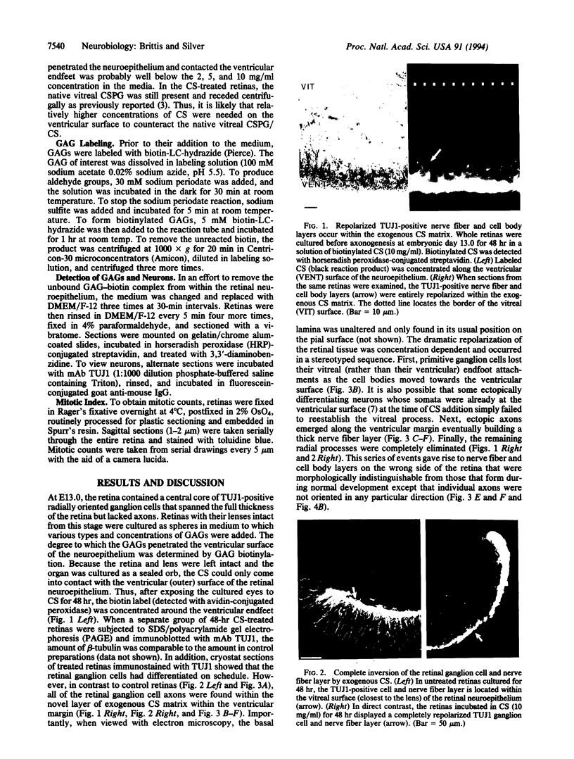
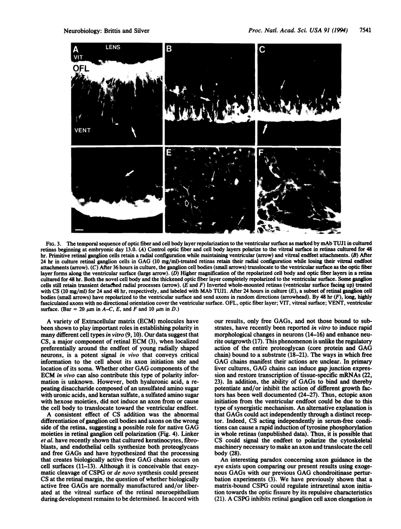
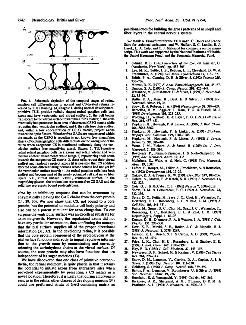
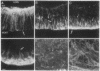
Prior to forming an axon, retinal ganglion cells retain a primitive radial configuration while maintaining ventricular and vitreal endfeet attachments. During their subsequent differentiation, ganglion cells polarize their cell body and axon only along the vitreal surface. When the ventricular surfaces of intact retinas in organ culture were exposed to free chondroitin sulfate (CS) in solution, both the cell body and nerve fiber layers were repolarized to the opposite side of the neuroepithelium. However, the basal lamina remained in its usual position. Thus, the ability to initiate an axon is not restricted to the vitreal endfoot region of differentiating neurons, and in addition, the radial position at which the axon emerges can be mediated by the location and concentration of the extracellular CS milieu.
Full text
PDF
Images in this article


Selected References
These references are in PubMed. This may not be the complete list of references from this article.
- Barcellos-Hoff M. H., Aggeler J., Ram T. G., Bissell M. J. Functional differentiation and alveolar morphogenesis of primary mammary cultures on reconstituted basement membrane. Development. 1989 Feb;105(2):223–235. doi: 10.1242/dev.105.2.223. [DOI] [PMC free article] [PubMed] [Google Scholar]
- Bicknese A. R., Sheppard A. M., O'Leary D. D., Pearlman A. L. Thalamocortical axons extend along a chondroitin sulfate proteoglycan-enriched pathway coincident with the neocortical subplate and distinct from the efferent path. J Neurosci. 1994 Jun;14(6):3500–3510. doi: 10.1523/JNEUROSCI.14-06-03500.1994. [DOI] [PMC free article] [PubMed] [Google Scholar]
- Brittis P. A., Canning D. R., Silver J. Chondroitin sulfate as a regulator of neuronal patterning in the retina. Science. 1992 Feb 7;255(5045):733–736. doi: 10.1126/science.1738848. [DOI] [PubMed] [Google Scholar]
- Cole G. J., McCabe C. F. Identification of a developmentally regulated keratan sulfate proteoglycan that inhibits cell adhesion and neurite outgrowth. Neuron. 1991 Dec;7(6):1007–1018. doi: 10.1016/0896-6273(91)90345-z. [DOI] [PubMed] [Google Scholar]
- Damon D. H., D'Amore P. A., Wagner J. A. Sulfated glycosaminoglycans modify growth factor-induced neurite outgrowth in PC12 cells. J Cell Physiol. 1988 May;135(2):293–300. doi: 10.1002/jcp.1041350217. [DOI] [PubMed] [Google Scholar]
- Dow K. E., Mirski S. E., Roder J. C., Riopelle R. J. Neuronal proteoglycans: biosynthesis and functional interaction with neurons in vitro. J Neurosci. 1988 Sep;8(9):3278–3289. doi: 10.1523/JNEUROSCI.08-09-03278.1988. [DOI] [PMC free article] [PubMed] [Google Scholar]
- Dunlop S. A. Early development of retinal ganglion cell dendrites in the marsupial Setonix brachyurus, quokka. J Comp Neurol. 1990 Mar 15;293(3):425–447. doi: 10.1002/cne.902930307. [DOI] [PubMed] [Google Scholar]
- Goldberg S. Polarization of the avian retina. Ocular transplantation studies. J Comp Neurol. 1976 Aug 1;168(3):379–391. doi: 10.1002/cne.901680305. [DOI] [PubMed] [Google Scholar]
- Hay E. D. Matrix-cytoskeletal interactions in the developing eye. J Cell Biochem. 1985;27(2):143–156. doi: 10.1002/jcb.240270208. [DOI] [PubMed] [Google Scholar]
- Jackson R. L., Busch S. J., Cardin A. D. Glycosaminoglycans: molecular properties, protein interactions, and role in physiological processes. Physiol Rev. 1991 Apr;71(2):481–539. doi: 10.1152/physrev.1991.71.2.481. [DOI] [PubMed] [Google Scholar]
- Lafont F., Rouget M., Triller A., Prochiantz A., Rousselet A. In vitro control of neuronal polarity by glycosaminoglycans. Development. 1992 Jan;114(1):17–29. doi: 10.1242/dev.114.1.17. [DOI] [PubMed] [Google Scholar]
- Lee M. K., Tuttle J. B., Rebhun L. I., Cleveland D. W., Frankfurter A. The expression and posttranslational modification of a neuron-specific beta-tubulin isotype during chick embryogenesis. Cell Motil Cytoskeleton. 1990;17(2):118–132. doi: 10.1002/cm.970170207. [DOI] [PubMed] [Google Scholar]
- Morest D. K. The pattern of neurogenesis in the retina of the rat. Z Anat Entwicklungsgesch. 1970;131(1):45–67. doi: 10.1007/BF00518815. [DOI] [PubMed] [Google Scholar]
- Newgreen D. F., Scheel M., Kastner V. Morphogenesis of sclerotome and neural crest in avian embryos. In vivo and in vitro studies on the role of notochordal extracellular material. Cell Tissue Res. 1986;244(2):299–313. doi: 10.1007/BF00219205. [DOI] [PubMed] [Google Scholar]
- Oakley R. A., Tosney K. W. Peanut agglutinin and chondroitin-6-sulfate are molecular markers for tissues that act as barriers to axon advance in the avian embryo. Dev Biol. 1991 Sep;147(1):187–206. doi: 10.1016/s0012-1606(05)80017-x. [DOI] [PubMed] [Google Scholar]
- Oohira A., Matsui F., Katoh-Semba R. Inhibitory effects of brain chondroitin sulfate proteoglycans on neurite outgrowth from PC12D cells. J Neurosci. 1991 Mar;11(3):822–827. doi: 10.1523/JNEUROSCI.11-03-00822.1991. [DOI] [PMC free article] [PubMed] [Google Scholar]
- Piepkorn M., Hovingh P., Linker A. Glycosaminoglycan free chains. External plasma membrane components distinct from the membrane proteoglycans. J Biol Chem. 1989 May 25;264(15):8662–8669. [PubMed] [Google Scholar]
- Piepkorn M., Hovingh P., Linker A. Proteoglycan and glycosaminoglycan free chain expression in keratinocytes, endothelium, and mesenchymal cells. Biochem Biophys Res Commun. 1991 Sep 30;179(3):1281–1288. doi: 10.1016/0006-291x(91)91712-l. [DOI] [PubMed] [Google Scholar]
- Piepkorn M., Hovingh P., Linker A. Topography of proteoglycan and glycosaminoglycan free chain expression in 3T3 fibroblasts and human keratinocytes. J Invest Dermatol. 1992 Oct;99(4):386–389. doi: 10.1111/1523-1747.ep12616098. [DOI] [PubMed] [Google Scholar]
- Price L. K., Choi H. U., Rosenberg L., Stanley E. R. The predominant form of secreted colony stimulating factor-1 is a proteoglycan. J Biol Chem. 1992 Feb 5;267(4):2190–2199. [PubMed] [Google Scholar]
- Ruoslahti E., Yamaguchi Y. Proteoglycans as modulators of growth factor activities. Cell. 1991 Mar 8;64(5):867–869. doi: 10.1016/0092-8674(91)90308-l. [DOI] [PubMed] [Google Scholar]
- Snow D. M., Lemmon V., Carrino D. A., Caplan A. I., Silver J. Sulfated proteoglycans in astroglial barriers inhibit neurite outgrowth in vitro. Exp Neurol. 1990 Jul;109(1):111–130. doi: 10.1016/s0014-4886(05)80013-5. [DOI] [PubMed] [Google Scholar]
- Snow D. M., Letourneau P. C. Neurite outgrowth on a step gradient of chondroitin sulfate proteoglycan (CS-PG). J Neurobiol. 1992 Apr;23(3):322–336. doi: 10.1002/neu.480230311. [DOI] [PubMed] [Google Scholar]
- Snow R. L., Robson J. A. Ganglion cell neurogenesis, migration and early differentiation in the chick retina. Neuroscience. 1994 Jan;58(2):399–409. doi: 10.1016/0306-4522(94)90046-9. [DOI] [PubMed] [Google Scholar]
- Spray D. C., Fujita M., Saez J. C., Choi H., Watanabe T., Hertzberg E., Rosenberg L. C., Reid L. M. Proteoglycans and glycosaminoglycans induce gap junction synthesis and function in primary liver cultures. J Cell Biol. 1987 Jul;105(1):541–551. doi: 10.1083/jcb.105.1.541. [DOI] [PMC free article] [PubMed] [Google Scholar]
- Verna J. M., Fichard A., Saxod R. Influence of glycosaminoglycans on neurite morphology and outgrowth patterns in vitro. Int J Dev Neurosci. 1989;7(4):389–399. doi: 10.1016/0736-5748(89)90060-9. [DOI] [PubMed] [Google Scholar]
- Watanabe M., Rutishauser U., Silver J. Formation of the retinal ganglion cell and optic fiber layers. J Neurobiol. 1991 Jan;22(1):85–96. doi: 10.1002/neu.480220109. [DOI] [PubMed] [Google Scholar]
- Wolburg H., Willbold E., Layer P. G. Müller glia endfeet, a basal lamina and the polarity of retinal layers form properly in vitro only in the presence of marginal pigmented epithelium. Cell Tissue Res. 1991 Jun;264(3):437–451. doi: 10.1007/BF00319034. [DOI] [PubMed] [Google Scholar]