Abstract
Aim
The aim of this study was to evaluate the effect of using the lateral wall bone in sinus lifting two-dimensional reconstruction on bone augmentation.
Patients and Methods
Ten patients affected by class V or VI maxillary atrophy with less than 3 mm of residual horizontal ridge were selected. Using a piezo-ultrasonic surgery tip bony lateral wall was cut. To expose native bone to the bone graft, multiple perforations, made through the cortical plate of the recipient site with a round bur. Once the bony buccal wall was adjusted it was fixed away from the ridge with two 1.5 x 13 mm bone fixation screws. Deficiencies created between the bony buccal wall and the ridge was filled with a mineralized cortical bone. A pericardium membrane was then placed on the graft. A biopsy for histologic evaluation was made.
Results
The data analysis in bone volume changes reported significant differences between the anterior and posterior locations before and after grafting (p < 0.05). The biopsy shows mature cancellous bone with predominantly lamellar structure.
Conclusion
The use of the lateral wall bone in sinus lift surgery showed significant increase in bone volume.
Keywords: Sinus lift, Grafting, Atrophic maxilla, Bone reconstruction
Introduction
Subantral augmentation or sinus lift is a common surgical procedure used to increase the height of the sinus floor in the posterior maxilla to create adequate bone volume for dental implant placement [1]. This surgical technique consists of surgically accessing the sinus cavity. In the transalveolar or crestal approach, the sinus is accessed through an upward fracture of the sinus floor with an osteotome inserted into a partially prepared alveolar osteotomy [2] or a tooth extraction socket [3]. In the lateral approach, an osteotomy of the lateral buccal wall is performed to create a window into the maxillary sinus. Once the sinus has been accessed, the Schneiderian membrane is carefully elevated away from the sinus floor. Bone graft material is then placed between the local host bone and the elevated membrane to enable simultaneous or delayed placement of dental implants [1, 4]. The long-term success rate of dental implants increases when bone graft materials are replaced or surrounded by newly formed bone, which grows from the patient’s existing bone into the augmented area [5].
The osteoconductive activity of various bone substitutes has been assessed according to the quality and quantity of newly formed bone in the augmented area [6]. In contrast, several recent clinical studies have suggested that bone augmentation may be achieved simply by elevating the Schneiderian sinus membrane without placing any underlying bone graft materials [7], but additional data are needed before definitive conclusions can be drawn.
Guided bone regeneration has been successfully used to increase alveolar ridge width for dental implant placement [8]. However, in cases of severe or localized horizontal bone deficiencies, achieving sufficient soft tissue mobilization to ensure primary wound closure over the augmented area can be difficult. In such cases, a periosteal pocket flap design has been reported [9] to be a predictable alternative flap approach for the correction of severe or localized horizontal bone deficiencies [8].
The out-fracture osteotomy technique for sinus bone grafts, in which a complete 360-degree osteotomy, followed by bony window separation from the sinus membrane [10], has been used instead of the more common superior hinge or in-fracturing technique [1, 11]. Kahnberg et al. [12] demonstrated that, after release of the sinus mucosa, the bony window is fractured into the sinus cavity and subsequently forms a roof for the augmented sinus floor. The bony window has been used in some procedures to cover the grafted sinus in place of an absorbable barrier membrane [10]. In other procedures, the bony window has been crushed and mixed with the grafting material [13].
In cases where a lateral wall is greater than 2 mm in thickness, the thickness of the lateral wall can first be reduced by eroding cortical particulate from the cortical plate using a piezo-ultrasonic surgical osteoplasty insert or a manual bone scraper. The collected bone chips can be subsequently used as autogenous graft material [14].
The aim of this study was to evaluate the effect of using the lateral wall bone in sinus lifting 2-dimensional reconstruction on bone augmentation. The null hypothesis was that there was no significant bone change in bone volume after the described surgical technique.
Materials and Methods
This study was conducted in accordance with the Helsinki agreement for research on humans, and the study design was approved through the Institutional Review Board and Independent Ethics Committee of the School of Dentistry, Lebanese University, Beirut, Lebanon. Written informed consent was obtained from all participants in the study prior to treatment.
Patients consecutively treated from October 2009 to July 2010 were included in this study. Patients with a history of systemic or local contraindications, including uncontrolled diabetes, bruxism, smoking, active sinus pathology, or uncontrolled periodontal disease, were excluded from the study.
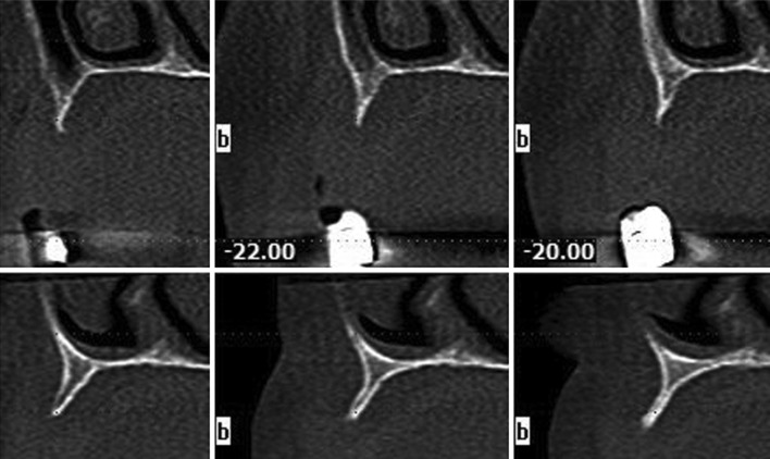
Preoperative radiographs, including panoramic radiography and cone beam computed tomography (CBCT) (Fig. 1), showed insufficient bone volume for the routine placement of implants in the posterior edentulous maxilla and deficiency of the bone width in the premolar region; therefore, sinus bone grafting using the lateral approach and 2-dimensional reconstruction (2DR) was indicated. The clinical and radiological findings were thoroughly discussed with all patients, and all available treatment options were explained. The patients provided written informed consent prior to surgery. Ten patients (five males and five females, mean age: 50 y/o), affected with class V or VI maxillary atrophy [4] with less than 3 mm of residual horizontal ridge [15] formed the study group.
Fig. 1.
CBCT of the atrophic maxilla class VI
Sinus Lift surgical technique surgery was performed using local analgesia (2 % articaine 1:100,000 adrenaline, 3M Espe, Seefeld, Germany). Prophylactic antibiotic treatment (2 g amoxicillin or 600 mg Dalacin C for patients allergic to penicillin) was prescribed 1 h before surgery, followed by 1 g/300 mg b.i.d for 5 days. Analgesic medication (600 mg ibuprofen, Abbott Healthcare Products Limited, UK) was also prescribed at 1 h before surgery, and the patients were advised to rinse their mouths daily with 0.12 % chlorhexidine gluconate oral rinse (PerioGard, Colgate-Palmolive, UK) during healing.
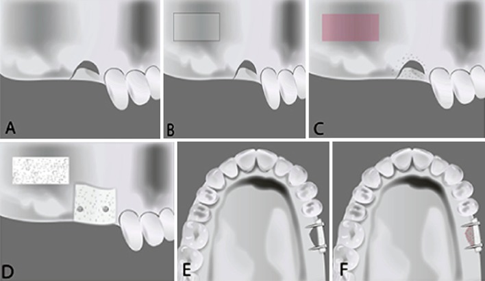
A horizontal incision was made along the crestal bone in the edentulous area, and vertical releasing incisions were made at each end of the crestal incision. A full-thickness mucoperiosteal flap was elevated to access the anterior bony wall of the maxillary sinus. The window of the lateral maxillary wall was designed according to the planned location(s) of the implant(s) and the anatomy of the maxillary sinus (Figs. 2, 3), and was prepared by cutting the bony lateral buccal wall using a piezo-ultrasonic (EMS, Nyon, Switzerland) device with a surgical tip (Tip SC, EMS, Nyon, Switzerland) designed to minimize the risk of unintended sinus membrane perforation. After completion of a rectangular osteotomy and mobilization of the lateral window, a small periosteal elevator was inserted into the osteotomy line and the bony window was carefully detached from the sinus membrane using an elevated motion. The Schneiderian membrane was initially elevated with a detached membrane tip (Tip SL3, EMS, Nyon, Switzerland) from the piezo-ultrasonic surgical unit, and then with sinus curettes.
Fig. 2.
Diagrammatic representation of the surgical technique: A Pre-operative situation. B Osteotomy of the buccal wall of the sinus and cortical perforation of the bony defect. C Elevation of the buccal wall. D Sinus grafted and the buccal bone fixed to the bony defect. E Occlusal view of the buccal bone fixed with screws to the defect. F The space between the buccal bone and the bony defect filled with bone substitute material
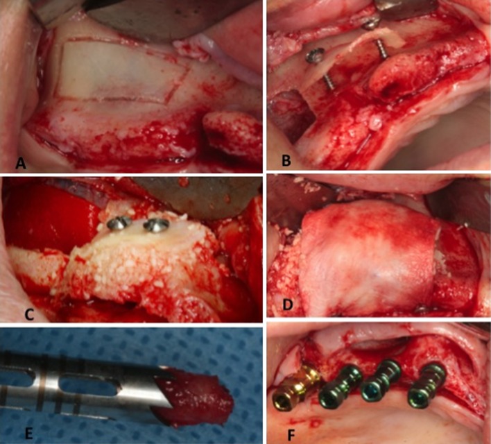
Fig. 3.
Clinical procedures: A Preparation of the buccal bony wall of the sinus. B Bony wall fixation away from the ridge using bone fixation screws. C Gap between the bony buccal wall and the ridge, filled with a mineralized cortical bone allograft. D Pericardium membrane placed on the graft and stabilized using four tacks. E The biopsy specimen was obtained from the augmented area using a trephine bur. F Implant placement in the augmented area and grafted sinus
Augmentation technique to induce bleeding and expose the native bone to the bone graft material
Multiple perforations were made through the cortical plate of the recipient site using a round bur. The sinus cavity was packed with a mineralized cortical bone allograft (MCBA) particulate (Puros Cortical Allograft, Zimmer Dental GmbH, Freiburg, Germany), and the excised bony buccal plate was adjusted and fixed away from the ridge using one anterior and one posterior bone screw (Salvin Dental Specialties, Inc., Charlotte, USA) (1.5 mm × 13 mm) (Fig. 3). Deficiencies of 0.25–1 mm created between the bony buccal plate and the ridge were filled with the MCBA material (Fig. 3).
A bovine pericardium membrane (CopiOs Pericardium Xenograft, Zimmer Dental GmbH) was placed over the graft site and four tacks were used to stabilize the membrane and protect the graft from invasion of the soft tissues (Fig. 3). The lateral window was covered with a collagen wound dressing (CollaTape, Zimmer Dental GmbH). Periosteal-releasing incisions were made, and the flaps were adjusted and closed using interrupted sutures (Vicryl 4/0, Ethicon Johnson & Johnson, Somerville, NJ, USA).
Clinical and radiologic follow-up
Patients underwent follow-up exams weekly during the first postoperative month, and monthly thereafter until re-entry surgery. Postoperative panoramic radiographs and CBCT scans were obtained. At 6 months postoperative, re-entry was performed using a similar flap design under local analgesia. After removing the fixation screws during implant placement, core biopsies were removed from the implant site using a trephine bur (outer diameter 3.5 mm, inner diameter 2.7 mm, and length 14 mm) for histological evaluation, and implants were placed in the augmented areas (Fig. 3). Implants (Tapered Screw-Vents, Zimmer Dental, Winterthur, Switzerland) (ranging from 11.5 to 13 mm in length and 4.1–4.7 mm in diameter), were placed according to the product’s instructions for use.
CBCT scans were performed before grafting and implant placement. The CBCT images were acquired using the i-CAT® classic system (Imaging Sciences International, Hatfield, PA, USA) with a voxel size of 0.2 mm and a 15 cm × 15 cm field of view (FOV). The exposure parameters were controlled by automatic exposure control. Overall, the exposure was performed at 120 kV and in a range of 18–48 mA. The CBCT images were exported from XoranCat® software in DICOM multi-file format and imported into ICATVision® software for carrying out the measurements.
Reproducibility of the paraxial cone beam cuts was assured using inter-premaxilla sutures perpendicular to the axial cuts as a reference for each patient. Two-dimensional paraxial slices with 0.40 mm slice thickness and 0.4 mm slice spacing were created in order to measure the buccal bone width using the re-slicing function of the software.
All CBCT measurements were made on standardized slices created at the level of bone screws.
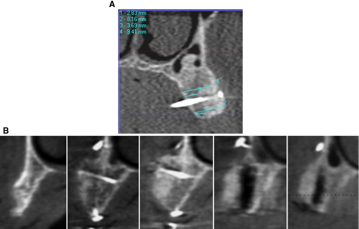
Buccal bone measurements of the grafted area were made parallel to the bone screws. Linear measurements of the horizontal augmentation area were recorded in millimeters at the crest, the middle and at the two fixation sites (Fig. 4).
Fig. 4.
A Paraxial cut of the CBCT showing the measurement. B Complete series of paraxial CBCT cuts of the same site during the complete treatment phase
Cemented definitive prostheses were delivered 2 months later. All implants were well integrated without any complications. Clinical and radiological follow up were performed after the first year of loading.
Specimen preparation for histological analysis after retrieval
The biopsy specimens were fixed in 4 % formaldehyde (Merck, Darmstadt, Germany), and dehydrated using a graded series of alcohol (70–100 % ethanol) for one week. Following dehydration, the specimens were infiltrated with a light-curing embedding resin (Technovit 9100, Heraeus Kulzer, Wehrheim, Germany) according to the manufacturer’s protocol. The specimens were prepared using the cutting/grinding method of Donath and Breuner [16]. The specimens were prepared in an apicocoronal direction parallel to the long axis and cut to a thickness of 150 μm using a cutting/grinding system (EXAKT Technologies, Inc., Oklahoma City, OK, USA). The cores were polished to a thickness of 45–65 μm using a micro grinding system with a series of polishing sandpaper disks from 800 to 2,400 grit, followed by a final polish with 0.3 μm alumina. The slides were stained with Giemsa-Paragon and cover-slipped for histological analysis using a light microscope equipped with a digital camera (Zeiss Axio Imager®; Carl Zeiss AG, Oberkochen, Germany).
Statistical analysis
A computer statistical software program (SPSS 18.0, SPSS Inc, Chicago, IL, USA) was used to evaluate bone levels before and after grafting at different locations (means and standard deviations). Changes in the bone volume were analyzed using Student’s t test (α = 0.05).
Results
Clinical evaluation
All patients healed without complications and were successfully treated with dental implants in the augmented sinus areas. Analysis of bone volume changes showed significant differences before and after grafting in both the anterior and posterior jaw locations (p < 0.05), but no significant differences was observed when anterior and posterior jaw locations were compared either before or after grafting (8.91 ± 1.16 and 8.13 ± 1.33, respectively). These data are reported in Table 1.
Table 1.
Bone volume changes before and after grafting (mm)
| Means and standard deviations | |||
|---|---|---|---|
| Jaw Region | Before grafting | After grafting | Percent (%) of change |
| Anterior | 2.52 ± 0.90a* | 8.13 ± 1.13b* | ±223 % |
| Posterior | 2.76 ± 0.97a | 8.91 ± 01.16b | ±223 % |
* Similar superscript letters indicate no significant difference (p < 0.05)
Histological findings
Biopsies showed mature cancellous bone with a predominantly lamellar structure. The well-vascularized intertrabecular spaces were filled with connective tissue and bone marrow, and there were no signs of acute inflammatory reactions (Fig. 5).
Fig. 5.
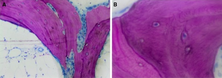
A The histological findings show mature cancellous bone with a predominantly lamellar structure (×40). B Higher magnification reveals a bone structure, with vital spongy bone trabeculae, enclosing numerous osteocytes within the lacunae (×100)
Higher magnifications revealed the bone structure, with new woven or lamellar bone and active osteocytes (Fig. 5).
Six months after surgery, the biopsies showed a composite formed with MCBA particles and newly formed bone trabeculae.
Discussion
The results of this study led to the rejection of the null hypothesis, stating that there will be no significant bone change after using the lateral wall bone in sinus lift.
Localized horizontal bone defects in the alveolar crest can be treated using different augmentation techniques and grafting protocols, depending on the morphology of the defect [17]. Slight horizontal defects can be solved using a single-stage augmentation procedure. In addition, a resorbable or non-resorbable membrane, with or without tenting screws, can be placed to prevent resorption and volume maintenance [17]. Hence, the reconstruction of large defects requires horizontal and/or vertical augmentation using autogenous bone grafts [17].
For the 2-dimensional reconstructions, the chin or retromolar regions were used to harvest intraoral bone grafts. Pikos [18] described the inlay technique, placing corticocancellous block grafts with accurate fit in the residual alveolar bone. Other studies have described a shell technique for 2-dimensional hard tissue grafting [19]. Thin cortical bone shells, harvested using a special diamond-cutting wheel from the retro molar region, were placed to reshape the alveolar crest and protect the particular bone, placed in the cavity between the shells, from resorption. These techniques required the recipient site to be augmented into a donor intra-oral site, resulting in two different surgical intervention surgeries. Despite the known complications of intra-oral bone harvesting, additional intra and postoperative stress for the patient have been observed, as demonstrated through patient statements and an increase in subjective post-operative swelling [13].
Different bone substitute materials, such as an allograft, have become established as alternatives to autogenous bone and serve as scaffolding for blood vessel invasion and hard tissue regeneration. These osteoconductive bone substitutes become integrated into the newly formed bone matrix [20]. In this study, we used MCBA based on encouraging documented reports of new bone formation [21]. Our histological results were consistent with those of Noumbissi et al. [22], Froum et al. [23] and Schmitt et al. [13]. Therefore, MCBA can be considered as a bone substitute material that is totally resorbed and replaced with autogenous bone, integrating well into the organism [23].
The described technique offers several clinical advantages as the entire sinus elevation and horizontal augmentation procedures were achieved in the same surgery without the necessity of exposing the patient to a second phase. Subsequently, the implants could be simultaneously placed in the grafted sinus and the augmented area. To improve the regeneration outcome of the augmented area, bone substitute materials can be separated from the surrounding connective tissue using barrier membranes, following the principle of guided bone regeneration [24].
Keeping the bone substitute isolated from the oral cavity prevents fibrous organization of the augmented material. Because of their compact origin, membranes from pericardial tissue show resorptive stability [25]. The compact interconnecting pericardium collagen might positively influence the resorption stability, supporting bone regeneration, particularly in larger augmentation volumes (resorbed without inflammation within 12 weeks) [25] .
The significant bone volume increase from 2.52 to 8.13 mm and from 2.76 to 8.91 mm observed after grafting may reflect the trans membrane penetration of blood vessels, which improves the blood supply of a graft, thereby encouraging tissue regeneration in bone reconstruction [26]. This hypothesis is supported through the basic principles of osteogenesis. It has been reported that osteogenetic cells develop from the undifferentiated mesenchymal progenitor cells of the bone marrow stroma and from the pericytes of smaller blood vessels in connective tissue [27]. Therefore, from a clinical point of view, several studies have recommended the perforation of the underlying compact bone in guided bone regeneration [28]. The opening of the bone marrow spaces facilitates improved bone regeneration due to increased rapid vascularization. However, other studies have not confirmed this observation [28]. The drawback of this technique is the risk of sinus membrane perforation during bony wall reflection or even bony wall fracture during fixation to the residual crest as previously described [29].
Conclusions
Within the limitations of this clinical study and the small number of cases observed, the following conclusions can be drawn:
The repositioning of the bony window of the lateral maxillary wall after osteotomy using a piezo-ultrasonic surgical device to reconstruct a 2-dimensional bony defect is a reproducible and reliable technique.
In most cases, the healing of the space between the repositioned window and the alveolar ridge was observed. The 6-month postoperative CBCT scan also confirmed the healing of bony defects.
The increase in the volume of the defects was significant and facilitated the placement of implants in an adequate position during a second phase.
The use of the bony wall to protect the bone particulate, thereby creating a box shape, prevented membrane collapse and provided an architectural design for the augmented area.
Acknowledgments
The authors thank Prof. Saad Khaillarah, Department of Histopathology, Faculty of Medicine, Lebanese University, for conducting a precise histological analysis and providing invaluable collaboration.
Ethical statemant
Authors received the ethical approval statement from 1374–5 Lebanese University.
Conflict of interest
None.
Footnotes
Disclaimers: The authors do not have any financial interests, either directly or indirectly, in the products or information listed in this paper.
References
- 1.Boyne PJ, James RA. Grafting of the maxillary sinus floor with autogenous marrow and bone. J Oral Surg. 1980;38:613–616. [PubMed] [Google Scholar]
- 2.Le Gall MG. Localized sinus elevation and osteocompression with single-stage tapered dental implants: technical note. Int J Oral Maxillofac Implants. 2004;19:431–437. [PubMed] [Google Scholar]
- 3.Kolhatkar S, Bhola M, Thompson-Sloan TN. Sinus floor elevation via the maxillary premolar extraction socket with immediate implant placement: a case series. J Periodontol. 2010;82:820–828. doi: 10.1902/jop.2010.100557. [DOI] [PubMed] [Google Scholar]
- 4.Hatano N, Shimizu Y, Ooya K. A clinical long-term radiographic evaluation of graft height changes after maxillary sinus floor augmentation with a 2:1 autogenous bone/xenograft mixture and simultaneous placement of dental implants. Clin Oral Implants Res. 2004;15:339–345. doi: 10.1111/j.1600-0501.2004.00996.x. [DOI] [PubMed] [Google Scholar]
- 5.Schlegel KA, Fichtner G, Schultze-Mosgau S, Wiltfang J. Histologic findings in sinus augmentation with autogenous bone chips vs. a bovine bone substitute. Int J Oral Maxillofac Implant. 2003;18:53–58. [PubMed] [Google Scholar]
- 6.Artzi Z, Nemcovsky CE, Tal H, Dayan D. Histopathological morphometric evaluation of 2 different hydroxyapatite-bone derivatives in sinus augmentation procedures: a comparative study in humans. J Periodontol. 2001;72:911–920. doi: 10.1902/jop.2001.72.7.911. [DOI] [PubMed] [Google Scholar]
- 7.Ellegaard B, Kolsen-Petersen J, Baelum V. Implant therapy involving maxillary sinus lift in periodontally compromised patients. Clin Oral Implants Res. 1997;8:305–315. doi: 10.1034/j.1600-0501.1997.080409.x. [DOI] [PubMed] [Google Scholar]
- 8.Urban IA, Nagursky H, Lozada JL, Nagy K (2103) Horizontal ridge augmentation with a collagen membrane and a combination of particulated autogenous bone and anorganic bovine bone-derived mineral: a prospective case series in 25 patients. Int J Periodontics Restorative Dent 33:2 99–307. doi:10.11607/prd.1407 [DOI] [PubMed]
- 9.Steigmann M, Salama M, Wang HL. Periosteal pocket flap for horizontal bone regeneration: a case series. Int J Periodontics Restorative Dent. 2012;32:311–320. [PubMed] [Google Scholar]
- 10.Lee JK. Outfracture osteotomy on lateral maxillary wall as a modified sinus graft techniques. J Oral Maxillofac Surg. 2012;68:1639–1641. doi: 10.1016/j.joms.2009.07.067. [DOI] [PubMed] [Google Scholar]
- 11.Wallace SS, Froum SJ, Cho SC, Elian N, Monteiro D, Kim BS, Tarnow DP. Sinus augmentation utilizing anorganic bovine bone (Bio-Oss) with absorbable and nonabsorbable membranes placed over the lateral window: histomorphometric and clinical analyses. Int J Periodontics Restorative Dent. 2005;25:551–559. [PubMed] [Google Scholar]
- 12.Kahnberg K-E, Ekestubbe A, Grondahl K, Nilsson P, Hirsch J-M. Sinus lifting procedure. I. One-stage surgery with bone transplants and implants. Clin Oral Implants Res. 2001;12:479–487. doi: 10.1034/j.1600-0501.2001.120508.x. [DOI] [PubMed] [Google Scholar]
- 13.Schmitt CM, Doering H, Schmidt T, Lutz R, Neukam FW, Schlegel KA. Histological results after maxillary sinus augmentation with Straumann® BoneCeramic, Bio-Oss®, Puros®, and autologous bone. A randomized controlled clinical trial. Clin Oral Implants Res. 2013;24:576–585. doi: 10.1111/j.1600-0501.2012.02431.x. [DOI] [PubMed] [Google Scholar]
- 14.Tarnow DP, Wallace SS, Testori T, Froum SJ, Motroni A, Prasad HS. Maxillary sinus augmentation using recombinant bone morphogenetic protein-2/acellular collagen sponge in combination with a mineralized bone replacement graft: a report of three cases. Int J Periodontics Restorative Dent. 2010;30:139–149. [PubMed] [Google Scholar]
- 15.Cawood J, Howell R. A classification of the edentulous jaws. Int J Oral Maxillofac Surg. 1988;17:234–236. doi: 10.1016/S0901-5027(88)80047-X. [DOI] [PubMed] [Google Scholar]
- 16.Donath K, Breuner G. A method for the study of undecalcified bones and teeth with attached soft tissues. The Säge-Schliff (sawing and grinding) technique. J Oral Pathol. 1982;11:318–326. doi: 10.1111/j.1600-0714.1982.tb00172.x. [DOI] [PubMed] [Google Scholar]
- 17.Jensen SS, Terheyden H. Bone augmentation procedures in localized defects in the alveolar ridge: clinical results with different bone grafts and bone-substitute materials. Int J Oral Maxillofac Implants. 2009;24(Suppl):218–236. [PubMed] [Google Scholar]
- 18.Pikos MA. Block autografts for localized ridge augmentation: Part II. The posterior mandible. Implant Dent. 2000;9:67–75. doi: 10.1097/00008505-200009010-00012. [DOI] [PubMed] [Google Scholar]
- 19.Khoury F, Khoury C (2006) Mandibular bone block grafts: diagnosis, instrumentation, harvesting techniques and surgical procedures. In: Khoury F, Antoun H, Missika P (eds). Bone augmentation in oral implantology. Quintessence, Chicago, pp 115–212
- 20.Garofalo GS. Autogenous, allogenetic and xenogenetic grafts for maxillary sinus elevation: literature review, current status and prospects. Minerva Stomatol. 2007;56:373–392. [PubMed] [Google Scholar]
- 21.Minichetti JC, D’Amore JC, Hong AY, Cleveland DB. Human histologic analysis of mineralized bone allograft (Puros) placement before implant surgery. J Oral Implantol. 2004;30:74–82. doi: 10.1563/0.693.1. [DOI] [PubMed] [Google Scholar]
- 22.Noumbissi SS, Lozada JL, Boyne PJ, Rohrer MD, Clem D, Kim JS, Prasad H. Clinical, histologic, and histomorphometric evaluation of mineralized solvent-dehydrated bone allograft (Puros) in human maxillary sinus grafts. J Oral Implantol. 2005;31:171–179. doi: 10.1563/1548-1336(2005)31[171:CHAHEO]2.0.CO;2. [DOI] [PubMed] [Google Scholar]
- 23.Froum SJ, Wallace SS, Elian N, Cho SC, Tarnow DP. Comparison of mineralized cancellous bone allograft (Puros) and anorganiz bovine bone matrix (Bio-Oss) for sinus augmentation: histomorphometry at 26 to 32 seeks after grafting. Int J Periodontics Restorative Dent. 2006;26:543–551. [PubMed] [Google Scholar]
- 24.Dahlin C, Linde A, Gottlow J, Nyman S. Healing of bone defects by guided tissue regeneration. Plast Reconstr Surg. 1988;81:672–676. doi: 10.1097/00006534-198805000-00004. [DOI] [PubMed] [Google Scholar]
- 25.Rothamel D, Ferrari D, Fienitz T, Smeets R, Dreiseidler T, Ritter L, Happe A, Zöller J. Biocompatibility and biodegradation of a native porcine pericardium membrane: results of in vitro and in vivo examinations. Int J Oral Maxillofac Implants. 2012;27:146–154. [PubMed] [Google Scholar]
- 26.Hardwick R, Scantlebury TV, Sanchez R, Whitley N, Ambruster J. Membrane design criteria for guided bone regeneration of the alveolar ridge. In: Buser D, Dahlin C, Schenk RK, editors. Guided bone regeneration in implant dentistry. Chicago: Quintessence; 1994. p. 101. [Google Scholar]
- 27.Long MW, Robinson JA, Ashcraft EA, Mann KG. Regulation of human bone marrow-derived osteoprogenitor cells by osteogenic growth factors. J Clin Invest. 1995;95:881–887. doi: 10.1172/JCI117738. [DOI] [PMC free article] [PubMed] [Google Scholar]
- 28.Buser D, Bragger U, Lang NP, Nyman S. Regeneration and enlargement of jaw bone using guided tissue regeneration. Clin Oral Implants Res. 1990;1:22–32. doi: 10.1034/j.1600-0501.1990.010104.x. [DOI] [PubMed] [Google Scholar]
- 29.Vercellotti T, De Paoli S, Nevins M. The piezoelectric bony window osteotomy and sinus membrane elevation: introduction of a new technique for simplification of the sinus augmentation procedure. Int J Periodontics Restorative Dent. 2001;21:561–567. [PubMed] [Google Scholar]


