

Abstract
Introduction
Oroantral communications (OAC) are probable surgical complications of dentoalveolar procedures. OACs 2 mm in diameter or smaller are likely to close spontaneously without the need for any surgical intervention. However, OACs 3 mm in diameter or larger, or OACs associated with maxillary or periodontal inflammation, may persist, and surgical closure is recommended. Various surgical techniques have been suggested for the closure of oroantral defects.
Case Details
We have found the technique of two layer closure with buccal fat pad (BFP) and buccal mucoperiosteum quite useful for closure of chronic Oroantral fistula (OAF) and this article reports a case of OAF in the left first molar region of a 50 year old male, which has been closed successfully with this technique.
Conclusion
Buccal fat pad is a pedicled locally available flap which has its own blood supply and hence can be used with great efficacy in closure of OAF. This paper aims to elaborate the surgical details of this technique and its usefulness in closure of chronic OAF.
Keywords: Oro antral fistula, Two layer closure, Buccal fat pad, Oro antral communication, Buccal advancement flap
Introduction
Oroantral communications (OAC) are uncommon surgical complications of dentoalveolar procedures [1]. An oroantral fistula is a pathological condition in which the oral and antral cavities have a permanent communication by means of a fibrous connective tissue fistula coated by epithelium.
OACs 2 mm in diameter or smaller are likely to close spontaneously without the need for surgical intervention [1]. However, OACs 3 mm in diameter or larger, or OACs associated with maxillary sinusitis or periodontal inflammation, may persist, and surgical closure is recommended. Various surgical techniques have been suggested for the closure of oral defects such as primary closure, buccal mucosal graft, split thickness skin graft, allogenic graft, regional rotational flap, distant flap, mucoperiosteal flaps (vestibular, palatine, lingual or combined), bone grafts, or buccal fat pad grafts (Bichat ball) [1]. The type and size of the defect determine the technique to be used.
The use of the buccal fat pad (BFP) as a grafting source in the closure of intra-oral defects has gained popularity in the last quarter of twentieth century. Its use as a pedicle graft for oral reconstruction was first reported by Egyedi in 1977 [2].The buccal flap technique can be satisfactorily employed in the treatment of small and medium-sized communications provided there is clear sinus lavage through OAF and proper antral regime has been maintained 4–5 days prior to the surgery. Studies have proved through series of cases that the use of buccal pad fat in the closure of OAC has significantly reduced the failure rate of the treatment. The aim of this case report presentation is to elaborate the surgical technique for the use of buccal pad fat in the closure of oro-antral fistula.
Case Report
A 50 year old male patient came to the maxillofacial department OP complaining of drainage through the nose while taking water in the mouth. He had undergone extraction of upper right first molar elsewhere, 11 days earlier due to caries. On clinical examination the socket appeared traumatic and margins of the socket were not healed properly and communication to antrum of size 5 × 5 mm was noted (Fig. 1). OPG revealed an oroantral communication at the extraction socket (Fig. 2). PNS view showed generalized haziness of the right antrum which was suggestive of sinusitis (Fig. 3). The treatment plan was to do surgical closure after following proper antral regime and till clear lavage was achieved.
Fig. 1.
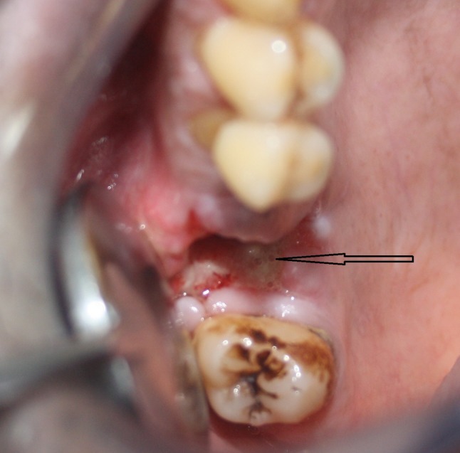
Oro antral fistula seen at upper right first molar traumatic extraction site
Fig. 2.
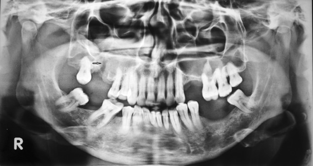
OPG showing the communication between maxillary sinus and extraction socket. (Note the arrow)
Fig. 3.

PNS view skull showing right maxillary sinus haziness
Surgical Technique in Detail
The procedure was done under local anesthesia. The area to be surgically closed was anesthetized by posterior superior alveolar nerve block or buccal infiltration and greater palatine nerve block anesthesia. The fistulous tract around the socket was excised using No. 11 blade. For a successful surgical closure the extracted socket was curetted to remove any granulation tissue. The buccal and palatal margins of the fistula were freshened with No 15 blade. The palatal margin was freshened in a semilunar shape for easier adaptation of the buccal flap to the palatal margin. An extended trapezoidal flap from the socket margin to the buccal vestibule was made. Once the incision was placed flap was raised and periosteal scoring was done to advance and release the mucoperiosteal flap. At the posterior most point of the flap using mosquito forceps the periosteum was teased open upwards till a bright yellow lobulated mass was seen popping out. The lobule was gently teased out till sufficient amount to close the oroantral fistula was available to reach the palatal margin (Fig. 4). Care was taken not to rupture the capsule of the BFP. The the BFP was anchored to the palatal margin using 3-‘0’ non cutting vicryl rapid (Fig. 5). After this the buccal mucoperiosteal flap was raised and reapproximated to cover the BFP and flap was sutured to palatal margin using horizontal mattress suture (Fig. 6). Usually the posterior release of mucoperiosteal flap need not be closed as it will self approximate on sliding. The anterior release was closed with 3-‘0’ vicryl rapid. Patient is then under strict follow up and the antral regime was continued for five more days.
Fig. 4.

Buccal fat pad being teased out from the buccal vestibule
Fig. 5.

The buccal fat pad being advanced and sutured to the palatal margin
Fig. 6.

The buccal mucoperiosteal flap sutured to the palatal margin and covering the buccal fat pad
Discussion
The BFP as an anatomic element was first mentioned by Heister in 1732 and was described by Bichat in 1802 [3]. The BFP is an anatomically rounded and biconvex structure that is of great importance in the facial contour. It is an adipose tissue surrounded by a thin capsule and located inside both masticatory spaces in the oromaxillofacial region. The BFP has a central body with four extensions: pterygopalatine, temporal, pterygoid, and buccal [4]. The central body and buccal extension account for approximately 50 % of the BFP and are the most clinically significant portions. The BFP has a 10-mL volume and a thickness of 6 mm and an approximate weight of 9.3 g [4]. The BFP is surrounded by a thin fibrous capsule. Blood supply is provided by the vestibular and deep branches of the maxillary artery, the transverse facial branches of the superficial temporal artery and branches of the facial artery. The rich blood supply may explain the high success rate. It also may be one reason for the quick epithelialization of the fat. The BFP is a mass of specialized fatty tissue called syssarcosis, a fat that enhances muscular motion. It is distinct from the subcutaneous fat and shows marked similarity to the orbital fat. It can easily cover small to medium sized defects of about 4 cm in diameter. When properly dissected and mobilized the BFP provides 7 × 4 × 3 cm of a pedicled graft.
The physiology of buccal fat tissue is not totally clarified. However, it is thought that the BFP is closely associated with the muscles of mastication. It plays an important role in masticatory function especially in the infant during suckling. Its size diminishes as the infant grows with the accompanying growth of the surrounding facial structures. In the adult, the BFP enhances inter-muscular motion and resembles orbital fat in appearance and function.
The histological nature of the healing process of the BFP was first reported by Samman et al. [5]. He stated that no fat cells were seen in sections taken from healed sites, indicating at least partial fibrosis of the fat tissue. Fujimura et al. [6] showed that BFP started to epithelialize in a week and completed its epithelialization within 6 weeks. Here we could see that complete epithelialization occurred after 2 months (Fig. 7).
Fig. 7.

Excellent healing of the closure site
The BFP structure can be used in the correction of several oral defects, such as fistulas and OAC; in reconstruction after tumor resection; in rehabilitation of cleft patients; in aesthetic corrections of the face; and in implant-graft coating [7]. The scope of defects that can be treated using the pedicled BFP flap varies according to the patient’s morphology, as this structure varies in dimension from one individual to another [8].
Oral defect closure using the BFP has been increasingly employed because it is a fast surgical procedure, is relatively easy to perform and has a high success rate. It is important to preserve the thin capsule of the BFP in order not to damage the small blood vessels.
To date, reported complications with the use of the BFP flap are haematoma, partial necrosis, excessive scarring, infection or facial nerve injury [9]. It must be kept in mind that BFP should be exposed by blunt dissection without causing any tension to pull it out [10]. The use of the BFP in patients with prior local radiotherapy, malar hypoplasia or thin cheeks are relative contraindications.
Conclusion
The closure of OAF with single layered buccal mucoperiosteal sliding flaps (Berger flap/Reherman flap) has been well documented in literature. Without much change in the surgical steps adding a second layer of BFP will enhance the success rate of closure. Here we have proved that the two layer closure technique is very much successful in the management of a chronic oro-antral fistula with sinusitis which was caused by traumatic extraction of first molar.
Conflict of interest
None.
References
- 1.Awang MN. Closure of oroantral fistula. Int J Oral Maxillofac Surg. 1988;17:110–115. doi: 10.1016/S0901-5027(88)80162-0. [DOI] [PubMed] [Google Scholar]
- 2.Egyedi P. Utilization of the buccal fat pad for closure of oro-antral and/or oro-nasal communications. J Maxillofac Surg. 1977;5:241. doi: 10.1016/S0301-0503(77)80117-3. [DOI] [PubMed] [Google Scholar]
- 3.Bichat FMX (1801) Anatomie Generale: Appliquee a la Physiologie et la Medecine. Paris. Cited in Stuzin JM, Wagstrom L, Kawamoto HK, Baker TJ, Wolfe A (1990) The anatomy and clinical applications of the buccal fat pad. Plast Reconstr Surg 85:29 [DOI] [PubMed]
- 4.Saha J, Pathak H, Poddar RN. Use of buccal fat pad to repair intraoral defects. J Indian Dent Assoc. 2011;1(1):1–5. [Google Scholar]
- 5.Samman N, Cheung LK, Tideman H. The buccal fat pad in oral reconstruction. Int J Oral Maxillofac Surg. 1993;22:2–6. doi: 10.1016/S0901-5027(05)80346-7. [DOI] [PubMed] [Google Scholar]
- 6.Fujimura N, Nagura H, Enomoto S. Grafting of the buccal fat pad into palatal defects. J Craniomaxillofac Surg. 1990;18:219–222. doi: 10.1016/S1010-5182(05)80415-9. [DOI] [PubMed] [Google Scholar]
- 7.Batra H, Jindal G, Kaur S. Evaluation of different treatment modalities for closure of oro-antral communications and formulation of a rational approach. J Maxillofac Oral Surg. 2010;9(1):13–18. doi: 10.1007/s12663-010-0006-y. [DOI] [PMC free article] [PubMed] [Google Scholar]
- 8.Abad-Gallegos M, Figueiredo R, Rodriguez-Baeza A, Gay-Escoda C. Use of Bichat’s buccal fat pad for the sealing of orosinusal communicatins: a presentation of 8 cases. Med Oral Patol Oral Cir Bucal. 2011;6(2):e215–e219. doi: 10.4317/medoral.16.e215. [DOI] [PubMed] [Google Scholar]
- 9.Scott P, Fabbroni G, Mitchell DA. The buccal fat pad in the closure of oro-antral communications: an illustrated guide. Dent Update. 2004;31:363–366. doi: 10.12968/denu.2004.31.6.363. [DOI] [PubMed] [Google Scholar]
- 10.Nezafati S, Vafaii A, Ghojazedeh M. Comparison of pedicled buccal fat pad flap with buccal flap for closure of oro-antral communication. Int J Oral Maxillofac Surg. 2012;41:624–628. doi: 10.1016/j.ijom.2011.11.011. [DOI] [PubMed] [Google Scholar]