

Abstract
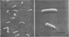
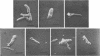
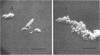
The morphology of cells of the H37Ra strain of Mycobacterium tuberculosis exposed to 0.5 μg of isonicotinic acid hydrazide (isoniazid) per ml was examined by scanning electron microscopy (SEM). Cells that were exposed to isoniazid for 3 h showed no detectable change, whereas cells exposed to the drug for 24 h exhibited diverse morphological features. From our examination of these SEM pictures, we have reconstructed the probable sequence of morphological changes to be as follows: (i) the wrinkling of the cell surface was ascribed as the earliest observable change, (ii) the cell surface then became very rough and ragged, (iii) eventually the cytoplasmic material was extruded from the cell, (iv) this event produced a collapsed cell, (v) the cells began to fragment, (vi) the fragmented cells then coalesced to form an amorphous mass of cell debris.
Full text
PDF


Images in this article
Selected References
These references are in PubMed. This may not be the complete list of references from this article.
- BARCLAY W. R., EBERT R. H., KOCHWESER D. Mode of action of isoniazid. Am Rev Tuberc. 1953 Apr;67(4):490–496. doi: 10.1164/art.1953.67.4.490. [DOI] [PubMed] [Google Scholar]
- BLOCH H. Studies on the virulence of tubercle bacilli; isolation and biological properties of a constituent of virulent organisms. J Exp Med. 1950 Feb;91(2):197-218, pl. doi: 10.1084/jem.91.2.197. [DOI] [PMC free article] [PubMed] [Google Scholar]
- Bartlett G. A. Scanning electron microscope: potentials in the morphology of microorganisms. Science. 1967 Dec 8;158(3806):1318–1319. doi: 10.1126/science.158.3806.1318. [DOI] [PubMed] [Google Scholar]
- Greenwood D., O'Grady F. Antibiotic-induced surface changes in microorganisms demonstrated by scanning electron microscopy. Science. 1969 Mar 7;163(3871):1076–1078. doi: 10.1126/science.163.3871.1076. [DOI] [PubMed] [Google Scholar]
- Imaeda T., Kanetsuna F., Galindo B. Ultrastructure of cell walls of genus Mycobacterium. J Ultrastruct Res. 1968 Oct;25(1):46–63. doi: 10.1016/s0022-5320(68)80059-0. [DOI] [PubMed] [Google Scholar]
- Kanetsuna F. Chemical analyses of mycobacterial cell walls. Biochim Biophys Acta. 1968 Apr 16;158(1):130–143. doi: 10.1016/0304-4165(68)90080-9. [DOI] [PubMed] [Google Scholar]
- Klainer A. S., Perkins R. L. Antibiotic-induced alterations in the surface morphology of bacterial cells: a scanning-beam electron miscroscopy study. J Infect Dis. 1970 Oct;122(4):323–328. doi: 10.1093/infdis/122.4.323. [DOI] [PubMed] [Google Scholar]
- Lederer E. The mycobacterial cell wall. Pure Appl Chem. 1971;25(1):135–165. doi: 10.1351/pac197125010135. [DOI] [PubMed] [Google Scholar]
- MIDDLEBROOK G. Sterilization of tubercle bacilli by isonicotinic acid hydrazide and the incidence of variants resistant to the drug in vitro. Am Rev Tuberc. 1952 Jun;65(6):765–767. [PubMed] [Google Scholar]
- Takayama K., Wang L., David H. L. Effect of isoniazid on the in vivo mycolic acid synthesis, cell growth, and viability of Mycobacterium tuberculosis. Antimicrob Agents Chemother. 1972 Jul;2(1):29–35. doi: 10.1128/aac.2.1.29. [DOI] [PMC free article] [PubMed] [Google Scholar]
- Wang L., Takayama K. Relationship between the uptake of isoniazid and its action on in vivo mycolic acid synthesis in Mycobacterium tuberculosis. Antimicrob Agents Chemother. 1972 Dec;2(6):438–441. doi: 10.1128/aac.2.6.438. [DOI] [PMC free article] [PubMed] [Google Scholar]
- Winder F. G., Collins P. B. Inhibition by isoniazid of synthesis of mycolic acids in Mycobacterium tuberculosis. J Gen Microbiol. 1970 Sep;63(1):41–48. doi: 10.1099/00221287-63-1-41. [DOI] [PubMed] [Google Scholar]