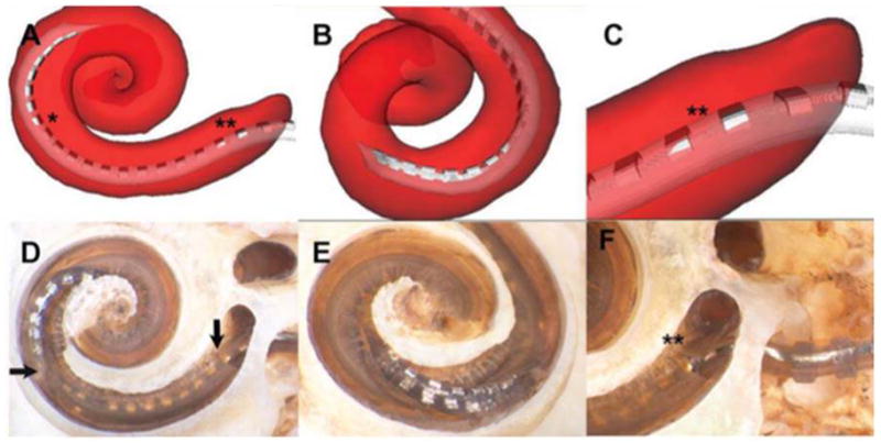
Figure 2.

Insertional trauma with array crossing basilar membrane at 170°. In this specimen, the image reconstruction shows that the array initially occupied the scala tympani (ST) after insertion through the cochleostomy but crossed the basilar membrane at approximately 170° to enter the scala vestibuli (SV) (asterisk on A). Microdissection verified this finding, illustrating a breach in the basilar membrane (horizontal arrow, D), distal to which the apical electrode contacts are inside the SV. B and E depict a rotated view of the point of basilar membrane crossing. The reconstruction also suggested subtle insertional trauma in the area of the hook (double asterisk, A and C); this was also verified upon microdissection, with a small fracture of the osseous lamina shown in D (vertical arrow) and F (double asterisk).
