Abstract
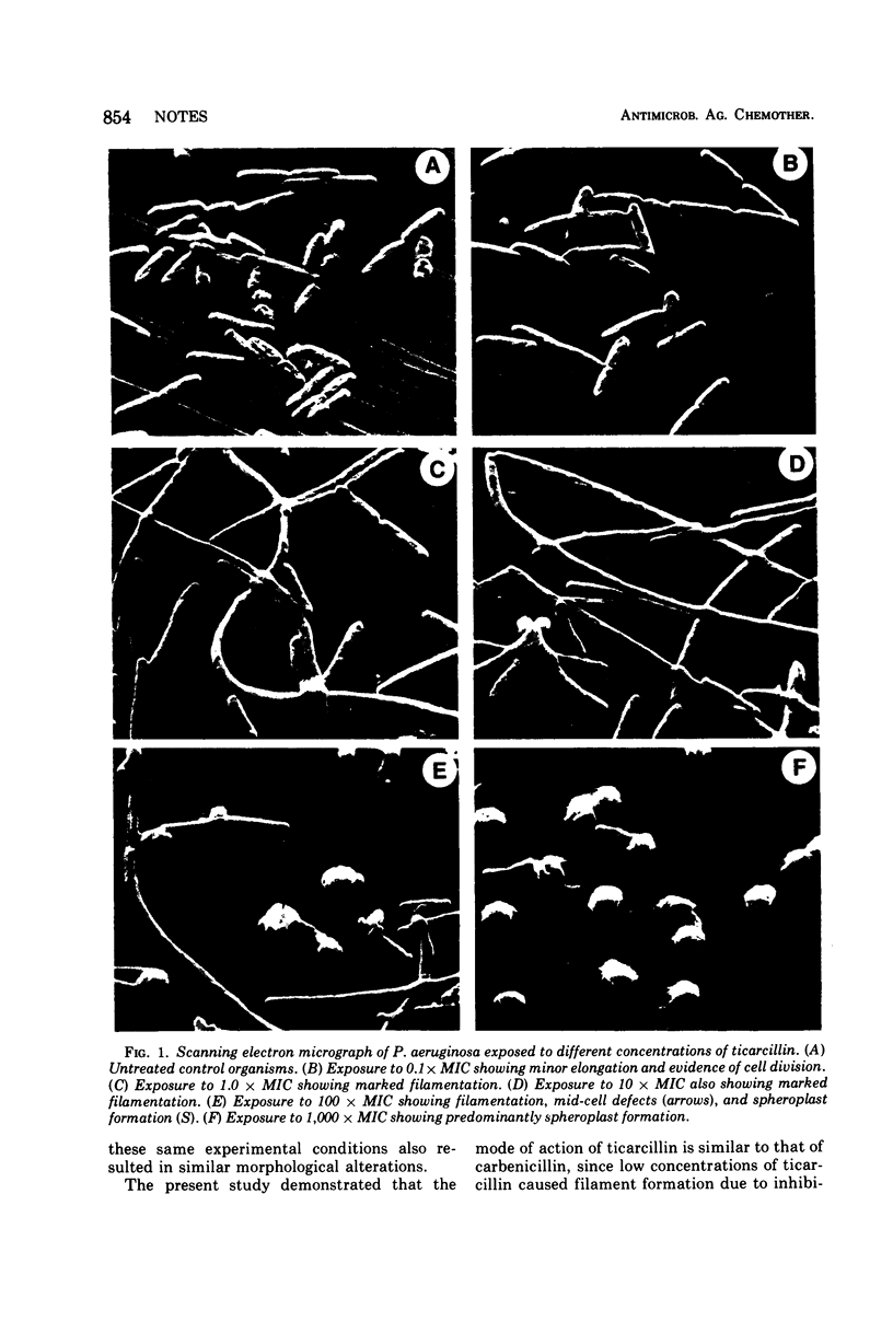
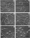
Pseudomonas aeruginosa was exposed to 0.1, 1.0, 10, 100, and 1,000 times the minimal inhibitory concentration of ticarcillin in vitro and subsequently examined with the scanning electron microscope. The morphological alterations observed were filamentation, mid-cell defects, and spheroplast formation, and these alterations were dependent upon the drug concentration.
Full text
PDF


Images in this article
Selected References
These references are in PubMed. This may not be the complete list of references from this article.
- Bodey G. P., Deerhake B. In vitro studies of alpha-carboxyl-3-thienylmethyl penicillin, a new semisynthetic penicillin. Appl Microbiol. 1971 Jan;21(1):61–65. doi: 10.1128/am.21.1.61-65.1971. [DOI] [PMC free article] [PubMed] [Google Scholar]
- Gardner W. G., Prior R. B., Perkins R. L. Fluid and pharmacological dynamics in a subcutaneous chamber implanted in rats. Antimicrob Agents Chemother. 1973 Aug;4(2):196–197. doi: 10.1128/aac.4.2.196. [DOI] [PMC free article] [PubMed] [Google Scholar]
- Prior R. B., Perkins R. L. Artifacts induced by preparation for scanning electron microscopy, in Proteus mirabilis exposed to carbenicillin. Can J Microbiol. 1974 May;20(5):794–795. doi: 10.1139/m74-122. [DOI] [PubMed] [Google Scholar]


