Abstract
A 20-year-old male was evaluated for winging of the scapula and an enlarging axillary mass of 4 months’ duration. Imaging demonstrated a multiloculated cystic lesion that extended into the axilla and superiorly displaced the brachial plexus and axillary vessels surrounding an exostotic mass arising from the scapula. Surgery confirmed the mass to be a benign osteochondroma with a reactive bursa. The long thoracic nerve was intact and the serratus anterior muscle contracted normally with nerve stimulation. The scapular winging resolved completely following resection of the osteochondroma, and shoulder and arm function remained normal. A literature review of causes of pseudo-winging of the scapula was performed. Scapular osteochondroma is a rarely reported cause of scapula winging.
Background
Winging of the scapula occurs when the medial border of the scapula is more prominent than normal. Classically, this is caused by injury or dysfunction of the long thoracic nerve with subsequent paralysis of the serratus anterior muscle. Winging of the scapula due to other causes is known as pseudo-winging. Multiple causes of pseudo-winging have been reported in the literature secondary to shoulder girdle instability, displaced fractures and malunions, and bony tumors [3, 8]. Osteochondroma of the scapula is a rare cause of scapular pseudo-winging. Here, we present a review of the literature and a case report of an isolated osteochondroma of the scapula which presented with pseudo-winging and a mass.
Case Report
A 20-year-old right-hand-dominant male was referred for evaluation of left scapula winging and an enlarging painless axillary mass for 4 months. He had some catching and clunking in his left shoulder with range of motion but no weakness or sensory changes of his left upper extremity. He was otherwise healthy except for a remote history of repair of a congenital heart defect through a median sternotomy.
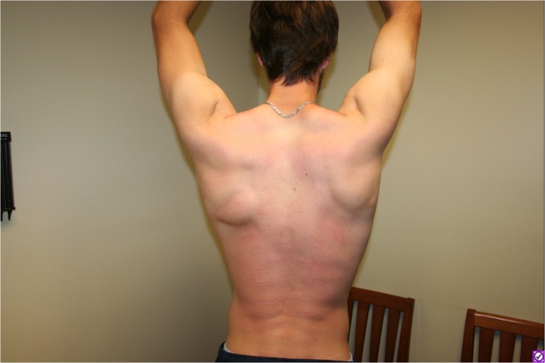
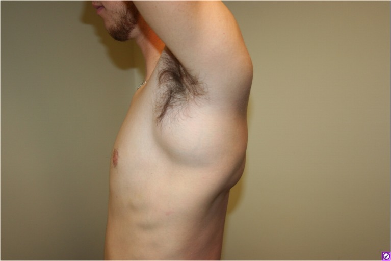
On physical exam, the scapula was elevated and displaced laterally (Fig. 1). There was a prominent soft, cystic mass in the upper axilla, which became more prominent with shoulder abduction (Fig. 2). Strength of the shoulder girdle muscles was normal, including the serratus anterior muscle. Exam of the left arm and hand showed normal motor and sensory function.
Fig. 1.
Posterior view of left scapula showing elevation laterally. A portion of the bursa protrudes beneath the inferior border of the scapula
Fig. 2.
Large mass is visible within the axilla
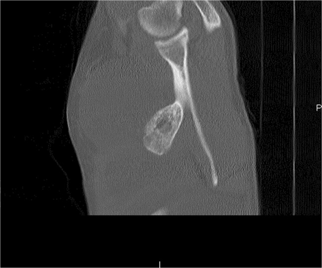
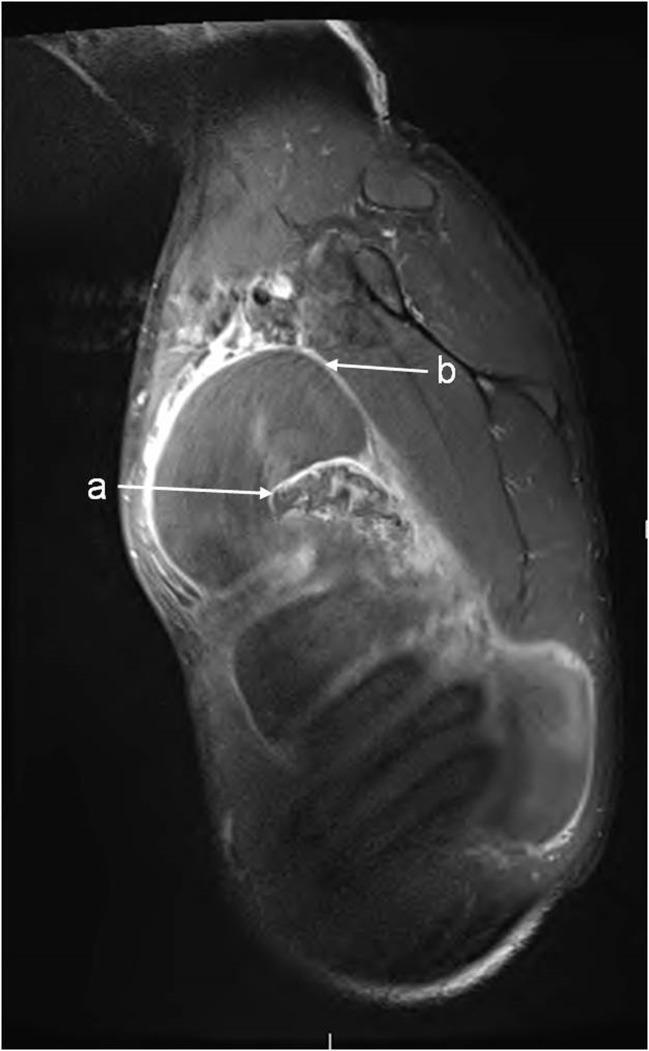
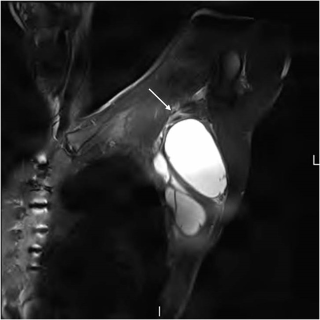
Computed tomography (CT) imaging demonstrated an exostotic lesion arising from the ventral surface of the scapula, which was abutting the rib cage (Fig. 3). Magnetic resonance (MR) showed an 18 × 10 × 8-cm fluid-filled, multiloculated sac surrounding the lesion, which displaced the serratus anterior muscle from the chest wall and the anterior border of the latissimus dorsi muscle laterally. The mass abutted the brachial plexus and axillary vessels with some displacement superiorly without evidence of compression of the vessels (Fig. 4). The inferior portion of the brachial plexus did appear to be flattened (Fig. 5). These findings were consistent with a large reactive bursa.
Fig. 3.
Sagittal CT scan shows the exostosis on the ventral surface of the scapula
Fig. 4.
T1 MRI sagittal view (a) bony exostosis of the scapula and (b) superior portion of the bursa sac near the brachial plexus
Fig. 5.
T2 MRI coronal view showing fluid-filled mass adjacent to the neurovascular structures of the axilla. Arrow denotes brachial plexus superior to mass
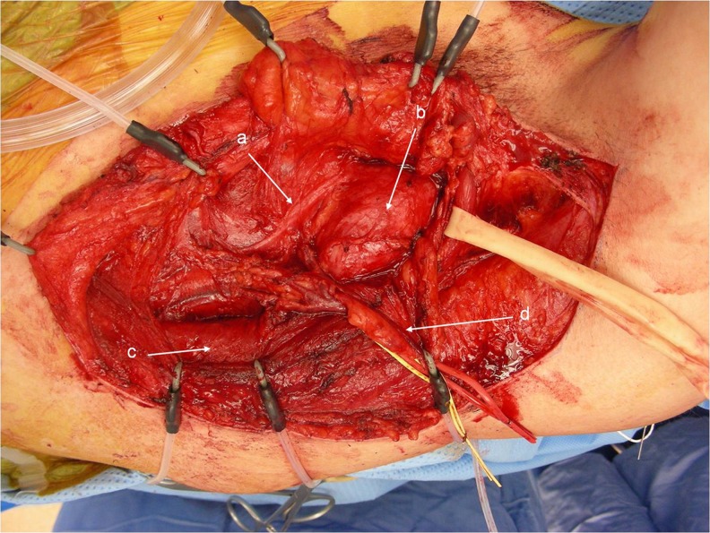
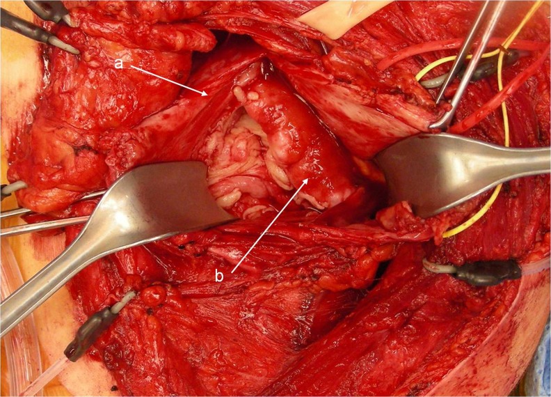
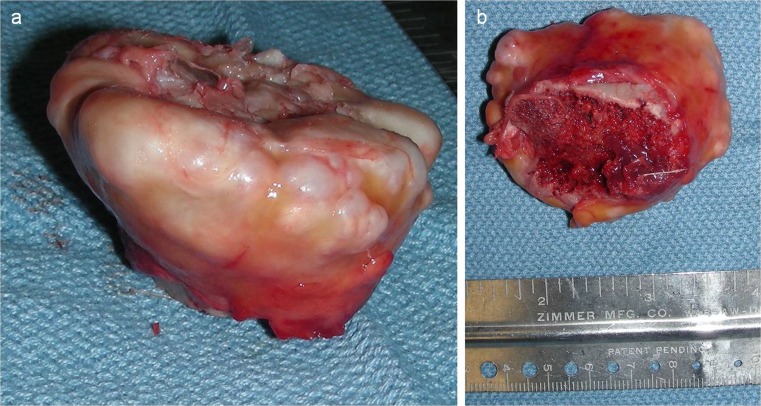
Surgery was undertaken using a team approach with the orthopedic oncologist (DH). An incision was made along the anterior border of the latissimus dorsi muscle. The thoracodorsal neurovascular bundle was found to be displaced medially from the muscle. This was dissected free. The long thoracic nerve was identified on the serratus anterior muscle (Fig. 6). With these two nerves protected, the bursa was entered and the serous fluid drained (Fig. 7). The mass, which had a prominent cartilaginous cap, was excised from the inferior border of the scapula in two pieces (Fig. 8a, b). The size of the mass was 6 × 6 × 10 cm. Resorption of the underlying ribs from pressure was noted. A portion of the bursa was resected, but to avoid injury to the neurovascular structures, there was no attempt to remove the superior extension into the axilla. Once the lesion was removed, a reactive bursa was unlikely to reform. Histologic analysis revealed benign osteochondroma with cartilage cap thickness of 0.4 cm. The surrounding capsule was reported as reactive synovium.
Fig. 6.
Operative photograph exposing (a) long thoracic nerve displaced laterally, (b) the exostosis covered by surrounding bursa, (c) the latissimus dorsi muscle, and (d) thoracodorsal neurovascular bundle (retracted)
Fig. 7.
(a) Wall of the bursa has been opened and retracted; (b) the mass protruding from within the bursa sac
Fig. 8.
a Bony exostosis following excision. b Smooth cartilaginous cap is consistent with a benign mass
Postoperative drainage was minimal and drains were removed 1 week later. Shoulder function and position were normal 1 year later with no recurrence of the bursa. He is currently followed clinically for symptoms of tumor recurrence without need for imaging surveillance.
Review of Literature and Discussion
Pseudo-winging and classic winging of the scapula differ only in their etiology. True winging of the scapula is caused by serratus anterior muscle palsy. Any other cause of winging deformity such as tumor, glenohumeral instability, fracture, paralysis of the trapezius muscle, or others is considered pseudo-winging of the scapula [3].
Osteochondroma accounts for 35 % of benign bone tumors [10]. Only 4.0–4.6 % of scapula tumors are osteochondroma [9, 5]. In osteochondromas occurring on the ventral surface of the scapula, pseudo-winging may result from the mass effect causing protrusion of the scapula. Osteochondroma of the scapula causing deformity was first described in 1914. McWilliams described a case of “adventitious bursa” surrounding a scapular exostosis in an 18-year-old female [7]. A subscapular mass, when presenting as a scapular deformity or as winging, may lead to the misdiagnosis of serratus anterior palsy. Clinically, differentiating between true scapular winging and pseudo-winging may be difficult.
In osteochondroma, progressive prominence of the scapula is common, as the ventral exostosis presses against the thoracic rib cage. Presentation is typically in children or those near skeletal maturity, as osteochondroma growth often mirrors skeletal growth. There may be complaints of catching, clicking, or grinding with shoulder mobility. Thorough physical exam is essential. In winging secondary to long thoracic nerve paralysis, the deformity is worse with motion such as extending the outstretched arms forward or pushing against a wall; it is less noticeable at rest. With pseudo-winging, the deformity is generally prominent at rest and less affected by motion. Patients with pseudo-winging due to subscapular exostosis have notable deformity that remains while at rest. Winging secondary to trapezius muscle paralysis can be diagnosed by physical exam with confirmation by electromyography [8]. Glenohumeral instability and at times malunion of either the scapula or humerus likely present with a history of trauma and may require additional imaging studies.
Appropriate diagnosis of bony exostosis of the scapula includes CT with possible adjunctive MR or ultrasound. Though rare, osteochondroma has the potential to undergo malignant transformation to chondrosarcoma, which occurs in 0.4–2.0 % of solitary osteochondromas [1]. Cartilage cap thickness greater than 2 cm is worrisome for malignant transformation. CT and MRI imaging are both capable of measuring the cartilage cap thickness consistently, but in the presence of a reactive bursa, false-positive interpretation by CT has been reported in two cases. MR and ultrasound are good adjuncts for differentiating thickened cartilage cap from presence of a bursa. When 2 cm of cap thickness is used as the cutoff, the sensitivities and specificities of MR are 100 and 98 %, and those of CT, 100 and 95 %, respectively [2]. Benign bone tumors such as osteochondromas may be staged using the Enneking staging system for tumors of the musculoskeletal system. This system categorizes benign bone tumors as 1 (latent), 2 (active), or 3 (aggressive) based on radiographic features and growth behavior [6]. In this case, the tumor could be classified as latent because it was not actively growing, as osteochondromas typically cease growth when the patient reaches skeletal maturity. Deformity and painful abutment of the ribs were indications for removal of this patient’s tumor.
Our surgical approach in this case was from an inferior and lateral approach. Common convention with this type of subscapular tumor is to approach it through a dorsal incision along the medial border of the scapula [9]. With the bursa displacing surrounding structures, our approach allowed direct visualization and protection of the thoracodorsal nerve, artery, and vein, as well as the long thoracic nerve (Fig. 6). Once these structures were retracted safely, we were able to easily enter the bursa sac and excise the tumor.
Unique to this case is the very large size of the reactive bursa within the axilla. This is thought to be a reaction to the mass grinding on the rib cage repeatedly with movement in a physically active patient. One series of eight patients with scapular osteochondroma reported that four were accompanied by a reactive bursa [4]. The ventral surface of the scapula is most commonly involved, and typically, the bursa is more localized. Involvement of the thoracodorsal and long thoracic nerves by an axillary bursa is very rare in the literature. Aggressive resection of the bursa may have led to injury to the brachial plexus and is unnecessary, as the bursa should not recur once there is no longer irritation of the underlying ribs. Other authors have reported success with partial resection of the bursa [10]. Review of the literature confirms that in rare cases, scapular osteochondroma of the ventral scapula surface produces pseudo-winging by displacement by bony mass effect or formation of a reactive subscapular bursa and should be considered in the differential diagnosis when winging and a mass are present.
Acknowledgments
We thank Beth Kaczmarek of The Medical Wordsmith, Inc., for her assistance in the preparation of this manuscript.
Grant Support
No grants were used for the completion of this work.
Conflict of Interest
Nicholas A. Flugstad declares that he has no conflict of interest. James R. Sanger declares that he has no conflict of interest. Donald A. Hackbarth has reported all possible conflicts of interest.
Statement of Human and Animal Rights
All procedures followed were in accordance with the ethical standards of the responsible committee on human experimentation (institutional and national) and with the Helsinki Declaration of 1975, as revised in 2008. Informed consent was obtained from the patient.
References
- 1.Ahmed AR, Tan TS, Unni KK, et al. Secondary chondrosarcoma in osteochondroma: report of 107 patients. Clin Orthop Rel Res. 2003;411:193–206. doi: 10.1097/01.blo.0000069888.31220.2b. [DOI] [PubMed] [Google Scholar]
- 2.Bernard SA, Murphey MD, Flemming DJ, et al. Improved differentiation of benign osteochondromas from secondary chondrosarcomas with standardized measurement of cartilage cap at CT and MR imaging. Radiology. 2010;255(3):857–65. doi: 10.1148/radiol.10082120. [DOI] [PubMed] [Google Scholar]
- 3.Cooley LH, Torg JS. “Pseudowinging” of the scapula secondary to subscapular osteochondroma. Clin Orthop Rel Res. 1982;162:119–24. [PubMed] [Google Scholar]
- 4.Frost NL, Parada SA, Manoso MW, et al. Scapular osteochondromas treated with surgical excision. Orthopedics. 2010;33(11):804. doi: 10.3928/01477447-20100924-09. [DOI] [PubMed] [Google Scholar]
- 5.Galate JF, Blue JM, Gaines RW. Osteochondroma of the scapula. Mo Med. 1995;92(2):95–7. [PubMed] [Google Scholar]
- 6.Enneking WF, Spanier SS, Goodman MA. A system for the surgical staging of musculoskeletal sarcomas. Clin Orthop Relat Res. 1980;153:106–20. [PubMed] [Google Scholar]
- 7.McWilliams CA. Subscapular exostosis. JAMA. 1914;63(17):1473–4. [Google Scholar]
- 8.Martin RM, Fish DE. Scapular winging: anatomical review, diagnosis, and treatments. Current Rev Musculoskelet Med. 2008;1:1–11. doi: 10.1007/s12178-007-9000-5. [DOI] [PMC free article] [PubMed] [Google Scholar]
- 9.Mohsen MS, Moosa NK, Kumar P. Osteochondroma of the scapula associated with winging and large bursa formation. Med Princ Pract. 2006;15:387–90. doi: 10.1159/000094275. [DOI] [PubMed] [Google Scholar]
- 10.Unni K, Inwards C. Dahlin’s bone tumors: general aspects and data on 10,165 cases. 6. Philadelphia: Wollters Kluwer Health/Lippincott Williams & Wilkins; 2010. [Google Scholar]


