Abstract
Iatrogenic hepatopancreaticobiliary injuries occur after various types of surgical and nonsurgical procedures. Symptomatically, these injuries may lead to a variety of clinical presentations, including tachycardia and hypotension from hemobilia or hemorrhage. Iatrogenic injuries may be identified during the intervention, immediately afterwards, or have a delayed presentation. These injuries are categorized into nonvascular and vascular injuries. Nonvascular injuries include biliary injuries such as biliary leak or stricture, pancreatic injury, and the development of fluid collections such as abscesses. Vascular injuries include pseudoaneurysms, arteriovenous fistulas, dissection, and perforation. Imaging studies such as ultrasound, computed tomography, magnetic resonance imaging, and digital subtraction angiography are critical for proper diagnosis of these conditions. In this article, we describe the clinical and imaging presentations of these iatrogenic injuries and the armamentarium of minimally invasive procedures (percutaneous drainage catheter placement, balloon dilatation, stenting, and coil embolization) that are useful in their management.
Keywords: hepatobiliary, hepatopancreaticobiliary, bile duct injury, iatrogenic injuries, interventional radiology
Objectives: Upon completion of this article, the reader will be able to identify the imaging findings and role of minimally invasive interventional techniques in the management of iatrogenic hepatobiliary injuries.
Accreditation: This activity has been planned and implemented in accordance with the Essential Areas and Policies of the Accreditation Council for Continuing Medical Education (ACCME) through the joint providership of Tufts University School of Medicine (TUSM) and Thieme Medical Publishers, New York. TUSM is accredited by the ACCME to provide continuing medical education for physicians.
Credit: Tufts University School of Medicine designates this journal-based CME activity for a maximum of 1 AMA PRA Category 1 Credit™. Physicians should claim only the credit commensurate with the extent of their participation in the activity.
Injury to the hepatopancreaticobiliary system is an important complication of surgical, interventional, and endoscopic interventions. These injuries may result in substantial morbidity and mortality. Surgical procedures that may be complicated by hepatopancreaticobiliary injuries include cholecystectomy, pancreatic surgery, gastrointestinal surgery (e.g., Roux-en-Y hepaticojejunostomy), hepatic resection, and orthotopic liver transplantation. Nonsurgical procedures with the potential to cause injuries include liver or pancreatic biopsy, endoscopic retrograde cholangiopancreatography (ERCP), percutaneous transhepatic cholangiography (PTHC), transjugular intrahepatic portosystemic shunt (TIPS) placement, cholecystostomy tube placement, percutaneous or laparoscopic ablation of liver tumors, and transarterial chemoembolization (TACE)/radioembolization. Injuries may manifest during the procedures, in the immediate postoperative period, or even months to years later.
Iatrogenic hepatopancreaticobiliary injuries are divided into two main categories: nonvascular and vascular. Management of many of these injuries requires a multidisciplinary approach that is determined by multiple factors, including the type of injury, its location, the patient's clinical condition, and the experience of the interventional radiologist and hepatobiliary surgeon.1 2 Reviewed here are methods of diagnosis and management of iatrogenic injuries to the hepatopancreaticobiliary system.
Nonvascular Injuries
Bile Duct Injury
Etiology
Laparoscopic cholecystectomy is one of the most common causes of iatrogenic biliary injuries. The advent of laparoscopic cholecystectomy has reduced healing times and overall complication rates as compared with open cholecystectomy, but the rate of injury to biliary structures has increased in the laparoscopic era.1 2 3 The reported incidence of iatrogenic bile duct injury is 0.1 to 0.2% with open cholecystectomy and 0.4 to 0.6% with laparoscopic cholecystectomy.1 3 4 5 6 7 A large Swedish study identified older age (particularly older than 70 years) and male sex as risk factors for iatrogenic bile duct injury.8 Inflammation around the gallbladder, obesity, and anatomic anomalies of the bile ducts and hepatic arteries also result in an increased risk.9 The classic laparoscopic injury involves mistaking the common bile duct for the cystic duct due to an anomalous insertion of the cystic duct into the common hepatic duct. Resection of part of the common bile duct, common hepatic duct (Fig. 1), or right hepatic arterial injury may result from this error. Clip ligation of the common bile duct may result in biliary obstruction (Fig. 2).1 2 3 4 5 6 7 9 Laparoscopic cholecystectomy is also associated with a greater risk of cystic duct leak than open cholecystectomy.2 6
Fig. 1.
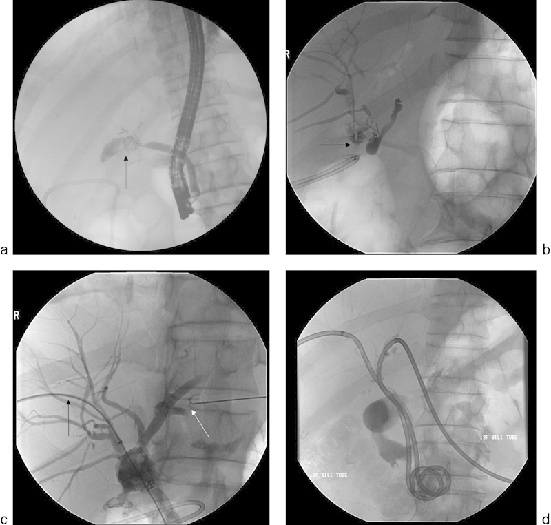
A 52-year-old man underwent laparoscopic cholecystectomy and presented with persistent moderately severe abdominal pain 1 week after the cholecystectomy. Cross-sectional imaging (not shown) revealed a fluid collection in the surgical bed. (a) A fluoroscopic image obtained during ERCP demonstrates contrast extravasation from a blind-ending common bile duct (arrow), consistent with disruption. (b) PTHC was performed for biliary drainage. Cholangiographic image demonstrates disruption of the right hepatic ducts (arrow). There is no opacification of the common bile duct. (c) Fluoroscopic image showing placement of a wire across the disrupted right hepatic duct (black arrow) into the common bile duct. Cannulation of the left hepatic duct (white arrow) also demonstrates left hepatic ductal disruption. (d) Bilateral biliary drainage catheters were placed into the duodenum.
Fig. 2.
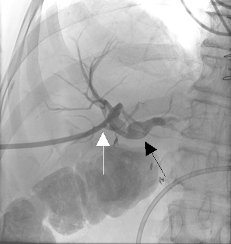
A 37-year-old woman presented with progressively increasing jaundice and abdominal pain after she underwent laparoscopic cholecystectomy. A single fluoroscopic image obtained during PTHC shows complete occlusion of the common hepatic duct (black arrow) and drainage by an external biliary drainage catheter (white arrow).
Anastomotic biliary strictures may complicate surgical procedures such as liver transplantation and Roux-en-Y hepaticojejunostomy after common bile duct injury.10 Less commonly, the strictures are the result of ischemia caused by hepatic artery thrombosis or stenosis.11 Complications from stricture formation can include cholangitis and secondary biliary cirrhosis.1 12 13
Biliary duct injury, most commonly seen as biloma formation from bile leak, may also occur after surgical procedures such as hepatic resection and nonsurgical procedures such as ERCP, PTHC, or percutaneous biopsy. ERCP may result in additional complications such as perforation of the pancreatic duct or duodenum (Fig. 3).
Fig. 3.

A 54-year-old man with jaundice, abdominal pain, and chronic pancreatitis underwent ERCP and pancreatic stent placement. During cannulation of the common bile duct, the patient became hypoxemic and the procedure was terminated, and an emergent CT scan was performed. (a) Axial CT image demonstrates diffuse subcutaneous emphysema, pneumoperitoneum, and pneumoretroperitoneum (white arrows). The patient underwent exploratory laparotomy, which revealed a malpositioned pancreatic stent (white circle) and perforated duodenum. (b) Postoperatively, biliary fluid drained from the incision site. PTHC showed extravasation from the distal common bile duct (black arrow). (c) An internal/external biliary drainage catheter was placed (black arrow), with the pigtail in the duodenum.
Clinical Presentation
Iatrogenic injuries may be identified during surgery but are more often recognized in the postoperative period.1 14 Jaundice is the most common sign of a biliary stricture; however, patients may also present with fever and epigastric pain. Often, however, only nonspecific symptoms such as malaise, anorexia, nausea, or abdominal discomfort occur. If a postsurgical drain is still in place, patients with bile leaks will generally have bile in the closed suction drain. If this drain becomes occluded, a biloma or abscess may develop.7 9 13
Imaging
Radiological studies are critical to both the diagnosis and management of iatrogenic biliary complications, as these studies are used to identify and define the extent of injury and assist in planning management. Cholescintigraphy using hydroxy iminodiacetic acid (HIDA) scan is a sensitive study for identifying the presence of bile leaks but has limited utility in planning management because of its inability to provide detailed anatomic information. Computed tomography (CT) and ultrasound are noninvasive, effective methods for detecting dilated biliary ducts; CT is particularly useful in identifying fluid collections and vascular injuries.4 7 9 13 15 Magnetic resonance cholangiopancreatography (MRCP) is a noninvasive method for defining biliary tract anatomy proximal and distal to an injury.13 16 17 MRCP can demonstrate fluid collections and MR imaging with intravenous gadolinium contrast administration can identify an arterial injury. Hepatobiliary contrast agents (e.g., gadoxetate disodium) permit detection of bile leakage in nearly all patients with this complication and can accurately localize the lesion in more than 80% of cases. Improved imaging protocols have reduced the time needed to acquire MRCP images.15 18
ERCP is invasive and is only able to delineate anatomy distal to the site of biliary injury. Other techniques, such as MRCP, better depict upstream ducts in patients with biliary stricture or disruption. For example, ERCP is insensitive in detecting ligated/transected anomalous right hepatic bile ducts because of its retrograde approach. Additionally, ERCP cannot be used in patients with a biliary enteric anastomosis, as the anastomosis cannot typically be reached via an endoscope. The main advantage of ERCP is its therapeutic capability in placing stents and drainage catheters.9 13
Cholangiography via a surgical or percutaneous catheter can be performed to precisely define injuries. This technique is particularly useful in determining the full extent of an injury when the injury is identified during surgery. Opacification of the bile ducts by injection of contrast through an existing biliary catheter commonly identifies the site of injury, with contrast extravasation occurring at the site.13 14 This technique can accurately delineate proximal duct injuries, transections/ligations of the common duct, and injuries to an anomalous right hepatic bile duct. PTHC is invasive, with an approximate 2% risk of significant complications.19 PTHC can be both diagnostic and therapeutic, and is most useful in patients who require biliary tract decompression with placement of a percutaneous transhepatic biliary drain.4 7 13 19
Management
Initial management of the acutely ill patient with suspected iatrogenic injury to the biliary ductal system should focus on hemodynamic stabilization. Administration of intravenous fluids; blood products, appropriate electrolytes and antibiotics is essential.4 13 CT is commonly used to guide initial management, as this imaging modality reliably identifies and facilitates drainage of abscesses or bilomas. Characterization of the injury often requires cholangiography.7 Interventional approaches for managing iatrogenic biliary injuries include percutaneous transhepatic biliary drainage (PTBD) with internal/external or external drain placement (Figs. 1 2 3). PTHC permits placement of drainage catheters in peripheral ducts; central duct puncture is avoided because of the risk of arterial or portal venous bleeding. Injuries to the common duct generally require a single drain, whereas hilar injuries may require bilateral drain placement (Fig. 1). In cases of cholangitis, initial placement of an external drain proximal to the obstruction reduces the potential for septic complications via manipulation of catheters inserted across the stricture.4 13 PTBD allows for decompression of the biliary tree while providing bridging therapy for surgical management.1 4 7 13
Postoperative biliary strictures can be managed by placement of biliary drainage catheters with or without transhepatic balloon dilatation. Bile leaks/fistulas can be managed solely with PTBD to properly direct biliary drainage and allow the tract to resolve (Fig. 4).6 9 Occasionally, bile leaks can be successfully managed with embolization (Fig. 5). PTHC/PTBD can also be used to manage surgical complications such as anastomotic dehiscence (Fig. 6) or ischemic cholangiopathy (Fig. 7). Complications of PTBD include sepsis, hemobilia, hemorrhage, bile leak, and acute pancreatitis.19
Fig. 4.
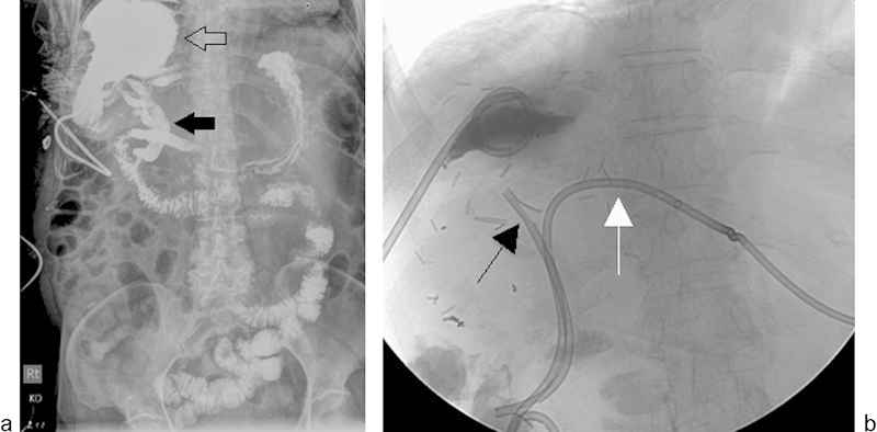
A 72-year-old woman underwent right hepatectomy and resection of the caudate lobe for hepatocellular carcinoma. On postoperative day 2, a large fluid collection was identified in the surgical bed. The collection was drained by a 12 French drainage catheter placed under CT guidance (not shown). Large volume drainage from the catheter persisted for ∼2 months. (a) Fluoroscopic injection of the large right subphrenic fluid collection cavity (open arrow) shows fistulous communication with the biliary system (solid arrow). (b) Additional drainage by placement of an endoscopic biliary stent (black arrow) and left internal–external biliary drain (white arrow) resulted in complete resolution of communication after 6 weeks. Separate contrast injection of the right upper quadrant abscess drainage catheter and biliary catheter revealed no residual communication between the right perihepatic fluid collection and the bile ducts (not shown).
Fig. 5.
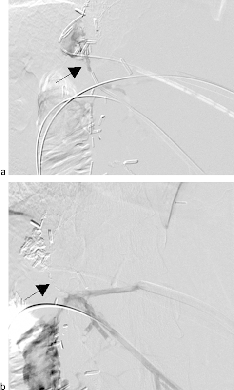
Images from a 50-year-old woman who underwent trisegmentectomy for cholangiocarcinoma. The patient presented with persistent leakage of bile despite PTHC and PTBD (a) Cholangiogram of a segment 3 duct through the sheath demonstrated active leakage of contrast in the perihepatic space (arrow). (b) After glue (n-butyl cyanoacrylate) injection, there was complete obliteration of the fistulous tract (arrow).
Fig. 6.

A 53-year-old man underwent left hepatectomy with Roux-en-Y hepaticojejunostomy for metastatic rectal carcinoma. The patient then underwent PTHC for persistent bile drainage from his surgical drain. (a) PTHC shows leakage of contrast at the hepaticojejunostomy site as a result of partial anastomotic dehiscence (arrow). (b) Fluoroscopic image shows placement of the external/internal biliary drainage catheter. (c) Tube cholangiogram shows resolution of the leak after 8 weeks.
Fig. 7.
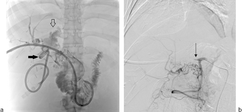
A 38-year-old woman with carcinoid tumor metastases to the liver underwent extended left hepatectomy, hepatic artery thrombectomy with interposition graft, and choledochocholedochostomy. The patient had persistent bile leak from the T-tube tract and underwent PTHC. (a) PTHC shows complete loss of integrity of the common bile duct (open arrow), which is replaced by debris. A pigtail catheter is seen in the liver draining a biloma (solid arrow). (b) Digital subtraction angiography of the proper hepatic artery performed via a right common femoral artery approach demonstrates complete occlusion of the hepatic artery (arrow).
Surgical management of iatrogenic biliary injuries varies depending on the type, severity, and location of the injury, as well as the patient's general health. Strictures of the common bile duct or distal common hepatic duct are easier to repair than more proximal injuries.20 The Bismuth classification of biliary strictures, which is based on the most distal level of healthy biliary mucosa for anastomosis, has historically been used to determine the most appropriate method of repair and to predict outcome.21 This classification system, however, originated in the era of open surgery and does not include the entire spectrum of injuries. Laparoscopic injuries to the bile duct are not only more frequent but also typically more severe than those associated with open cholecystectomy. Strasberg et al22 modified Bismuth's classification to include laparoscopic bile duct injuries, and many others have proposed more comprehensive classification systems.1 20
In one study, 43% of referrals for repair of iatrogenic biliary ductal injuries were for problems identified at the time of surgery.23 The indications for surgical repair included obstruction in 43% of patients, leak in 25%, and both in 5%. Other studies have demonstrated that up to 50% of injuries are diagnosed at the time of injury.2 4 Operative management of these injuries generally involves the creation of an anastomosis between the upstream bile structure and the jejunum (hepaticojejunostomy); this is the only option when there is a complete transection of a biliary duct. Furthermore, complex strictures and anatomic variants may leave surgery as the only option.7 9 13 In one series, successful surgical repair of iatrogenic injury to the biliary tract was achieved in more than 90% of patients.12 Failure to achieve adequate repair can lead to continued bile leak, intra-abdominal abscesses, or cholangitis, which may require a permanent indwelling biliary catheter.7 12 Even surgical repair is commonly associated with additional delayed complications; therefore, endoscopists and interventional radiologists have been increasingly involved in the management of these injuries.7 9 13
Endoscopic methods for managing biliary strictures include dilatation and stenting. Multiple endoscopic procedures are often required and if technically feasible may be combined with surgical repair. Long-term outcomes with these combined approaches are excellent.4 12 24
Pancreatic Injury
Etiology
Iatrogenic pancreatic injuries occur after placement of peripancreatic drains, biopsies, and surgical procedures such as pancreatic necrosectomy and pancreaticoduodenectomy (Whipple procedure).25 Improvements in pancreaticoduodenectomy procedural techniques have reduced mortality to less than 5%; however, complication rates are still reported to range from 20 to 60%.26 Postoperative complications include hemorrhage, pseudocyst, abscess, biloma, and fistula formation. When these complications are present, mortality rates are significantly increased; patients requiring surgical reintervention for management of these complications have mortality rates ranging from 13 to 60%.26 27 Pancreatic fistulas occur from damage to the pancreatic duct resulting in an abnormal communication with drainage of pancreatic fluid. Pancreatic fistulas and leaks have been reported to occur in an average of 12.9% of patients after pancreaticoduodenectomy.25 28
Clinical Presentation
Patients with pancreatic injury present with a variety of symptoms including pain, fever, nausea, weight loss, and persistent pancreatitis. Hypotension may be present in patients who have pancreatic hemorrhage, and elevated white blood cell count may be present in those with abscess formation.25
Imaging
Complications resulting in pancreatic injury are often first assessed with CT imaging (Fig. 8a, b). ERCP can be used to identify perforation of the pancreatic duct, with this injury demonstrating extravasation of contrast after cannulation. Additionally, pancreatic fistula tracts can be opacified during ERCP to determine the path of communication.25 MRCP can be used as an alternative to ERCP in the diagnosis of pancreatic duct injury, and secretin-stimulated MRCP can assist in the diagnosis of duct disruption and detection of pancreatic duct leaks.29
Fig. 8.
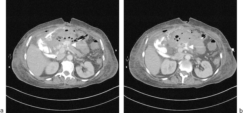
A 73-year-old woman who underwent pylorus-preserving pancreaticoduodenectomy for pancreatic intraductal mucinous adenoma presented with abdominal pain. (a and b) Axial contrast-enhanced CT images show contrast leak (arrow) at the pancreaticojejunostomy anastomosis with adjacent fluid collection.
Management
Endoscopic techniques such as stent placement are the primary form of management for pancreatic ductal injuries in patients with normal pancreatic anatomy. Interventional radiologic techniques for the treatment of pancreatic injuries are mostly limited to management of postoperative fluid collections, usually with the placement of drainage catheters (discussed in the next section).25 Such techniques have played a significant role in reducing postoperative morbidity and mortality. In one study, interventional radiological procedures reduced the need for additional surgical intervention to 2.5%.26
Postoperative Fluid Collection
Etiology
Fluid collections, including seromas, hematomas, bilomas, and abscesses, are noted relatively commonly after surgical procedures. These may be caused by violation of intra-abdominal structures, disruption of the biliary tree secondary to direct damage, faulty anastomoses, or unsterile technique.4 7 9 13
Clinical Presentation
Fever and other signs of infection and abdominal pain are the most frequent manifestations of a biloma or abscess formation. Fatigue, anorexia, nausea, and other nonspecific symptoms should alert the clinician to the possibility of iatrogenic complications in a patient who has undergone an abdominal procedure.4 7 9 13
Imaging and Management
Bilomas and abscesses are often treated with drainage and placement of indwelling percutaneous catheters (Fig. 4a, b). Access is often guided by CT, ultrasound, or less commonly, fluoroscopic imaging. Complications resulting from drain placement include sepsis, hemorrhage, and peritonitis.13 30 If a biliary leak is identified as the underlying cause of the fluid collection, simultaneous decompression of the biliary system may be needed.13 31
Gallbladder Injury
Etiology
Injury to the gallbladder may occur during any hepatobiliary intervention because of the gallbladder's close proximity to the liver and its association with the biliary tree. The risk of bile leak is greatest at the fundus because of its poor vascular supply. Iatrogenic perforation of the gallbladder can occur during laparoscopic cholecystectomy from laceration secondary to grasper traction or electrocautery dissection. Male sex, obesity, inflammatory changes of the gallbladder, and difficulty with hilar dissection are all associated with an increased risk of perforation during cholecystectomy. In a single institutional study, 36% (512 of 1,412 patients) of laparoscopic cholecystectomies were complicated by gallbladder perforation.32 Another study demonstrated a 25.5% incidence of perforation.33 Operating time and duration of hospital admission were significantly longer in patients with gallbladder perforation than those without.32 33 Gallbladder perforation can result in delayed infection distant to the gallbladder fossa due to dropped gallstones. The estimated incidence of abscess formation caused by dropped stones after the laparoscopic approach is ∼0.3%.34
TACE can also lead to ischemic cholecystitis secondary to nontarget embolization.35 36 In one study, bland embolization of the right hepatic artery resulted in nontarget cystic artery embolization in 22 of 135 patients (16%)37; however, in another study that reviewed 355 TACE cases, the incidence of acute cholecystitis was lower (∼4.9%).38 Many operators believe the likelihood of causing clinically significant cholecystitis post-TACE to be more of the order of 1 to 2%. An uncommon cause of iatrogenic acute cholecystitis is radiation-induced cholecystitis. In a recently published series of 133 patients who underwent Y-90 radioembolization for primary and secondary hepatocellular malignancies, the incidence of clinically significant radiation-induced cholecystitis was only 0.8%.39
Clinical Presentation
Clinically, perforation of the gallbladder results in spillage of bile and gallstones into the peritoneal cavity with accompanying infection, leading to abdominal pain and fever as well as often noting signs or symptoms of peritonitis.
Imaging
Imaging of gallbladder injuries often demonstrates inflammatory changes of the gallbladder, including mural thickening, abnormal contrast enhancement on CT scan, and pericholecystic fluid collections. Fluid collections most often occur in the subdiaphragmatic or subhepatic spaces. CT may demonstrate adjacent areas of fat stranding and inflammatory changes. In the presence of gallbladder perforation, cholelithiasis may remain within the gallbladder or spilled into the peritoneal cavity.40
Management
Management of gallbladder injury includes cholecystectomy or percutaneous decompression. Acute cholecystitis occurring after nontarget embolization can usually be managed conservatively but may occasionally require cholecystostomy tube placement.36 37 Cholecystostomy tube placement is associated with a risk of hemorrhage, biloma formation, peritonitis, and sepsis.19
A rare cause of injury to the hepatobiliary system includes radiofrequency ablation of liver tumors that can lead to seeding of tumors along the instrumentation tract (Fig. 9). In patients undergoing radiofrequency ablation of liver tumors adjacent to the gallbladder, needle decompression of the gallbladder may reduce the risk of injury.41 Saline infusion technique can also help to separate structures, thereby reducing the risk of this type of injury.42
Fig. 9.
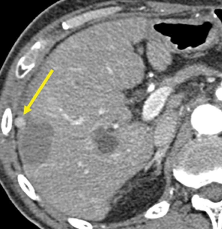
A 56-year-old man underwent radiofrequency ablation of hepatocellular carcinoma involving the right lobe of the liver. At a 3-month follow-up, contrast-enhanced CT showed a hyperenhancing nodule adjacent to the ablation zone, representing tumor seeding along the needle track (arrow).
Vascular Injuries
Iatrogenic vascular injuries can occur during manipulation of hepatic arteries, hepatic veins, or portal veins.
Pseudoaneurysms
Etiology
Pseudoaneurysms, also known as false aneurysms, develop from damage to an arterial wall resulting in a rupture that is contained by surrounding tissues.43 44 In a study at the Mayo Clinic of postoperative hepatic artery pseudoaneurysms, most occurred after hepatic (65%), biliary (30%), and pancreatic (5%) surgical and nonsurgical procedures, and were diagnosed an average of 5.7 months after the initial intervention. For pseudoaneurysms arising from the hepatic artery, the right hepatic artery was the site of 79% of pseudoaneurysms, with the remaining 21% occurring in the left hepatic, common hepatic, and cystic arteries combined.45 The presence of bile is known to damage blood vessels, so simultaneous injury to biliary structures can delay healing of an injured artery and predispose that artery to the formation of pseudoaneurysms.46 Cases of pseudoaneurysm after endoscopic stenting of the bile duct have also been reported.47
Clinical Presentation
Clinical presentation of pseudoaneurysms includes hemobilia, hematemesis, and abdominal pain. In the report from the Mayo Clinic, hemobilia occurred in 76% of patients with pseudoaneurysms, hematemesis in 59%, and abdominal pain in 35%. Two patients (11.7%) were hemodynamically unstable, and two (11.7%) were asymptomatic. Rupture occurred in 76% of all patients.45
Imaging
CT of pseudoaneurysms typically demonstrates a focal collection of intravenous contrast on arterial-phase images with washout on later phase imaging (Fig. 10). Imaging of pseudoaneurysms may also demonstrate an associated thrombus. Digital subtraction angiography of pseudoaneurysms demonstrates a contrast blush that extends beyond the normal contour of the vessel. Contrast may further extravasate if the pseudoaneurysm has ruptured.44
Fig. 10.
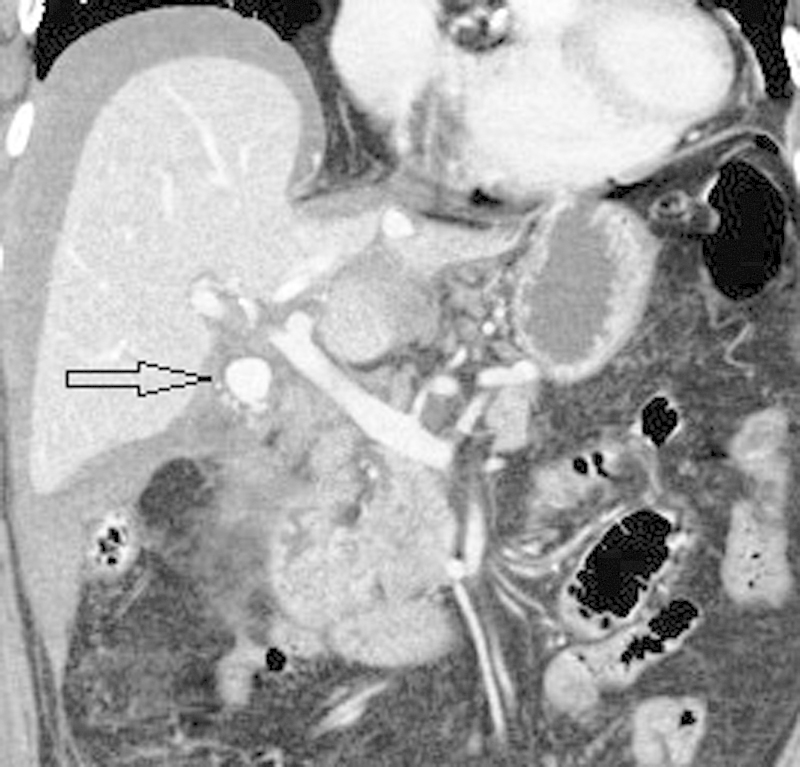
An 82-year-old woman presented to the emergency room with sudden onset of abdominal pain, tachycardia, and hypotension 3 days after laparoscopic cholecystectomy. Contrast-enhanced coronal CT scan image shows a loculated collection of contrast measuring ∼1.7 × 1.5 cm in the gallbladder fossa (arrow) representing a pseudoaneurysm. There is high-density perihepatic fluid due to intra-abdominal hemorrhage. The pseudoaneurysm was successfully treated by coil embolization of the cystic artery.
Management
Pseudoaneurysms carry a significant risk of rupturing and therefore require immediate treatment, typically with placement of a stent graft or embolization.11 43 44 45 46 47 48 Embolization was successful for 86% of the patients in whom this treatment was attempted in the Mayo Clinic series. Preservation of arterial flow by placement of an endograft is desirable, if technically feasible. Operative intervention may be required for patients in whom embolization fails; however, mortality rates are higher in these patients.45 Ultrasound-guided thrombin injection has also been used successfully to treat an iatrogenic hepatic artery pseudoaneurysm after percutaneous transhepatic portal embolization, when subsequent embolization of the hepatic artery may have caused hepatic infarction.49 Deployment of a covered stent into the artery covering the origin of the bleeding can result in successful exclusion of the pseudoaneurysm.11 44 48
Hematoma
Etiology
Several surgical scenarios place patients at high risk for parenchymal injury and subsequent bleeding. There is risk of hemorrhagic injury to the liver during trocar placement and while traction is being implemented for gallbladder removal.50 51 Parenchymal injury causing hemorrhage may also occur during interventional procedures such as TIPS placement, PTHC, biopsy, and cholecystostomy tube placement (Fig. 11). As many as one-third of patients experience transcapsular puncture during TIPS procedures, and 1 to 2% of these patients progress to develop intraperitoneal hemorrhage.52 Subcapsular hematomas occur when hemorrhage is present within an intact liver capsule, resulting in mass effect on the liver parenchyma.50
Fig. 11.
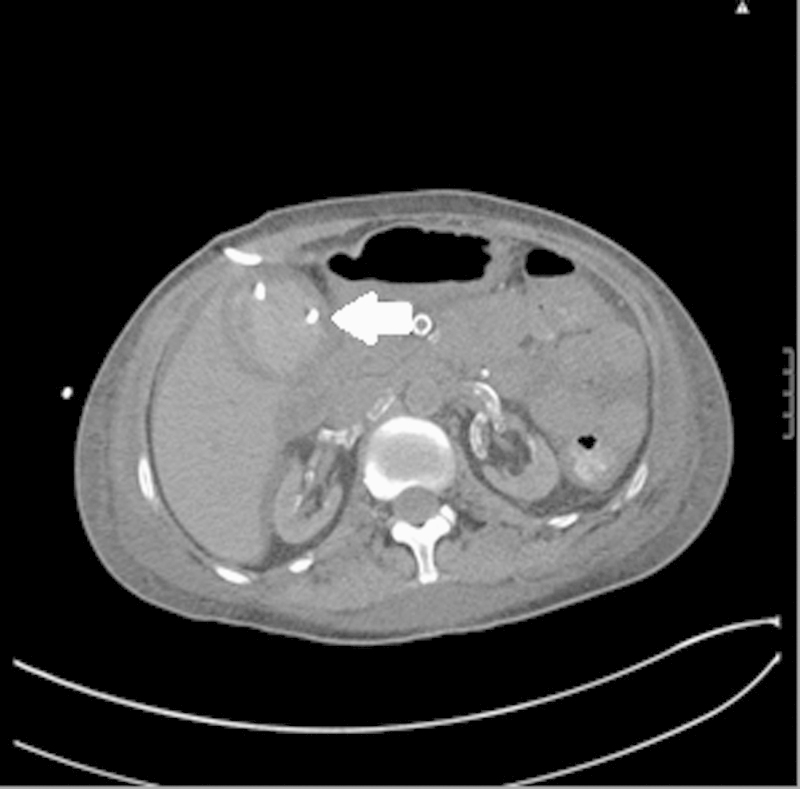
A 35-year-old woman underwent percutaneous cholecystostomy tube placement for acute cholecystitis due to multiple comorbidities preventing cholecystectomy. Following cholecystostomy tube placement, the patient became mildly tachycardic and complained of severe abdominal pain without a significant change in blood pressure. Axial noncontrast CT image of the abdomen reveals a hyperdense fluid collection within the gallbladder (arrow) after cholecystostomy tube placement, representing acute hemorrhage/hematoma.
Imaging and Management
Hematomas are visualized as a hyperdense collection on CT imaging. A drain should not be placed unless the hematomas are secondarily infected and liquefied. The dual blood supply to a native liver by the hepatic artery and portal vein make infarction uncommon.11 If branch arteries are disrupted, persistent bleeding may be treated by selective embolization using microcoils, n-butyl cyanoacrylate, Gelfoam, or particles to occlude the feeding artery (Fig. 12).
Fig. 12.
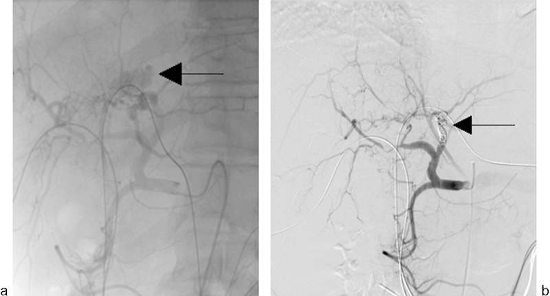
A 37-year-old man with unresectable cholangiocarcinoma and bilateral internal/external biliary drainage catheters presented with hemodynamic instability and bright red blood from the left internal/external biliary catheter 2.5 months after catheter placement. (a) Emergency hepatic arteriogram revealed active extravasation from a major branch of the left hepatic artery (arrow). (b) The left hepatic artery was successfully embolized using 0.018 microcoils (arrow).
Other Vascular injuries
Arterial Dissection
Hepatic artery dissections (Fig. 13) occur during manipulation of intravascular catheters and wires, resulting in separation of the vascular intima from the remainder of the vessel wall.53 As the dissection propagates along the length of the vessel, the dissection flap can restrict flow or result in obstruction of branch arteries originating from the involved artery. Imaging of these dissections typically demonstrates an intimal flap with contrast opacification of the true lumen. The false lumen may opacify with contrast or thrombose, resulting in narrowing of the vessel lumen. Treatment of vascular dissections typically involves angioplasty of the intimal flap and placement of a stent to restore patency.
Fig. 13.
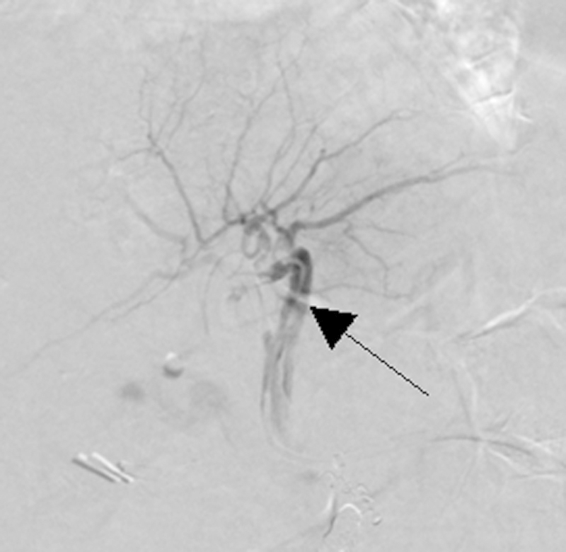
Image from a 62-year-old woman with unresectable hepatocellular carcinoma. Digital subtraction angiography performed during embolotherapy of the hepatic artery shows a non–flow-limiting intimal dissection of the left hepatic artery due to catheter manipulation (arrow).
Nontarget Embolization
Nontarget embolization of the vascular structures is a potential complication of endovascular procedures involving embolization. Nontarget embolization from particle embolization is typically treated conservatively, although occasionally surgery may be required. However, nontarget embolization of coils can often be managed by coil retrieval using a microsnare or exclusion of the coil using an endograft (Fig. 14).
Fig. 14.
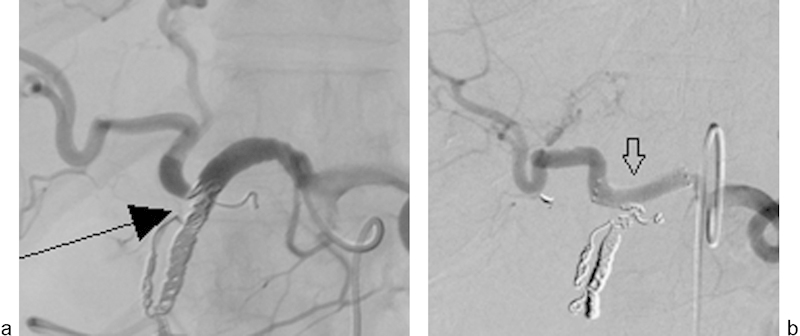
A 64-year-old woman with hepatocellular carcinoma underwent gastroduodenal artery embolization before hepatic radioembolization. (a) A common hepatic angiogram shows displacement of the last coil (arrow) into the common hepatic artery, resulting in spasm and mechanical obstruction. (b) A digital subtraction angiogram shows successful exclusion of the displaced coil by a 5-mm-diameter covered endograft (arrow).
Vascular Stenosis and Thrombosis
Vascular stenosis and thrombosis can involve the hepatic artery, hepatic veins, or portal veins (Fig. 15). These complications may occur at vascular anastomoses or after catheter manipulation. Procedures that involve transarterial catheterization can result in localized inflammation and intimal hyperplasia, resulting in arterial stenosis or occlusion.11 Hepatic vein stenosis may occur after TIPS placement,52 and all hepatobiliary vessels are at risk for occlusive disease after transplantation.11 First-line treatment for stenoses and strictures is percutaneous transluminal balloon angioplasty, which is followed by stenting when there is more than 30% residual stenosis or a persistent pressure gradient. In the management of acute thrombosis, intravascular tissue plasminogen activator (t-PA) plus mechanical thrombectomy can be used to relieve the obstruction. Chronic occlusion of vessels, however, may require surgical intervention.
Fig. 15.
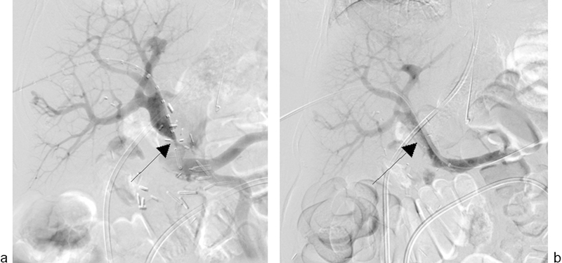
A 58-year-old woman underwent pancreaticoduodenectomy and partial portal vein resection repaired by a Gore-Tex patch for pancreatic adenocarcinoma. Follow-up ultrasound revealed a severe stenosis at the site of reconstruction. (a) Transhepatic portogram shows severe stenosis at the site of portal vein reconstruction (arrow). (b) A 16-mm-diameter Wallstent (Boston Scientific Inc., Marlborough, MA) was deployed across the stenosis using a transhepatic approach. Portogram after wall stent placement shows a patent portal vein with mild residual stenosis at the site of stenosis (arrow).
Vascular Transection or Rupture
Transection or perforation of a hepatobiliary vessel during a surgical or interventional procedure is a rare complication that is often diagnosed at the time of injury. Imaging, when performed, demonstrates contrast extravasation in these cases. Interventional techniques such as stent graft placement across the injured segment or coil embolization of the acutely injured artery are useful for managing most of the vascular injuries, but a transection is typically a surgical emergency. The right hepatic artery's location directly behind the common hepatic duct, at the typical level of biliary transection, makes this artery particularly susceptible to iatrogenic injury.1 7 54
Vascular Fistulas
Etiology
A fistula occurs when there is an abnormal connection, typically between an artery and vein. Hepatic arteriovenous fistulas are abnormal connections between the hepatic arterial and portal or hepatic venous branches. Iatrogenic causes of hepatic arteriovenous fistulas include liver biopsy, transhepatic cholangiogram with or without drain placement, and surgery.55
Clinical Presentation
The clinical presentation of vascular fistulas depends on the types of vessels that are abnormally communicating, etiology of the fistula, and degree of flow through the shunt. Portal hypertension may develop when the hepatic artery communicates with the portal vein, while arterial communication with a hepatic vein may lead to increased venous blood return to the heart and eventual right-sided heart failure. Gastrointestinal bleeding due to hemobilia is common, as injury to the artery and vein makes the adjacent biliary duct prone to ischemic injury.55
Imaging and Management
Imaging of fistulas demonstrates simultaneous opacification of both vessels with early venous filling (Fig. 16). Ultrasound with Doppler and CT are generally used to initially evaluate patients with suspected arterioportal fistulas.55 Small fistulas are often asymptomatic and may resolve on their own, while symptomatic vascular fistulas can be treated with injection of sclerosants, placement of a covered stent, embolization, or surgical repair.
Fig. 16.
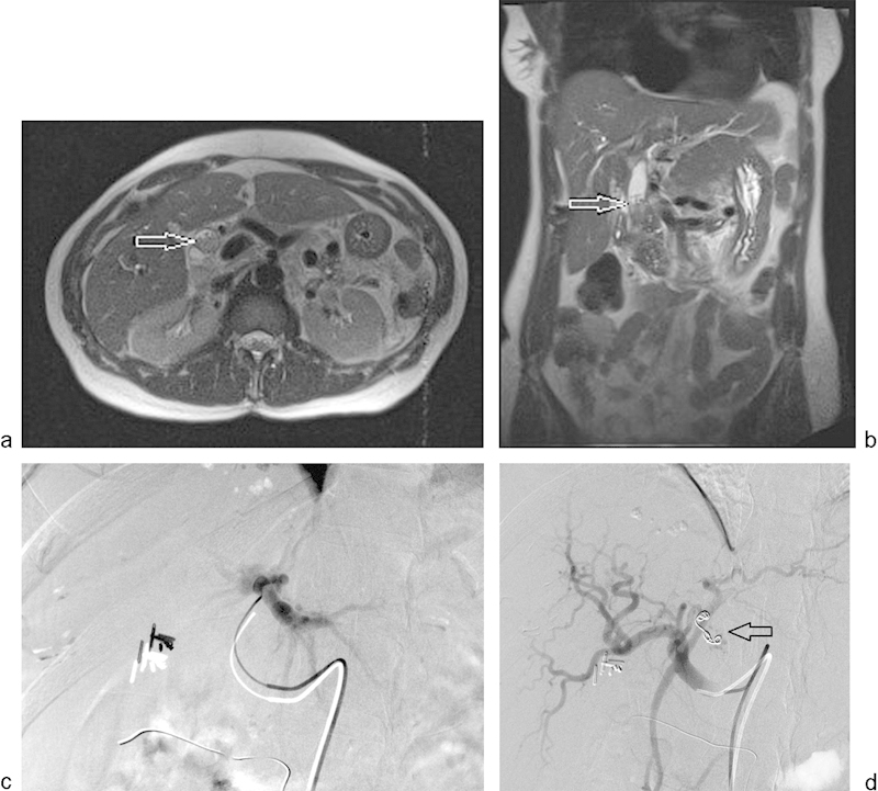
A 41-year-old woman underwent percutaneous biopsy of the left lobe of the liver and presented to the emergency room with upper gastrointestinal hemorrhage and decreasing hemoglobin level. Upper gastrointestinal endoscopy revealed blood oozing from the papilla. (a and b) T2-weighted MRI images/MRCP revealed dilated common bile duct and amorphous material in the common bile duct representing acute hemorrhage (arrow). No imaging abnormality was seen at the biopsy site. (c and d) Selective left hepatic angiogram performed via a right common femoral approach revealed an arterioportal fistula that was successfully embolized using two 0.0184-mm hydrocoils (arrow). On follow-up, no further evidence of clinical gastrointestinal hemorrhage was reported.
Conclusion
Injuries to the hepatopancreaticobiliary system may occur after a variety of surgical and nonsurgical procedures. Injuries to vascular structures can lead to hemorrhage or ischemia. Nonvascular injuries often result in fluid collections. Multidisciplinary treatment strategies are integral to the management of these complications. Imaging studies, temporizing procedures, and definitive treatments continue to evolve. As new technologies become available, physicians must negotiate new complications and explain the risks to patients who might otherwise assume that newer techniques are safer than the more traditional methods.
Acknowledgment
The authors thank Megan Griffiths, scientific/medical writer, Imaging Institute, Cleveland Clinic, for assistance in editing the article.
References
- 1.Lau W Y, Lai E C, Lau S H. Management of bile duct injury after laparoscopic cholecystectomy: a review. ANZ J Surg. 2010;80(1–2):75–81. doi: 10.1111/j.1445-2197.2009.05205.x. [DOI] [PubMed] [Google Scholar]
- 2.Nuzzo G, Giuliante F, Giovannini I. et al. Advantages of multidisciplinary management of bile duct injuries occurring during cholecystectomy. Am J Surg. 2008;195(6):763–769. doi: 10.1016/j.amjsurg.2007.05.046. [DOI] [PubMed] [Google Scholar]
- 3.Flum D R, Cheadle A, Prela C, Dellinger E P, Chan L. Bile duct injury during cholecystectomy and survival in Medicare beneficiaries. JAMA. 2003;290(16):2168–2173. doi: 10.1001/jama.290.16.2168. [DOI] [PubMed] [Google Scholar]
- 4.Saad N, Darcy M. Iatrogenic bile duct injury during laparoscopic cholecystectomy. Tech Vasc Interv Radiol. 2008;11(2):102–110. doi: 10.1053/j.tvir.2008.07.004. [DOI] [PubMed] [Google Scholar]
- 5.Roslyn J J, Binns G S, Hughes E F, Saunders-Kirkwood K, Zinner M J, Cates J A. Open cholecystectomy. A contemporary analysis of 42,474 patients. Ann Surg. 1993;218(2):129–137. doi: 10.1097/00000658-199308000-00003. [DOI] [PMC free article] [PubMed] [Google Scholar]
- 6.MacFadyen B V Jr, Vecchio R, Ricardo A E, Mathis C R. Bile duct injury after laparoscopic cholecystectomy. The United States experience. Surg Endosc. 1998;12(4):315–321. doi: 10.1007/s004649900661. [DOI] [PubMed] [Google Scholar]
- 7.McPartland K J Pomposelli J J Iatrogenic biliary injuries: classification, identification, and management Surg Clin North Am 20088861329–1343., ix [DOI] [PubMed] [Google Scholar]
- 8.Waage A, Nilsson M. Iatrogenic bile duct injury: a population-based study of 152 776 cholecystectomies in the Swedish Inpatient Registry. Arch Surg. 2006;141(12):1207–1213. doi: 10.1001/archsurg.141.12.1207. [DOI] [PubMed] [Google Scholar]
- 9.Jabłońska B, Lampe P. Iatrogenic bile duct injuries: etiology, diagnosis and management. World J Gastroenterol. 2009;15(33):4097–4104. doi: 10.3748/wjg.15.4097. [DOI] [PMC free article] [PubMed] [Google Scholar]
- 10.Lee A Y, Gregorius J, Kerlan R K Jr, Gordon R L, Fidelman N. Percutaneous transhepatic balloon dilation of biliary-enteric anastomotic strictures after surgical repair of iatrogenic bile duct injuries. PLoS ONE. 2012;7(10):e46478. doi: 10.1371/journal.pone.0046478. [DOI] [PMC free article] [PubMed] [Google Scholar]
- 11.Caiado A HM, Blasbalg R, Marcelino A S. et al. Complications of liver transplantation: multimodality imaging approach. Radiographics. 2007;27(5):1401–1417. doi: 10.1148/rg.275065129. [DOI] [PubMed] [Google Scholar]
- 12.Lillemoe K D, Melton G B, Cameron J L. et al. Postoperative bile duct strictures: management and outcome in the 1990s. Ann Surg. 2000;232(3):430–441. doi: 10.1097/00000658-200009000-00015. [DOI] [PMC free article] [PubMed] [Google Scholar]
- 13.Thompson C M, Saad N E, Quazi R R, Darcy M D, Picus D D, Menias C O. Management of iatrogenic bile duct injuries: role of the interventional radiologist. Radiographics. 2013;33(1):117–134. doi: 10.1148/rg.331125044. [DOI] [PubMed] [Google Scholar]
- 14.Törnqvist B, Strömberg C, Persson G, Nilsson M. Effect of intended intraoperative cholangiography and early detection of bile duct injury on survival after cholecystectomy: population based cohort study. BMJ. 2012;345:e6457. doi: 10.1136/bmj.e6457. [DOI] [PMC free article] [PubMed] [Google Scholar]
- 15.Aduna M, Larena J A, Martín D, Martínez-Guereñu B, Aguirre I, Astigarraga E. Bile duct leaks after laparoscopic cholecystectomy: value of contrast-enhanced MRCP. Abdom Imaging. 2005;30(4):480–487. doi: 10.1007/s00261-004-0276-2. [DOI] [PubMed] [Google Scholar]
- 16.Fulcher A S, Turner M A. Orthotopic liver transplantation: evaluation with MR cholangiography. Radiology. 1999;211(3):715–722. doi: 10.1148/radiology.211.3.r99jn17715. [DOI] [PubMed] [Google Scholar]
- 17.Chaudhary A, Negi S S, Puri S K, Narang P. Comparison of magnetic resonance cholangiography and percutaneous transhepatic cholangiography in the evaluation of bile duct strictures after cholecystectomy. Br J Surg. 2002;89(4):433–436. doi: 10.1046/j.0007-1323.2002.02066.x. [DOI] [PubMed] [Google Scholar]
- 18.Kamaoui I, Milot L, Durieux M, Ficarelli S, Mennesson N, Pilleul F. [Value of MRCP with Mangafodipir Trisodium (Teslascan) injection in the diagnosis and management of bile leaks] J Radiol. 2007;88(12):1881–1886. doi: 10.1016/s0221-0363(07)78366-x. [DOI] [PubMed] [Google Scholar]
- 19.Saad W E, Wallace M J, Wojak J C, Kundu S, Cardella J F. Quality improvement guidelines for percutaneous transhepatic cholangiography, biliary drainage, and percutaneous cholecystostomy. J Vasc Interv Radiol. 2010;21(6):789–795. doi: 10.1016/j.jvir.2010.01.012. [DOI] [PubMed] [Google Scholar]
- 20.Lau W Y, Lai E C. Classification of iatrogenic bile duct injury. Hepatobiliary Pancreat Dis Int. 2007;6(5):459–463. [PubMed] [Google Scholar]
- 21.Bismuth H, Majno P E. Biliary strictures: classification based on the principles of surgical treatment. World J Surg. 2001;25(10):1241–1244. doi: 10.1007/s00268-001-0102-8. [DOI] [PubMed] [Google Scholar]
- 22.Strasberg S M, Hertl M, Soper N J. An analysis of the problem of biliary injury during laparoscopic cholecystectomy. J Am Coll Surg. 1995;180(1):101–125. [PubMed] [Google Scholar]
- 23.Walsh R M Henderson J M Vogt D P Brown N Long-term outcome of biliary reconstruction for bile duct injuries from laparoscopic cholecystectomies Surgery 20071424450–456., discussion 456–457 [DOI] [PubMed] [Google Scholar]
- 24.Misra S, Melton G B, Geschwind J F, Venbrux A C, Cameron J L, Lillemoe K D. Percutaneous management of bile duct strictures and injuries associated with laparoscopic cholecystectomy: a decade of experience. J Am Coll Surg. 2004;198(2):218–226. doi: 10.1016/j.jamcollsurg.2003.09.020. [DOI] [PubMed] [Google Scholar]
- 25.Fazel A. Postoperative pancreatic leaks and fistulae: the role of the endoscopist. Tech Gastrointest Endosc. 2006;8(2):92–98. [Google Scholar]
- 26.Sanjay P, Kellner M, Tait I S. The role of interventional radiology in the management of surgical complications after pancreatoduodenectomy. HPB (Oxford) 2012;14(12):812–817. doi: 10.1111/j.1477-2574.2012.00545.x. [DOI] [PMC free article] [PubMed] [Google Scholar]
- 27.Standop J, Glowka T, Schmitz V. et al. Operative re-intervention following pancreatic head resection: indications and outcome. J Gastrointest Surg. 2009;13(8):1503–1509. doi: 10.1007/s11605-009-0905-8. [DOI] [PubMed] [Google Scholar]
- 28.Veillette G, Dominguez I, Ferrone C. et al. Implications and management of pancreatic fistulas following pancreaticoduodenectomy: the Massachusetts General Hospital experience. Arch Surg. 2008;143(5):476–481. doi: 10.1001/archsurg.143.5.476. [DOI] [PMC free article] [PubMed] [Google Scholar]
- 29.Gillams A R, Kurzawinski T, Lees W R. Diagnosis of duct disruption and assessment of pancreatic leak with dynamic secretin-stimulated MR cholangiopancreatography. AJR Am J Roentgenol. 2006;186(2):499–506. doi: 10.2214/AJR.04.1775. [DOI] [PubMed] [Google Scholar]
- 30.Gervais D A, Brown S D, Connolly S A, Brec S L, Harisinghani M G, Mueller P R. Percutaneous imaging-guided abdominal and pelvic abscess drainage in children. Radiographics. 2004;24(3):737–754. doi: 10.1148/rg.243035107. [DOI] [PubMed] [Google Scholar]
- 31.Beart R W Jr, Putnam C W, Starzl T E. Use of a U tube in the treatment of biliary disease. Surg Gynecol Obstet. 1976;142(6):912–914. [PMC free article] [PubMed] [Google Scholar]
- 32.Hui T T, Giurgiu D I, Margulies D R, Takagi S, Iida A, Phillips E H. Iatrogenic gallbladder perforation during laparoscopic cholecystectomy: etiology and sequelae. Am Surg. 1999;65(10):944–948. [PubMed] [Google Scholar]
- 33.Zubair M, Habib L, Mirza M R, Channa M A, Yousuf M. Iatrogenic gall bladder perforations in laparoscopic cholecystectomy: an audit of 200 cases. Mymensingh Med J. 2010;19(3):422–426. [PubMed] [Google Scholar]
- 34.Morrin M M, Kruskal J B, Hochman M G, Saldinger P F, Kane R A. Radiologic features of complications arising from dropped gallstones in laparoscopic cholecystectomy patients. AJR Am J Roentgenol. 2000;174(5):1441–1445. doi: 10.2214/ajr.174.5.1741441. [DOI] [PubMed] [Google Scholar]
- 35.Malagari K, Pomoni M, Spyridopoulos T N. et al. Safety profile of sequential transcatheter chemoembolization with DC Bead™: results of 237 hepatocellular carcinoma (HCC) patients. Cardiovasc Intervent Radiol. 2011;34(4):774–785. doi: 10.1007/s00270-010-0044-3. [DOI] [PubMed] [Google Scholar]
- 36.Sakamoto I, Aso N, Nagaoki K. et al. Complications associated with transcatheter arterial embolization for hepatic tumors. Radiographics. 1998;18(3):605–619. doi: 10.1148/radiographics.18.3.9599386. [DOI] [PubMed] [Google Scholar]
- 37.Shah R P, Brown K T. Hepatic arterial embolization complicated by acute cholecystitis. Semin Intervent Radiol. 2011;28(2):252–257. doi: 10.1055/s-0031-1280675. [DOI] [PMC free article] [PubMed] [Google Scholar]
- 38.Wagnetz U, Jaskolka J, Yang P, Jhaveri K S. Acute ischemic cholecystitis after transarterial chemoembolization of hepatocellular carcinoma: incidence and clinical outcome. J Comput Assist Tomogr. 2010;34(3):348–353. doi: 10.1097/RCT.0b013e3181caaea3. [DOI] [PubMed] [Google Scholar]
- 39.Sag A A, Savin M A, Lal N R, Mehta R R. Yttrium-90 radioembolization of malignant tumors of the liver: gallbladder effects. AJR Am J Roentgenol. 2014;202(5):1130–1135. doi: 10.2214/AJR.13.10548. [DOI] [PubMed] [Google Scholar]
- 40.Thurley P D, Dhingsa R. Laparoscopic cholecystectomy: postoperative imaging. AJR Am J Roentgenol. 2008;191(3):794–801. doi: 10.2214/AJR.07.3485. [DOI] [PubMed] [Google Scholar]
- 41.Fernandes D D Shyn P B Silverman S G Gallbladder needle decompression during radiofrequency ablation of an adjacent liver tumour Can Assoc Radiol J 201263(3, Suppl):S37–S40. [DOI] [PubMed] [Google Scholar]
- 42.Han Y, Hao Y Z, Cai J Q. et al. [Ultrasound-guided assistant infusion technique for percutaneous radiofrequency ablation of liver cancer] Zhonghua Gan Zang Bing Za Zhi. 2012;20(4):266–269. doi: 10.3760/cma.j.issn.1007-3418.2012.04.008. [DOI] [PubMed] [Google Scholar]
- 43.Yamakado K, Nakatsuka A, Tanaka N, Takano K, Matsumura K, Takeda K. Transcatheter arterial embolization of ruptured pseudoaneurysms with coils and n-butyl cyanoacrylate. J Vasc Interv Radiol. 2000;11(1):66–72. doi: 10.1016/s1051-0443(07)61284-6. [DOI] [PubMed] [Google Scholar]
- 44.Saad N E, Saad W E, Davies M G, Waldman D L, Fultz P J, Rubens D J. Pseudoaneurysms and the role of minimally invasive techniques in their management. Radiographics. 2005;25 01:S173–S189. doi: 10.1148/rg.25si055503. [DOI] [PubMed] [Google Scholar]
- 45.Tessier D J, Fowl R J, Stone W M. et al. Iatrogenic hepatic artery pseudoaneurysms: an uncommon complication after hepatic, biliary, and pancreatic procedures. Ann Vasc Surg. 2003;17(6):663–669. doi: 10.1007/s10016-003-0075-1. [DOI] [PubMed] [Google Scholar]
- 46.Madanur M A, Battula N, Sethi H, Deshpande R, Heaton N, Rela M. Pseudoaneurysm following laparoscopic cholecystectomy. Hepatobiliary Pancreat Dis Int. 2007;6(3):294–298. [PubMed] [Google Scholar]
- 47.Watanabe M, Shiozawa K, Mimura T. et al. Hepatic artery pseudoaneurysm after endoscopic biliary stenting for bile duct cancer. World J Radiol. 2012;4(3):115–120. doi: 10.4329/wjr.v4.i3.115. [DOI] [PMC free article] [PubMed] [Google Scholar]
- 48.Paci E, Antico E, Candelari R, Alborino S, Marmorale C, Landi E. Pseudoaneurysm of the common hepatic artery: treatment with a stent-graft. Cardiovasc Intervent Radiol. 2000;23(6):472–474. doi: 10.1007/s002700010107. [DOI] [PubMed] [Google Scholar]
- 49.Tokue H, Takeuchi Y, Sofue K, Arai Y, Tsushima Y. Ultrasound-guided thrombin injection for the treatment of an iatrogenic hepatic artery pseudoaneurysm: a case report. J Med Case Reports. 2011;5:518. doi: 10.1186/1752-1947-5-518. [DOI] [PMC free article] [PubMed] [Google Scholar]
- 50.Minaya Bravo A M, González González E, Ortíz Aguilar M, Larrañaga Barrera E. Two rare cases of intrahepatic subcapsular hematoma after laparoscopic cholecystectomy. Indian J Surg. 2010;72(6):481–484. doi: 10.1007/s12262-010-0128-y. [DOI] [PMC free article] [PubMed] [Google Scholar]
- 51.Fusco M A, Scott T E, Paluzzi M W. Traction injury to the liver during laparoscopic cholecystectomy. Surg Laparosc Endosc. 1994;4(6):454–456. [PubMed] [Google Scholar]
- 52.Fidelman N, Kwan S W, LaBerge J M, Gordon R L, Ring E J, Kerlan R K Jr. The transjugular intrahepatic portosystemic shunt: an update. AJR Am J Roentgenol. 2012;199(4):746–755. doi: 10.2214/AJR.12.9101. [DOI] [PubMed] [Google Scholar]
- 53.Lin T S, Chiang Y C, Chen C L. et al. Intimal dissection of the hepatic artery following transarterial embolization for hepatocellular carcinoma: an intraoperative problem in adult living donor liver transplantation. Liver Transpl. 2009;15(11):1553–1556. doi: 10.1002/lt.21888. [DOI] [PubMed] [Google Scholar]
- 54.Gupta N, Solomon H, Fairchild R, Kaminski D L. Management and outcome of patients with combined bile duct and hepatic artery injuries. Arch Surg. 1998;133(2):176–181. doi: 10.1001/archsurg.133.2.176. [DOI] [PubMed] [Google Scholar]
- 55.Kumar A, Ahuja C K, Vyas S. et al. Hepatic arteriovenous fistulae: role of interventional radiology. Dig Dis Sci. 2012;57(10):2703–2712. doi: 10.1007/s10620-012-2331-0. [DOI] [PubMed] [Google Scholar]


