Abstract
Translation of poliovirus and hepatitis C virus (HCV) RNAs is initiated by recruitment of 40S ribosomes to an internal ribosome entry site (IRES) in the mRNA 5′ untranslated region. Translation initiation of these RNAs is stimulated by noncanonical initiation factors called IRES trans-activating factors (ITAFs). The La autoantigen is such an ITAF, but functional evidence for the role of La in poliovirus and HCV translation in vivo is lacking. Here, by two methods using small interfering RNA and a dominant-negative mutant of La, we demonstrate that depletion of La causes a dramatic reduction in poliovirus IRES function in vivo. We also show that 40S ribosomal subunit binding to HCV and poliovirus IRESs in vitro is inhibited by a dominant-negative form of La. These results provide strong evidence for a function of the La autoantigen in IRES-dependent translation and define the step of translation which is stimulated by La.
Translation initiation in eukaryotes is generally dependent on a structure termed the cap (m7GpppN, where m is a methyl group and N is any nucleotide), which is present at the 5′ends of all nuclear transcribed mRNAs. The cap recruits the 40S ribosomal subunit through its interaction with the cap binding protein complex, eIF4F, which forms a bridge between the mRNA and the 40S ribosomal subunit through eIF3 (19, 27). Following its recruitment to the mRNA 5′end, the 40S ribosomal subunit in association with several initiation factors, is thought to migrate along the 5′ untranslated region (5′UTR) until an initiation codon is encountered (27). However, in a sizeable fraction of eukaryotic cellular mRNAs (9, 25, 70), the 40S ribosomal subunit is recruited to the mRNA via an alternative mechanism using an internal ribosome binding site (IRES). IRESs were first discovered in picornavirus mRNAs (poliovirus and encephalomyocarditis virus), which are naturally uncapped (38, 54), and subsequently identified in many cellular mRNAs (reviewed in references 25 and 70; see the Internal Ribosome Entry Site database [7] for an updated list). Strikingly, cellular IRESs are mainly found in mRNAs that are translated under stress conditions (31). These conditions include hypoxia, apoptosis, serum starvation (26, 29, 32, 43, 45), and mitosis (10, 58). Thus, control of IRES-dependent translation plays a critical role in regulation of cell growth and survival.
To understand the mechanism of control of IRES function, it is imperative to study the mode by which the 40S ribosome subunit is recruited to the IRES. To this end, we and others undertook the identification and characterization of cellular proteins that interact with the poliovirus IRES. The studies of Meerovitch et al. led to the identification of the first IRES trans-acting factor (ITAF), the La autoantigen (47, 48). Subsequently, La was shown to bind to the hepatitis C virus (HCV) IRES near the initiation codon and to stimulate HCV RNA translation in a rabbit reticulocyte lysate at low concentrations (1, 2). The HCV IRES-La interaction has been recapitulated in a Saccharomyces cerevisiae three-hybrid system (57). In addition, La binds to cellular IRESs, such as X-linked inhibitor of apoptosis (XIAP) (30) and BiP (42).
Initiation of poliovirus translation from the correct initiation site in a rabbit reticulocyte lysate is feeble, and aberrant products are readily detected (8, 14), possibly due to limiting amounts of one or more translation factors. Interestingly, the amount of La protein present in reticulocyte lysate is very low compared to extracts from nucleated cells (48), and the addition of recombinant La enhances poliovirus RNA translation and eliminates the production of aberrant proteins (48, 65). It was thus concluded that La protein is a bona fide ITAF. However, the physiological significance of these studies was questioned (35, 36), because of the large amounts of recombinant La that had been used (10-fold more recombinant La than the amount of La protein present in HeLa cell extract). Thus, the possibility that La is not a physiological ITAF but rather mimics a genuine poliovirus ITAF, owing to its RNA binding properties was raised (35, 36, 72). To address this issue, we studied the requirement of La autoantigen for poliovirus translation in vivo. Using small interfering RNA (siRNA) and a dominant-negative mutant of La (LaDN), we demonstrate that La is required for optimal translation of poliovirus. In addition, we demonstrate that LaDN inhibits the initiation of formation of 48S ribosome complexes on HCV and poliovirus mRNAs.
MATERIALS AND METHODS
Cell culture and viruses.
HeLa CLL2 cells (American Type Culture Collection) were cultured under standard conditions in Dulbecco's modified Eagle's medium (DMEM) supplemented with 10% fetal bovine serum (FBS) and antibiotics. Cells were passaged by a 1:7 dilution before reaching confluency to maintain exponential growth. Poliovirus type 1 (Mahoney) was produced after transfection of HeLa cells with the T7 RNA polymerase transcripts of pPV1 as described previously (55). Adenovirus dl309 (39) was kindly provided by P. Branton (McGill University) and HCV-poliovirus chimera was obtained from F. Lahser and E. Wimmer (44, 77). Poliovirus was propagated in HeLa cells, and the virus titer was determined by a plaque assay as described previously (61).
siRNA transfection and bicistronic reporter cotransfection.
Target sequences for siRNA were designed by using the Ambion web-based criteria and purchased from Dharmacon. The positions and sequences of the siRNAs used in the present study are given in Table 1. One day prior to transfection, the cells were trypsinized, resuspended in medium without antibiotics, and transferred to 24-well plates at a density of 5 × 10⁵ cells per well in a volume of 500 μl. Transfection experiments were performed using Lipofectamine 2000. For each transfection, 3 μl of siRNA duplex (20 μM annealed duplex from Dharmacon) was mixed with 50 μl of OPTIMEM (Invitrogen). In a separate tube, 3 μl of Lipofectamine 2000 per reaction mixture was added to 50 μl of OPTIMEM and incubated for 5 min at room temperature. Both solutions were mixed and incubated for an additional 20 min at room temperature to allow the formation of complexes. The solutions were then added to the cells in 24-well plates. Cells were incubated at 37°C in the presence of the transfection solution for 24 h, washed with DMEM, and used for virus infection. For Western blotting and metabolic radiolabeling experiments, cells were lysed directly in 24-well plates with 1× Laemmli sample buffer. The lysates were incubated at 95°C for 5 min, and an equal amount of protein was resolved on sodium dodecyl sulfate (SDS)-15% polyacrylamide gels. For in vivo translation experiments, HeLa cells at 90% confluency were cotransfected in 24-well plates using Lipofectamine 2000 as described above. Briefly, 500 ng of bicistronic reporter pcDNA3-RLuc-PolioIRES-FLuc (56) and 3 μl of indicated siRNA duplex (20 μM annealed duplex from Dharmacon) were used. Cell extracts were prepared in passive lysis buffer (Promega) 20 h after transfection and assayed for Renilla reniformis luciferase (RLuc) and firefly luciferase (FLuc) activity in a Lumat LB9507 bioluminometer (EG&G Bertold) using a dual-luciferase reporter assay system (Promega) according to the manufacturer's instructions.
TABLE 1.
Positions and sequences of the siRNAs used for gene expression knockdown in this study
| Synthetic siRNA | Positions in the ORF | siRNA sequencea |
|---|---|---|
| La85-siRNA | 85-103 | 5′-UUUGCCACGGGACAAGUUU dTdT-3′ |
| 3′-dTdTAAACGGUGCCCUGUUGAAA-5′ | ||
| Nuc-siRNAb | 110-128 | 5′-AGGAGUCUCUCCUUCGUGGdTdT-3′ |
| 3′-dTdTUCCUCAGAGAGGAAGCACC-5′ | ||
| 4E-T inverted | 935-953 | 5′-AAGAAGAUGAUAGCAGUGGAGTdT-3′ |
| 3′-dTdTUUCUUCUACUAUCGUCACCUC-5′ |
dT, deoxyribosylthymine.
Nuc-siRNA, siRNA against nucleolin.
Virus infections and metabolic radiolabeling.
HeLa cells were first transfected with siRNA and then infected with the Mahoney strain of poliovirus type 1 or adenovirus type dl309 at a multiplicity of infection (MOI) of 5 PFU per cell. Following virus adsorption at room temperature for 30 min, cells were incubated in methionine-free DMEM at 37°C. At different times postinfection indicated in the figure legends, the medium was replaced with medium containing 10 μCi of [35S]methionine per ml. After further incubation for 30 min at 37°C, the cells were washed with phosphate-buffered saline and lysed in 1× Laemmli sample buffer. Radiolabeled lysates were subjected to SDS-15% polyacrylamide gel electrophoresis (SDS-15% PAGE) as previously described (67).
LaDN mutant expression.
DNA transfection was performed using Lipofectamine Plus reagent (Gibco) according to the protocol provided by the manufacturer. Briefly, cells were seeded at a density of 5 × 10⁴ cells/ml in 24-well plates and transfected 24 h later in serum-free Opti-MEM medium (Gibco) with 500 ng of pcDNA3-myc-La226-348 (kindly provided by Martin Holcik) or 500 ng of control plasmid (pcDNA3-myc), 4 μl of Lipofectamine Plus reagent, and 2 μl of Lipofectamine 2000 per well. The transfection mixture was replaced 3 h later with fresh DMEM supplemented with 10% fetal bovine serum. Twenty-four hours after transfection, cells were washed with DMEM and then infected with poliovirus. Proteins were metabolically labeled as described above.
[3H]uridine labeling of viral RNA.
HeLa cells previously transfected with pcDNA3-myc-La226-348 and control plasmids were infected with poliovirus at a MOI of 20. At various times postinfection, cells were treated with 5 μg of actinomycin D per ml for 1 h prior to the addition of [3H]uridine for 1 h. Ten microliters of cytoplasmic extract was spotted onto Whatman 3MM filter paper, dried for 20 min at room temperature, incubated for 5 min with cold 10% trichloroacetic acid (TCA), and washed three times with cold 5% TCA. Radioactivity was counted using a Beckman scintillation counter.
In vitro translation.
Poliovirus RNA was translated in vitro using micrococcal nuclease-treated HeLa S3 cell extracts that had not been subjected to dialysis (4, 50). The composition of the in vitro translation reaction mixture was similar to that described previously for the Krebs-2 cell-free system (67). Translation reaction mixtures (30 μl) contained 15 μl of extract and the following components (final concentrations are given) as follows: 1 mM ATP, 0.2 mM GTP, 0.2 mM CTP, 0.2 mM UTP, 10 mM creatine phosphate, 0.1 mg of creatine phosphokinase per ml, 20 μM concentrations of all l-amino acids except for l-methionine, 0.5 mCi of [35S]methionine per ml, 25 mM HEPES-KOH (pH 7.3), 15 mM KCl, 125 mM potassium acetate, 3 mM MgCl2, 250 μM spermidine, and 15 μg of poliovirus (type 1, Mahoney) RNA per ml. Following incubation (34°C, 4 h), reactions were stopped by the addition of 2 volumes of 1.5× Laemmli sample buffer. Translation products were detected following SDS-15% PAGE and autoradiography. To assay for poliovirus replication, reaction mixtures were supplemented with 20 μM unlabeled methionine and incubated at 34°C for 16 h. Reaction mixtures were treated with a mixture of RNase A and RNase T1 (50), diluted with phosphate-buffered saline, and assayed for plaque formation on HeLa cell monolayers (61).
Plaque assay.
The plaque assay was performed as previously described (61). Two hundred fifty microliters of cell extract in which poliovirus was produced was used to infect HeLa monolayer cells (2 × 10⁶ cells in 60-mm-diameter plates). After 3 days of incubation at 37°C, plaques were detected by staining the cells with 1% crystal violet.
Ribosome binding assays.
HCV IRES-containing RNA was transcribed in vitro with T7 RNA polymerase using plasmid pHCV(40-372) NS′ (60) that had been linearized at the junction of the HCV and nonstructural (NS′) sequence by BamHI. Poliovirus RNA was obtained as described previously (64), and rabbit globin mRNA was purchased commercially (Invitrogen). RNAs were 3′ end labeled to specific activities of about 107 cpm/μg for HCV IRES and 5 × 10⁵ for globin and poliovirus mRNAs using [α-32P]cordycepin-5′-triphosphate (Perkin-Elmer) and yeast poly(A) polymerase as recommended by the manufacturer (Amersham Biosciences). Ribosome binding experiments were performed using a cycloheximide-supplemented HeLa S10 cell extract (41) in a total volume of 40 μl for 15 min at 34°C. Reaction mixtures contained 20 μl of micrococcal nuclease-treated HeLa S10 cell extract, 0.6 mM cycloheximide, 4 μl of a 10× master mix (10 mM ATP, 2 mM GTP, 100 mM creatine phosphate, 1 mg of creatine phosphokinase per ml, 19 unlabeled l-amino acids [lacking l-methionine; 0.2 mM each], and 125 mM HEPES-KOH [pH 7.3]) (68), 2 μl of l-methionine (0.4 mM), 4 μl of a salt solution (0.8 M KCl, 5 mM MgCl2, 2.5 mM spermidine), and HCV IRES (∼106 cpm) or poliovirus (∼105 cpm) RNA. Following incubation, samples were chilled on ice, diluted fourfold with ice-cold high-salt buffer, and analyzed on 5-ml 15 to 30% linear sucrose gradients as described previously (66).
Assembly of 48S ribosome initiation complexes on HCV IRES was measured in a similar way, except that the GTP analogue β,γ-imido-GTP (GMP-PNP) (1 mM) was substituted for GTP in the reaction mixture and a low-salt buffer (23) was used to prepare 10 to 30% sucrose gradients. Gradients were centrifuged for 180 min at 54,000 rpm using a Beckman SW55 rotor. Ribosome binding was calculated by two methods. The total area under the 80S or 48S ribosome initiation complex curve was measured using the histogram function of Adobe Photoshop. Alternatively, the graphs were printed out, and the areas under the curves were cut out and weighed. No significant differences between the two methods were noted.
RESULTS
Specific inhibition of poliovirus translation by siRNA against La autoantigen.
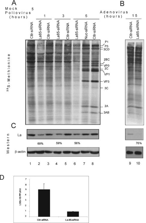
To examine the requirement of La for poliovirus translation in vivo, we used the RNA interference (RNAi) method (16, 17, 76). La-85-siRNA (named according to its position in the human La cDNA) was selected for use (from three different siRNA sequences), as it elicited the strongest level of La knockdown (see below). To examine the effect of La knockdown on poliovirus translation, cells were transfected with siRNA and then infected with type 1 poliovirus (Mahoney strain) at a MOI of 5. Cells were pulse-labeled with [35S]methionine at various times postinfection, and extracts were analyzed by SDS-15% PAGE. Infection with poliovirus resulted in a reduction of host cell protein synthesis (Fig. 1A) as expected (5, 75). Poliovirus proteins were conspicuous 5 h postinfection in cells transfected with a nonspecific siRNA (lane 8), or siRNA against nucleolin (lane 7). In sharp contrast, poliovirus proteins were barely detected at 5 h postinfection in cells that had been treated before with siRNA against La autoantigen (compare lane 6 to lanes 7 and 8). To further assess the specificity of the effect of the La knockdown on poliovirus translation, HeLa cells were infected with adenovirus, whose mRNAs are translated via a cap-dependent mechanism (reviewed in reference 12), which is not known to require La. La-85-siRNA failed to inhibit translation of adenovirus proteins (Fig. 1B, compare lanes 9 and 10), indicating that the La requirement is specific for poliovirus translation. The reduction of La protein by siRNA treatment was specific inasmuch as treatment of HeLa cells with La-85-siRNA (Fig. 1C, lanes 2, 4, and 6), but not with a nonspecific siRNA against the inverted sequence of 4E-T (15) (lanes 1, 3, 5, and 8) or an siRNA directed against nucleolin (lane 7), resulted in a reduction of endogenous La protein (>50%). Moreover, transfection with siRNA had no effect on the intracellular distribution of La protein (data not shown).
FIG. 1.
La knockdown diminishes translation and replication of poliovirus. Transfection of siRNA in HeLa cells (CLL2) was performed in 24-well plates. Twenty-four hours after transfection, cells were infected with poliovirus (A) or adenovirus (B) or mock infected, and protein synthesis was examined by pulse-labeling at various times as described in Materials and Methods. Cells were transfected with a nonspecific siRNA (control siRNA [Ctlr-siRNA]), La85-siRNA, or siRNA against nucleolin (Nuc-siRNA). (C) Western blot analysis of La protein. (D) Poliovirus yield is affected by La knockdown. A plaque assay was performed on a confluent monolayer of HeLa cells using serial dilutions of samples obtained from the lysates shown in panel A (5 h postinfection.).
To examine the effect of La knockdown on poliovirus yield, a plaque assay was performed 5 h postinfection. La-85-siRNA treatment resulted in a fivefold reduction in virus yield (Fig. 1D). Taken together, these data demonstrate that the La protein is required for efficient poliovirus infection in vivo.
La85-siRNA inhibits poliovirus IRES-driven protein synthesis.
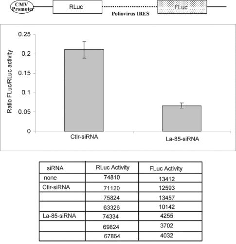
To determine whether the effect of La siRNA on viral protein synthesis in vivo is a consequence of inhibition of translation, the effect of La85-siRNA on in vivo expression of a bicistronic reporter mRNA, RLuc-PolioIRES-FLuc (56), was examined. The bicistronic mRNA contains the poliovirus IRES inserted between the RLuc and FLuc open reading frames (ORFs) (Fig. 2). Thus, translation of RLuc is cap dependent, whereas translation of the second cistron (FLuc) is IRES dependent. Cotransfection of HeLa cells with pcDNA3-RLuc-PolioIRES-FLuc and La-siRNA significantly reduced the FLuc/RLuc ratio (more than threefold) (Fig. 2). In striking contrast, the nonspecific siRNA (Ctlr-siRNA) failed to inhibit FLuc synthesis, indicating a specific reduction of IRES-dependent translation by La85-siRNA. Importantly, RLuc synthesis, which is cap dependent, did not vary significantly among cells transfected with La85-siRNA, cells transfected with a nonspecific siRNA (Ctlr-siRNA), or mock-transfected cells (Fig. 2), indicating that the decrease in IRES-dependent translation does not reflect a general decrease in translation. Taken together, these results bolster the conclusion that La protein enhances IRES activity in vivo.
FIG. 2.
IRES-driven translation of poliovirus RNA is inhibited by La85-siRNA. A schematic diagram of the bicistronic construct pcDNA3-RLuc-PolioIRES-FLuc is shown at the top of the figure (CMV, cytomegalovirus). The bicistronic construct pcDNA3-RLuc-PolioIRES-FLuc was cotransfected into HeLa cells with either La85-siRNA or control siRNA (Ctlr-siRNA). Twenty hours after transfection, cells were analyzed for FLuc and RLuc activities. The ratios of RLuc/FLuc activity are presented by the bars in the histogram. Absolute levels of RLuc and FLuc activities (in relative light units) are presented below the histogram.
A LaDN, La(226-348), inhibits poliovirus IRES activity in vitro.
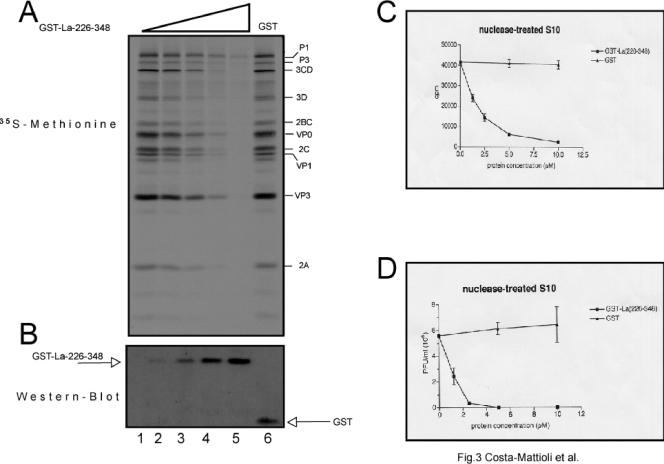
To provide further evidence for the importance of La in poliovirus translation in vivo, a LaDN was used. This mutant, La(226-348), consists of the C-terminal fragment of La protein (amino acids 226 to 348) (11), which was previously shown to inhibit La-stimulated translation of several viral and cellular mRNAs (11, 30, 69). First, we wished to examine whether the mutant form inhibits translation of poliovirus in a HeLa cell extract, as it was tested previously only in a reticulocyte lysate (11). The HeLa cell extract contains much larger amounts of La than the reticulocyte lysate does (48). In agreement with previous data (50), translation of poliovirus RNA in a HeLa cell extract yielded all the known authentic virus proteins (Fig. 3A, lane 1). When increasing amounts of glutathione S-transferase-tagged La(226-348) [GST-La(226-348)] (see Fig. 3B for expression) were added to the HeLa cell extract, a dramatic dose-dependent inhibition of poliovirus translation was evident (Fig. 3A and C, compare lane 1 to lane 5, >90% inhibition at the highest concentration). La(226-348) neither affected the spectrum of virus-specific proteins nor caused the appearance of any aberrant translation products (Fig. 3A). Craig et al. reported previously that in reticulocyte lysate, GST-La(226-348) preferentially inhibited the appearance of P1, compared to the synthesis of the products resulting from initiation at the “spurious” initiation sites (11). In contrast to GST-La(226-348), GST alone failed to inhibit poliovirus translation even at a very high concentration (10 μM; compare lanes 1 to 6). Consistent with its inhibition of translation, La(226-348) dramatically reduced virus yield. Indeed, La(226-348) at a concentration of 5 μM essentially abolished plaque formation (Fig. 3D). It is noteworthy that at this concentration of La(226-348), some residual virus translation (∼15%) was still detected (Fig. 3A, lane 5). Thus, the magnitude of inhibition at the level of protein synthesis is dramatically amplified at the downstream step of replication. The specific effect of La(226-348) was confirmed in an in vitro translation extract from HeLa cells using the bicistronic construct RLuc-poliovirus-Fluc mRNA in which only poliovirus IRES-driven translation was reduced (data not shown). Taken together, our data show that inactivation of La renders HeLa cell extract defective in supporting poliovirus translation and in turn virus replication.
FIG. 3.
Effects of LaDN on translation of poliovirus RNA and infectious virus synthesis in HeLa S10 cell extracts. (A) Effects of LaDN on poliovirus RNA translation in vitro. Translation reactions were performed in a total volume of 30 μl and contained HeLa S10 cell extract, poliovirus RNA (0.45 μg), [35S]methionine, and other components as described in Materials and Methods and previously (67). La(226-348) was added at the following final concentrations: 1.25 μM (lane 2), 2.5 μM (lane 3), 5 μM (lane 4), and 10 μM (lane 5). In lanes 1 and 6, reaction mixtures were supplemented with control buffer and GST (10 μM), respectively. Reaction mixtures were incubated at 34°C for 4 h, and the reactions were stopped by the addition of Laemmli sample buffer. Proteins were separated by SDS-15% PAGE, blotted onto a nitrocellulose membrane, and analyzed by autoradiography. The positions of the major virus proteins are indicated to the right of the gel. (B) The membrane from panel A was probed with antibodies against GST. The positions of GST-La(226-348) and GST are indicated at the sides of the gel. (C) TCA-insoluble radioactivity assay of 1-μl aliquots of the reaction mixtures from panel A. (D) Plaque assay for poliovirus infectivity. Reaction mixtures lacking [35S]methionine were programmed with poliovirus RNA for 16 h as described above for panel A. Virus titers were determined as described in Materials and Methods.
Poliovirus IRES-directed translation is impaired in cells transfected with La(226-348).
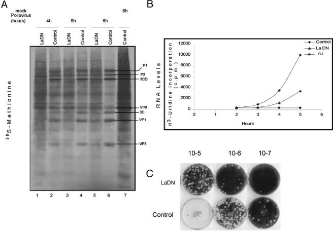
Next, the effect of La(226-348) on poliovirus IRES-mediated translation in cultured cells was examined. Cells were transfected with an expression plasmid encoding LaDN (pcDNA3-mycLa226-348) (30) and an empty vector as a control. Cells were subsequently infected with poliovirus (MOI of 20) and incubated for 30 min with [35S]methionine at different times after infection (Fig. 4A). In cells expressing LaDN, viral protein synthesis was inhibited, especially at early times after infection (compare lanes 1 and 2), compared to control, indicating that La(226-348) functions in a dominant-negative manner in vivo.
FIG. 4.
LaDN reduces poliovirus replication in vivo. (A) The kinetics of synthesis of poliovirus proteins was determined by pulse-labeling as described in Materials and Methods. Twenty-four hours after transfection with pcDNA3-myc-La226-348 (mutant) (LaDN) and pcDNA3-myc (control), cells were infected with poliovirus or mock infected and labeled with [35S]methionine. The positions of the major virus proteins are indicated to the right of the gel. (B) Kinetics of poliovirus RNA synthesis. At various times postinfection, control and LaDN-transfected cells were treated with 5 μg of actinomycin D per ml for 1 h and then labeled with [3H]uridine for 1 h. Noninfected cells (n.i) treated with actinomycin D are also shown. TCA-precipitated radioactivity was determined as described in Materials and Methods. (C) Plaque assays of lysates derived from LaDN- and control poliovirus-infected cells were performed as described in Materials and Methods.
We also wished to determine the effect of the LaDN on the processes downstream of translation, such as RNA synthesis and virus assembly. Cells were labeled with [3H]uridine for 1 h in the presence of actinomycin D. As expected, the synthesis of cellular RNA was inhibited in noninfected cells treated with actinomycin D (Fig. 4B). Viral RNA synthesis in the control cells was detected at 3 h postinfection compared with 4 h for cells expressing the LaDN mutant. Levels of viral RNA synthesis in cells expressing La(226-348) reached 33% of wild-type levels after 5 h (Fig. 4B). Thus, RNA synthesis is retarded by La(226-348), consistent with an inhibition of translation by the LaDN mutant. Moreover, La(226-348) caused a nearly 10-fold-lower yield of virus compared to the control (Fig. 4C). Taken together, the decrease in [3H]uridine and [35S]methionine incorporation indicate that the expression of La(226-348) affects translation and subsequently RNA replication and virus assembly.
HCV IRES-driven translation is impaired in La85-siRNA-treated cells.
Since HCV does not grow in cultured cells (3), we used a poliovirus-HCV(IRES-core) chimeric model (44, 77) to assess the requirement of La for HCV IRES activity in vivo. The poliovirus-HCV chimera (P/H 701-2A) contains the 5′ cloverleaf structure of poliovirus, followed by the HCV IRES (nucleotides 9 to 332), the first 369 nucleotides of the HCV core region, the entire poliovirus ORF, 3′UTR, and the poly(A) tail (77). In HeLa cells that were depleted of La by siRNA, the P/H 701-2A chimera grew to a titer that was approximately sevenfold lower than that for control cells (see Fig. S1C and D in the supplemental material). In addition, LaDN drastically inhibited translation of the poliovirus-HCV chimera mRNA in HeLa cell extracts (data not shown).
La protein promotes the initiation of formation of 48S and 80S ribosome complexes on poliovirus and HCV IRESs.
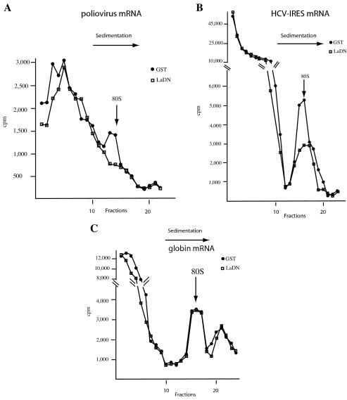
To further characterize La function in translation initiation of poliovirus mRNA, 80S ribosome binding assays were performed with poliovirus mRNA, and the effect of the La(226-348) dominant-negative mutant was examined. Initiation of formation of poliovirus 80S complexes, albeit inefficient, in agreement with earlier data (22), was inhibited by the addition of the La(226-348) dominant-negative mutant (Fig. 5A) (the position of the 80S ribosome initiation complex was determined by sedimenting a pure preparation of 80S ribosomes in a parallel tube). Thus, La plays a critical role in the initiation of formation of 80S ribosome complexes. Next, to determine the importance of La in the initiation of formation of 80S ribosome complexes on HCV IRES, the effect of the LaDN mutant on ribosome binding was examined. The addition of the GST-La(226-348) mutant resulted in a significant decrease (∼60%, three replicate samples) in viral RNA binding to ribosomes (Fig. 5B). In sharp contrast, binding of globin mRNA to ribosomes, which is not known to be dependent on La, was not affected by the LaDN mutant (Fig. 5C) (the second sedimenting complex [fractions 18 to 25] in Fig. 5C most likely represents disomes [24], but this complex has not been characterized further). To rule out the possibility that the ribosome binding inhibition seen by GST-LaDN is due to the GST tag, we cleaved the tag from the protein, and after purification, the untagged LaDN was tested for inhibiting the initiation of formation of 80S ribosome complexes. Untagged LaDN also inhibits HCV 80S ribosome binding in a dose-dependent manner (see Fig. S2 in the supplemental material). Thus, La protein is a specific ITAF for poliovirus and HCV IRESs but is not required for globin mRNA translation.
FIG. 5.
LaDN inhibits the initiation of formation of 80S ribosome complexes. RNA-ribosome binding assay was performed in a cycloheximide (0.6 mM)-treated HeLa cell extract (40 μl) with poliovirus (∼105 cpm) (A), HCV IRES-poliovirus chimera (∼106 cpm) (B), and rabbit globin (∼105 cpm) (C) 3′-end-labeled mRNAs. The extract was incubated with GST-La226-348 (4 μg) or GST (4 μg) at 34°C for 15 min and diluted with ice-cold high-salt buffer (66). Ribosome complexes were analyzed as described in Materials and Methods.
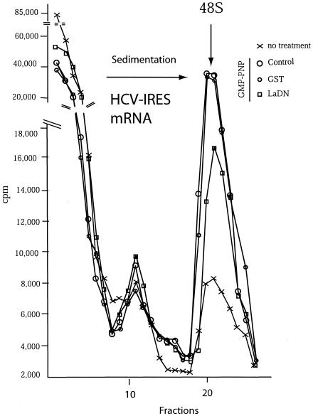
To determine whether inhibition of 80S initiation complex formation was a consequence of reduced 48S ribosome initiation complex formation, we performed ribosome binding studies on HCV mRNA in the presence of GMP-PNP. The GTP nonhydrolyzable analogue GMP-PNP competes with GTP for incorporation into the ternary complex (eIF2-GTP-tRNAi), thus inhibiting the release of eIF2 from the 40S subunit, and consequently preventing 60S ribosomal subunit joining (28). The addition of GMP-PNP caused the accumulation of 48S ribosome preinitiation complexes (compare 48S ribosome peaks in the presence [control and GST] and absence [no treatment] of GMP-PNP in Fig. 6; the sucrose gradient was 10 to 30% and the centrifugation time was 180 min to improve the resolution of the 48S mRNA ribosome complex). Consistent with the results seen with 80S ribosome initiation complex assembly on HCV IRES mRNA, GST-La(226-348) decreased (by ∼45%, two replicate samples) the binding of the 40S ribosomes to the HCV IRES (Fig. 6). Thus, the LaDN mutant inhibits the initiation of formation of complexes by preventing the recruitment of the 40S ribosomal subunit. In contrast, GST had no effect on 48S ribosome complex formation (Fig. 6). Similar results were obtained with the 48S ribosome initiation complex for poliovirus mRNA (data not shown). These results indicate that the La protein is directly involved in the initiation of formation of 48S ribosome complexes on poliovirus and HCV mRNAs.
FIG. 6.
LaDN inhibits the initiation of formation of 48S ribosome complexes. HCV IRES-labeled mRNA (∼106 cpm) was incubated for 10 min with a HeLa S10 cell extract and 4 μg of GST-La(226-348). GMP-PNP was included in the reaction mixtures where indicated. Samples were analyzed by sucrose gradient centrifugation (see Materials and Methods). Radioactivity was determined by scintillation counting.
DISCUSSION
In this study, a combination of genetic and biochemical methods consisting of RNAi and a LaDN mutant were used to demonstrate that La is required for IRES-dependent translation of poliovirus in vivo (Fig. 1, 2, and 4) and in vitro (Fig. 3). Adenovirus translation, which is not dependent on IRES function, was not affected by RNAi-mediated depletion of La (Fig. 1B). These results validate and extend earlier conclusions that the La protein binds to an internal region in the 5′UTR (47, 65) that is functionally important for poliovirus translation (46, 51, 53, 55). The La protein also interacts with both the 5′UTR and 3′UTR of HCV RNA (63) and was reported to stimulate HCV IRES-mediated translation (2, 13). In agreement with these findings, cells depleted of La by siRNA exhibited a decrease in virus yield of greater than sevenfold compared to control cells when infected with a poliovirus chimeric cDNA in which the poliovirus IRES is replaced by the HCV IRES (44, 77).
The physiological significance of earlier results demonstrating the importance of La in vitro was questioned because of the large amounts of La used in the earlier studies (35-37, 72). There are several possible explanations for this unusual requirement. First, the recombinant protein may have been rendered partially inactive during the purification process. Second, the effector domain (C-terminal region) of La is known to undergo phosphorylation (18), and this modification (which does not occur in the recombinant protein) could be important for optimal activity of the protein. Indeed, different RNAs are preferentially associated with the phosphorylated form of La compared to the nonphosphorylated form, and a fraction of the phosphorylated form of La is cytoplasmic (33). Finally, poliovirus causes the cleavage of La (62), which is then redistributed to the cytoplasm (34, 48), raising the possibility that cleaved La is more potent than wild-type La in translation.
What is the molecular mechanism by which La stimulates 40S ribosome recruitment? Our data demonstrate that a LaDN mutant protein inhibits 80S (Fig. 5) and 48S initiation complex formation (Fig. 6) on HCV and poliovirus mRNAs. The use of a highly fractionated system in combination with a toe-printing assay would allow further characterization of molecular mechanisms underlying this inhibition. Originally, it was suggested that La functions as an RNA chaperone to fold the IRES into a structure that can recruit initiation factors, such as eIF4G (6, 49, 65). Indeed, studies in yeast show that the yeast homologue of La protein (LHP1) functions as a molecular chaperone as it promotes the refolding of a mutant Met-tRNA (74). Alternatively, it is also possible that La stimulates translation indirectly by displacing an inhibitory protein from the IRES. In addition, La might aid in the recruitment of ribosomes in a more direct fashion, since human La protein has been reported to sediment with the 40S ribosomal subunit and to bind the 18S rRNA (52).
In summary, we determined that La functions to recruit the 40S ribosome subunit to the viral mRNA. In addition, we provided direct evidence based on in vivo studies that La protein is a bona fide ITAF for poliovirus and HCV. These results have important implications for studies on inhibition of virus replication in cells. Inhibition of poliovirus and HCV replication has been reported for siRNAs directly targeting poliovirus (21) or HCV RNA (40, 59, 71, 73). However, viruses are likely to evade siRNA by adaptive mutations of the target sequences (20). To circumvent this problem, it would be important to design siRNAs against cellular factors, such as La protein, which are required for optimal virus replication. We expect that the knowledge gained from this study will aid in the understanding of how cellular proteins may regulate IRES activity and might also be important in the design of new IRES-targeted antiviral strategies.
ADDENDUM IN PROOF
Pudi and colleagues recently reported that the La protein facilitates ribosomal assembly on the HCV RNA to influence IRES-mediated translation (R. Pudi, P. Srinivasan, and D. Das, J. Biol. Chem., 10 May 2004, posting date).
Supplementary Material
Acknowledgments
We thank Jerry Pelletier and Andrea Brueschke for critical reading of the manuscript, Karen Meerovitch for helpful discussions, Martin Holcik for providing the pcDNA-3-myc-La226-348 mutant, F. Lahser and E. Wimmer for the HCV-poliovirus chimera, and Colin Lister and Sandra Perrault for excellent assistance.
This work was supported by a grant from the Canadian Institute of Health Research (CIHR) to N.S., who is recipient of a CIHR Distinguished Scientific Award and a Howard Hughes Medical Institute International Scholarship. M.C.-M is a postdoctoral fellow supported by a CIHR fellowship.
Footnotes
Supplemental material for this article may be found at http://mcb.asm.org/.
REFERENCES
- 1.Ali, N., G. J. Pruijn, D. J. Kenan, J. D. Keene, and A. Siddiqui. 2000. Human La antigen is required for the hepatitis C virus internal ribosome entry site-mediated translation. J. Biol. Chem. 275:27531-27540. [DOI] [PubMed] [Google Scholar]
- 2.Ali, N., and A. Siddiqui. 1997. The La antigen binds 5′ noncoding region of the hepatitis C virus RNA in the context of the initiator AUG codon and stimulates internal ribosome entry site-mediated translation. Proc. Natl. Acad. Sci. USA 94:2249-2254. [DOI] [PMC free article] [PubMed] [Google Scholar]
- 3.Bartenschlager, R. 2002. Hepatitis C virus replicons: potential role for drug development. Nat. Rev. Drug Discov. 1:911-916. [DOI] [PubMed] [Google Scholar]
- 4.Barton, D. J., E. P. Black, and J. B. Flanegan. 1995. Complete replication of poliovirus in vitro: preinitiation RNA replication complexes require soluble cellular factors for the synthesis of VPg-linked RNA. J. Virol. 69:5516-5527. [DOI] [PMC free article] [PubMed] [Google Scholar]
- 5.Belsham, G. J., and R. J. Jackson. 2000. Translation initiation on picornavirus RNA, p. 869-900. In N. Sonenberg, J. W. B. Hershey, and W. C. Merrick (ed.), Translational control of gene expression. Cold Spring Harbor Laboratory Press, Cold Spring Harbor, N.Y.
- 6.Belsham, G. J., and N. Sonenberg. 2000. Picornavirus RNA translation: roles for cellular proteins. Trends Microbiol. 8:330-335. (Erratum, 8:472.) [DOI] [PubMed] [Google Scholar]
- 7.Bonnal, S., C. Boutonnet, L. Prado-Lourenco, and S. Vagner. 2003. IRESdb: the Internal Ribosome Entry Site database. Nucleic Acids Res. 31:427-428. [DOI] [PMC free article] [PubMed] [Google Scholar]
- 8.Brown, B. A., and E. Ehrenfeld. 1979. Translation of poliovirus RNA in vitro: changes in cleavage pattern and initiation sites by ribosomal salt wash. Virology 97:396-405. [DOI] [PubMed] [Google Scholar]
- 9.Carter, M. S., K. M. Kuhn, and P. Sarnow. 2000. Cellular Internal Ribosome Entry Site elements and the use of cDNA microarrays in their investigation, p. 615-636. In N. Sonenberg, J. W. B. Hershey, and W. C. Merrick (ed.), Translational control of gene expression. Cold Spring Harbor Laboratory Press, Cold Spring Harbor, N.Y.
- 10.Cornelis, S., Y. Bruynooghe, G. Denecker, S. Van Huffel, S. Tinton, and R. Beyaert. 2000. Identification and characterization of a novel cell cycle-regulated internal ribosome entry site. Mol. Cell 5:597-605. [DOI] [PubMed] [Google Scholar]
- 11.Craig, A. W., Y. V. Svitkin, H. S. Lee, G. J. Belsham, and N. Sonenberg. 1997. The La autoantigen contains a dimerization domain that is essential for enhancing translation. Mol. Cell. Biol. 17:163-169. [DOI] [PMC free article] [PubMed] [Google Scholar]
- 12.Cuesta, R., Q. Xi, and R. J. Schneider. 2001. Preferential translation of adenovirus mRNAs in infected cells. Cold Spring Harbor Symp. Quant. Biol. 66:259-267. [DOI] [PubMed] [Google Scholar]
- 13.Das, S., M. Ott, A. Yamane, W. Tsai, M. Gromeier, F. Lahser, S. Gupta, and A. Dasgupta. 1998. A small yeast RNA blocks hepatitis C virus internal ribosome entry site (HCV IRES)-mediated translation and inhibits replication of a chimeric poliovirus under translational control of the HCV IRES element. J. Virol. 72:5638-5647. [DOI] [PMC free article] [PubMed] [Google Scholar]
- 14.Dorner, A. J., B. L. Semler, R. J. Jackson, R. Hanecak, E. Duprey, and E. Wimmer. 1984. In vitro translation of poliovirus RNA: utilization of internal initiation sites in reticulocyte lysate. J. Virol. 50:507-514. [DOI] [PMC free article] [PubMed] [Google Scholar]
- 15.Dostie, J., M. Ferraiuolo, A. Pause, S. A. Adam, and N. Sonenberg. 2000. A novel shuttling protein, 4E-T, mediates the nuclear import of the mRNA 5′ cap-binding protein, eIF4E. EMBO J. 19:3142-3156. [DOI] [PMC free article] [PubMed] [Google Scholar]
- 16.Elbashir, S. M., J. Harborth, W. Lendeckel, A. Yalcin, K. Weber, and T. Tuschl. 2001. Duplexes of 21-nucleotide RNAs mediate RNA interference in cultured mammalian cells. Nature 411:494-498. [DOI] [PubMed] [Google Scholar]
- 17.Elbashir, S. M., W. Lendeckel, and T. Tuschl. 2001. RNA interference is mediated by 21- and 22-nucleotide RNAs. Genes Dev. 15:188-200. [DOI] [PMC free article] [PubMed] [Google Scholar]
- 18.Fan, H., A. L. Sakulich, J. L. Goodier, X. Zhang, J. Qin, and R. J. Maraia. 1997. Phosphorylation of the human La antigen on serine 366 can regulate recycling of RNA polymerase III transcription complexes. Cell 88:707-715. [DOI] [PubMed] [Google Scholar]
- 19.Gingras, A. C., B. Raught, and N. Sonenberg. 1999. eIF4 initiation factors: effectors of mRNA recruitment to ribosomes and regulators of translation. Annu. Rev. Biochem. 68:913-963. [DOI] [PubMed] [Google Scholar]
- 20.Gitlin, L., and R. Andino. 2003. Nucleic acid-based immune system: the antiviral potential of mammalian RNA silencing. J. Virol. 77:7159-7165. [DOI] [PMC free article] [PubMed] [Google Scholar]
- 21.Gitlin, L., S. Karelsky, and R. Andino. 2002. Short interfering RNA confers intracellular antiviral immunity in human cells. Nature 418:430-434. [DOI] [PubMed] [Google Scholar]
- 22.Golini, F., B. L. Semler, A. J. Dorner, and E. Wimmer. 1980. Protein-linked RNA of poliovirus is competent to form an initiation complex of translation in vitro. Nature 287:600-603. [DOI] [PubMed] [Google Scholar]
- 23.Gray, N. K., and M. W. Hentze. 1994. Iron regulatory protein prevents binding of the 43S translation pre-initiation complex to ferritin and eALAS mRNAs. EMBO J. 13:3882-3891. [DOI] [PMC free article] [PubMed] [Google Scholar]
- 24.Haghighat, A., S. Mader, A. Pause, and N. Sonenberg. 1995. Repression of cap-dependent translation by 4E-binding protein 1: competition with p220 for binding to eukaryotic initiation factor-4E. EMBO J. 14:5701-5709. [DOI] [PMC free article] [PubMed] [Google Scholar]
- 25.Hellen, C. U., and P. Sarnow. 2001. Internal ribosome entry sites in eukaryotic mRNA molecules. Genes Dev. 15:1593-1612. [DOI] [PubMed] [Google Scholar]
- 26.Henis-Korenblit, S., N. L. Strumpf, D. Goldstaub, and A. Kimchi. 2000. A novel form of DAP5 protein accumulates in apoptotic cells as a result of caspase cleavage and internal ribosome entry site-mediated translation. Mol. Cell. Biol. 20:496-506. [DOI] [PMC free article] [PubMed] [Google Scholar]
- 27.Hershey, J. W. B., and W. C. Merrick. 2000. Pathway and mechanism of initiation of protein synthesis, p. 33-88. In N. Sonenberg, J. W. B. Hershey, and W. C. Merrick (ed.), Translational control of gene expression. Cold Spring Harbor Laboratory Press, Cold Spring Harbor, N.Y.
- 28.Hinnebusch, A. G. 2000. Mechanism and regulation of initiator methionyl-tRNA binding to the ribosome, p. 185-244. In N. Sonenberg, J. W. B. Hershey, and W. C. Merrick (ed.), Translational control of gene expression. Cold Spring Harbor Laboratory Press, Cold Spring Harbor, N.Y.
- 29.Holcik, M., H. Gibson, and R. G. Korneluk. 2001. XIAP: apoptotic brake and promising therapeutic target. Apoptosis 6:253-261. [DOI] [PubMed] [Google Scholar]
- 30.Holcik, M., and R. G. Korneluk. 2000. Functional characterization of the X-linked inhibitor of apoptosis (XIAP) internal ribosome entry site element: role of La autoantigen in XIAP translation. Mol. Cell. Biol. 20:4648-4657. [DOI] [PMC free article] [PubMed] [Google Scholar]
- 31.Holcik, M., N. Sonenberg, and R. G. Korneluk. 2000. Internal ribosome initiation of translation and the control of cell death. Trends Genet. 16:469-473. [DOI] [PubMed] [Google Scholar]
- 32.Holcik, M., C. Yeh, R. G. Korneluk, and T. Chow. 2000. Translational upregulation of X-linked inhibitor of apoptosis (XIAP) increases resistance to radiation induced cell death. Oncogene 19:4174-4177. [DOI] [PubMed] [Google Scholar]
- 33.Intine, R. V., S. A. Tenenbaum, A. L. Sakulich, J. D. Keene, and R. J. Maraia. 2003. Differential phosphorylation and subcellular localization of La RNPs associated with precursor tRNA and translation-related mRNAs. Mol. Cell 12:1301-1307. [DOI] [PubMed] [Google Scholar]
- 34.Isoyama, T., N. Kamoshita, K. Yasui, A. Iwai, K. Shiroki, H. Toyoda, A. Yamada, Y. Takasaki, and A. Nomoto. 1999. Lower concentration of La protein required for internal ribosome entry on hepatitis C virus RNA than on poliovirus RNA. J. Gen. Virol. 80:2319-2327. [DOI] [PubMed] [Google Scholar]
- 35.Jackson, R. J. 1996. A comparative view of initiation site selection mechanisms, p. 71-112. In J. W. B. Hershey, M. B. Mathews, and N. Sonenberg (ed.), Translation control. Cold Spring Harbor Laboratory Press, Cold Spring Harbor, N.Y.
- 36.Jackson, R. J. 2002. Proteins involved in the function of picornavirus internal ribosomal entry sites, p. 171-186. In B. L. Semler and E. Wimmer (ed.), Molecular biology of picornaviruses. American Society for Microbiology, Washington, D.C.
- 37.Jackson, R. J., and A. Kaminski. 1995. Internal initiation of translation in eukaryotes: the picornavirus paradigm and beyond. RNA 1:985-1000. [PMC free article] [PubMed] [Google Scholar]
- 38.Jang, S. K., H. G. Krausslich, M. J. Nicklin, G. M. Duke, A. C. Palmenberg, and E. Wimmer. 1988. A segment of the 5′ nontranslated region of encephalomyocarditis virus RNA directs internal entry of ribosomes during in vitro translation. J. Virol. 62:2636-2643. [DOI] [PMC free article] [PubMed] [Google Scholar]
- 39.Jones, N., and T. Shenk. 1979. Isolation of adenovirus type 5 host range deletion mutants defective for transformation of rat embryo cells. Cell 17:683-689. [DOI] [PubMed] [Google Scholar]
- 40.Kapadia, S. B., A. Brideau-Andersen, and F. V. Chisari. 2003. Interference of hepatitis C virus RNA replication by short interfering RNAs. Proc. Natl. Acad. Sci. USA 100:2014-2018. [DOI] [PMC free article] [PubMed] [Google Scholar]
- 41.Khaleghpour, K., A. Kahvejian, G. De Crescenzo, G. Roy, Y. V. Svitkin, H. Imataka, M. O'Connor-McCourt, and N. Sonenberg. 2001. Dual interactions of the translational repressor Paip2 with poly(A) binding protein. Mol. Cell. Biol. 21:5200-5213. [DOI] [PMC free article] [PubMed] [Google Scholar]
- 42.Kim, Y. K., S. H. Back, J. Rho, S. H. Lee, and S. K. Jang. 2001. La autoantigen enhances translation of BiP mRNA. Nucleic Acids Res. 29:5009-5016. [DOI] [PMC free article] [PubMed] [Google Scholar]
- 43.Lang, K. J., A. Kappel, and G. J. Goodall. 2002. Hypoxia-inducible factor-1α mRNA contains an internal ribosome entry site that allows efficient translation during normoxia and hypoxia. Mol. Biol. Cell 13:1792-1801. [DOI] [PMC free article] [PubMed] [Google Scholar]
- 44.Lu, H. H., and E. Wimmer. 1996. Poliovirus chimeras replicating under the translational control of genetic elements of hepatitis C virus reveal unusual properties of the internal ribosomal entry site of hepatitis C virus. Proc. Natl. Acad. Sci. USA 93:1412-1417. [DOI] [PMC free article] [PubMed] [Google Scholar]
- 45.Marissen, W. E., and R. E. Lloyd. 1998. Eukaryotic translation initiation factor 4G is targeted for proteolytic cleavage by caspase 3 during inhibition of translation in apoptotic cells. Mol. Cell. Biol. 18:7565-7574. [DOI] [PMC free article] [PubMed] [Google Scholar]
- 46.Meerovitch, K., R. Nicholson, and N. Sonenberg. 1991. In vitro mutational analysis of cis-acting RNA translational elements within the poliovirus type 2 5′ untranslated region. J. Virol. 65:5895-5901. [DOI] [PMC free article] [PubMed] [Google Scholar]
- 47.Meerovitch, K., J. Pelletier, and N. Sonenberg. 1989. A cellular protein that binds to the 5′-noncoding region of poliovirus RNA: implications for internal translation initiation. Genes Dev. 3:1026-1034. [DOI] [PubMed] [Google Scholar]
- 48.Meerovitch, K., Y. V. Svitkin, H. S. Lee, F. Lejbkowicz, D. J. Kenan, E. K. Chan, V. I. Agol, J. D. Keene, and N. Sonenberg. 1993. La autoantigen enhances and corrects aberrant translation of poliovirus RNA in reticulocyte lysate. J. Virol. 67:3798-3807. [DOI] [PMC free article] [PubMed] [Google Scholar]
- 49.Meerovitch, K., and N. Sonenberg. 1993. Internal initiation of picornavirus RNA translation. Semin. Virol. 4:217-227. [Google Scholar]
- 50.Molla, A., A. V. Paul, and E. Wimmer. 1991. Cell-free, de novo synthesis of poliovirus. Science 254:1647-1651. [DOI] [PubMed] [Google Scholar]
- 51.Nicholson, R., J. Pelletier, S. Y. Le, and N. Sonenberg. 1991. Structural and functional analysis of the ribosome landing pad of poliovirus type 2: in vivo translation studies. J. Virol. 65:5886-5894. [DOI] [PMC free article] [PubMed] [Google Scholar]
- 52.Peek, R., G. J. Pruijn, and W. J. Van Venrooij. 1996. Interaction of the La (SS-B) autoantigen with small ribosomal subunits. Eur. J. Biochem. 236:649-655. [DOI] [PubMed] [Google Scholar]
- 53.Pelletier, J., M. E. Flynn, G. Kaplan, V. Racaniello, and N. Sonenberg. 1988. Mutational analysis of upstream AUG codons of poliovirus RNA. J. Virol. 62:4486-4492. [DOI] [PMC free article] [PubMed] [Google Scholar]
- 54.Pelletier, J., and N. Sonenberg. 1988. Internal initiation of translation of eukaryotic mRNA directed by a sequence derived from poliovirus RNA. Nature 334:320-325. [DOI] [PubMed] [Google Scholar]
- 55.Pilipenko, E. V., A. P. Gmyl, S. V. Maslova, Y. V. Svitkin, A. N. Sinyakov, and V. I. Agol. 1992. Prokaryotic-like cis elements in the cap-independent internal initiation of translation on picornavirus RNA. Cell 68:119-131. [DOI] [PubMed] [Google Scholar]
- 56.Poulin, F., A. C. Gingras, H. Olsen, S. Chevalier, and N. Sonenberg. 1998. 4E-BP3, a new member of the eukaryotic initiation factor 4E-binding protein family. J. Biol. Chem. 273:14002-14007. [DOI] [PubMed] [Google Scholar]
- 57.Pudi, R., S. Abhiman, N. Srinivasan, and S. Das. 2003. Hepatitis C virus internal ribosome entry site-mediated translation is stimulated by specific interaction of independent regions of human La autoantigen. J. Biol. Chem. 278:12231-12240. [DOI] [PubMed] [Google Scholar]
- 58.Pyronnet, S., L. Pradayrol, and N. Sonenberg. 2000. A cell cycle-dependent internal ribosome entry site. Mol. Cell 5:607-616. [DOI] [PubMed] [Google Scholar]
- 59.Randall, G., A. Grakoui, and C. M. Rice. 2003. Clearance of replicating hepatitis C virus replicon RNAs in cell culture by small interfering RNAs. Proc. Natl. Acad. Sci. USA 100:235-240. [DOI] [PMC free article] [PubMed] [Google Scholar]
- 60.Reynolds, J. E., A. Kaminski, H. J. Kettinen, K. Grace, B. E. Clarke, A. R. Carroll, D. J. Rowlands, and R. J. Jackson. 1995. Unique features of internal initiation of hepatitis C virus RNA translation. EMBO J. 14:6010-6020. [DOI] [PMC free article] [PubMed] [Google Scholar]
- 61.Rueckert, R. R., and M. A. Pallansch. 1981. Preparation and characterization of encephalomyocarditis (EMC) virus. Methods Enzymol. 78:315-325. [PubMed] [Google Scholar]
- 62.Shiroki, K., T. Isoyama, S. Kuge, T. Ishii, S. Ohmi, S. Hata, K. Suzuki, Y. Takasaki, and A. Nomoto. 1999. Intracellular redistribution of truncated La protein produced by poliovirus 3Cpro-mediated cleavage. J. Virol. 73:2193-2200. [DOI] [PMC free article] [PubMed] [Google Scholar]
- 63.Spangberg, K., L. Wiklund, and S. Schwartz. 2001. Binding of the La autoantigen to the hepatitis C virus 3′ untranslated region protects the RNA from rapid degradation in vitro. J. Gen. Virol. 82:113-120. [DOI] [PubMed] [Google Scholar]
- 64.Svitkin, Y. V., S. V. Maslova, and V. I. Agol. 1985. The genomes of attenuated and virulent poliovirus strains differ in their in vitro translation efficiencies. Virology 147:243-252. [DOI] [PubMed] [Google Scholar]
- 65.Svitkin, Y. V., K. Meerovitch, H. S. Lee, J. N. Dholakia, D. J. Kenan, V. I. Agol, and N. Sonenberg. 1994. Internal translation initiation on poliovirus RNA: further characterization of La function in poliovirus translation in vitro. J. Virol. 68:1544-1550. [DOI] [PMC free article] [PubMed] [Google Scholar]
- 66.Svitkin, Y. V., L. P. Ovchinnikov, G. Dreyfuss, and N. Sonenberg. 1996. General RNA binding proteins render translation cap dependent. EMBO J. 15:7147-7155. (Erratum, 16:896, 1997.) [PMC free article] [PubMed] [Google Scholar]
- 67.Svitkin, Y. V., and N. Sonenberg. 2003. Cell-free synthesis of encephalomyocarditis virus. J. Virol. 77:6551-6555. [DOI] [PMC free article] [PubMed] [Google Scholar]
- 68.Svitkin, Y. V., and N. Sonenberg. 2004. An efficient system for cap- and poly(A)-dependent translation in vitro. Methods Mol. Biol. 257:155-170. [DOI] [PubMed] [Google Scholar]
- 69.Trotta, R., T. Vignudelli, O. Candini, R. V. Intine, L. Pecorari, C. Guerzoni, G. Santilli, M. W. Byrom, S. Goldoni, L. P. Ford, M. A. Caligiuri, R. J. Maraia, D. Perrotti, and B. Calabretta. 2003. BCR/ABL activates mdm2 mRNA translation via the La antigen. Cancer Cell 3:145-160. [DOI] [PubMed] [Google Scholar]
- 70.Vagner, S., B. Galy, and S. Pyronnet. 2001. Irresistible IRES. Attracting the translation machinery to internal ribosome entry sites. EMBO Rep. 2:893-898. [DOI] [PMC free article] [PubMed] [Google Scholar]
- 71.Wilson, J. A., S. Jayasena, A. Khvorova, S. Sabatinos, I. G. Rodrigue-Gervais, S. Arya, F. Sarangi, M. Harris-Brandts, S. Beaulieu, and C. D. Richardson. 2003. RNA interference blocks gene expression and RNA synthesis from hepatitis C replicons propagated in human liver cells. Proc. Natl. Acad. Sci. USA 100:2783-2788. [DOI] [PMC free article] [PubMed] [Google Scholar]
- 72.Wolin, S. L., and T. Cedervall. 2002. The La protein. Annu. Rev. Biochem. 71:375-403. [DOI] [PubMed] [Google Scholar]
- 73.Yokota, T., N. Sakamoto, N. Enomoto, Y. Tanabe, M. Miyagishi, S. Maekawa, L. Yi, M. Kurosaki, K. Taira, M. Watanabe, and H. Mizusawa. 2003. Inhibition of intracellular hepatitis C virus replication by synthetic and vector-derived small interfering RNAs. EMBO Rep. 4:602-608. [DOI] [PMC free article] [PubMed] [Google Scholar]
- 74.Yoo, C. J., and S. L. Wolin. 1997. The yeast La protein is required for the 3′ endonucleolytic cleavage that matures tRNA precursors. Cell 89:393-402. [DOI] [PubMed] [Google Scholar]
- 75.Zamora, M., W. E. Marissen, and R. E. Lloyd. 2002. Poliovirus-mediated shut-off of host translation: an indirect effect, p. 313-320. In B. L. Semler and E. Wimmer (ed.), Molecular biology of picornaviruses. American Society for Microbiology, Washington, D.C.
- 76.Zamore, P. D., T. Tuschl, P. A. Sharp, and D. P. Bartel. 2000. RNAi: double-stranded RNA directs the ATP-dependent cleavage of mRNA at 21 to 23 nucleotide intervals. Cell 101:25-33. [DOI] [PubMed] [Google Scholar]
- 77.Zhao, W. D., E. Wimmer, and F. C. Lahser. 1999. Poliovirus/hepatitis C virus (internal ribosomal entry site-core) chimeric viruses: improved growth properties through modification of a proteolytic cleavage site and requirement for core RNA sequences but not for core-related polypeptides. J. Virol. 73:1546-1554. [DOI] [PMC free article] [PubMed] [Google Scholar]
Associated Data
This section collects any data citations, data availability statements, or supplementary materials included in this article.


