
Fig. 4.
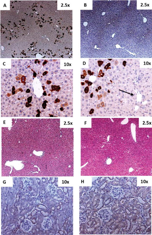
Heterogeneous distribution of PAH expression in mouse liver and kidney upon HTV injection of MCs. 77 μg or 53.7 pmol (3 3 × 10¹³ particles) of MC-DNA vector MC.PKU37 expressing an N-terminal Flag-tagged PAH was infused into the liver of 6 adult female PKU mice by way of hydrodynamic tail vein (HTV) injection. All treated mice were sacrificed on day 12. One PKU adult female PKU mouse was infused with saline only by way of HTV, and was sacrificed on day 12 as a negative control. Sections (A, B, C, and D) were stained with anti-Flag antibody followed by hematoxylin counterstain. Representative examples of a random field in 2.53× magnification in (A) a PKU mouse liver after delivery of MC.PKU37 demonstrating 16.7% ± 2.9* positively stained liver cells which were distributed heterogeneously, and (B) PKU mouse liver injected with saline only. PAH-expressing cells in a PKU mouse liver injected with MC.PKU37 were predominately expressed in the pericentral area (C,D), whereas hepatocytes around the portal triad (arrow in D) do not show any positive staining for PAH transgene expression. Liver sections were examined histologically by hematoxylin and eosin staining from (E) a PKU mouse liver infused with MC.PKU37, and (F) a negative control mouse liver. Kidney sections stained with anti-Flag antibody followed by hematoxylin counterstain were obtained from mice injected with (G) saline only or (H) MC.PKU37 (original magnification 10-fold). *The mean ± SD was calculated according to the percentage of the positively stained cells from five random fields in 2.5× magnification of each mouse.